

The DOI matches its Crossref record; 71 of the 71 references it cites were found in a registry or an index.
Sex-specific effects of microbiome perturbations on cerebral Aβ amyloidosis and microglia phenotypes
Open accessAbstract
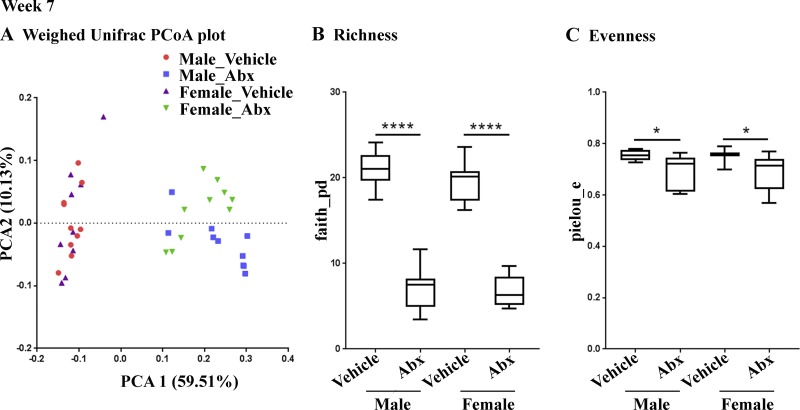
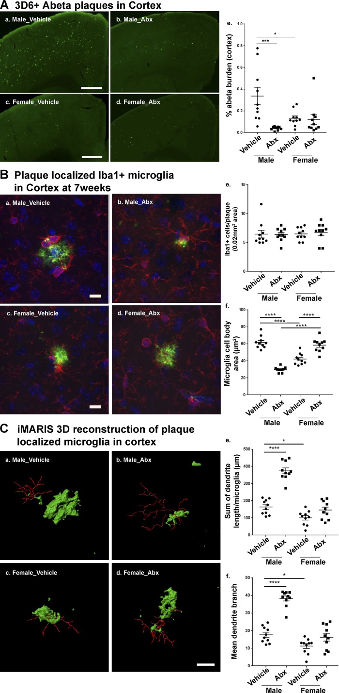
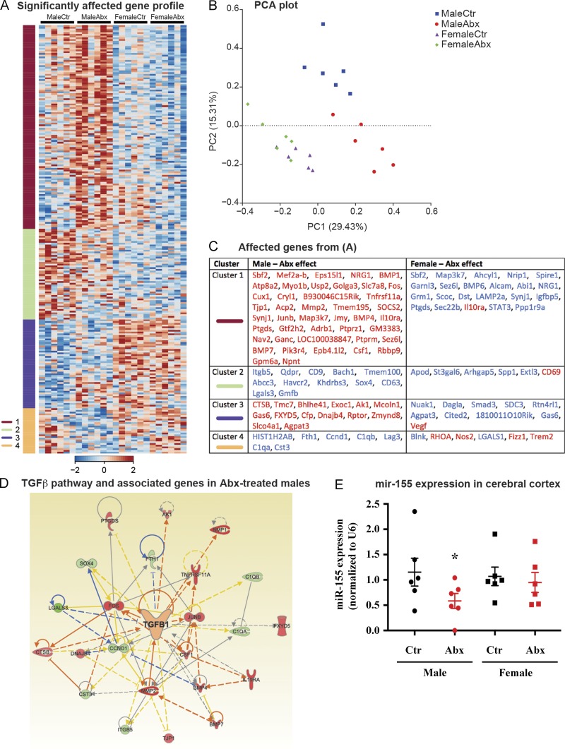
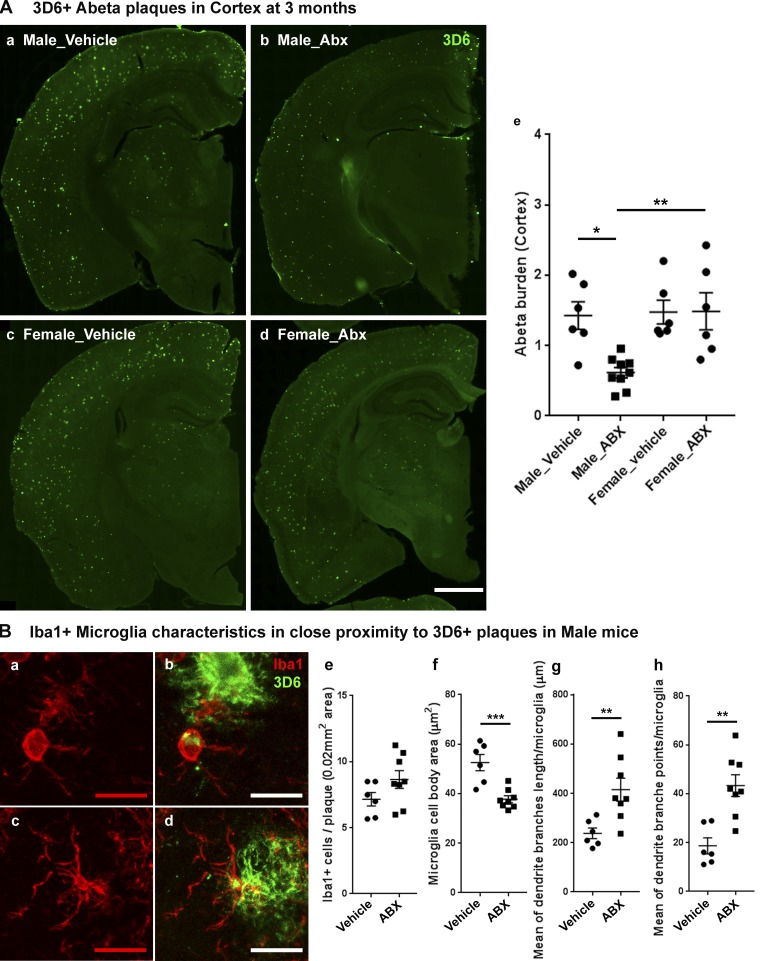
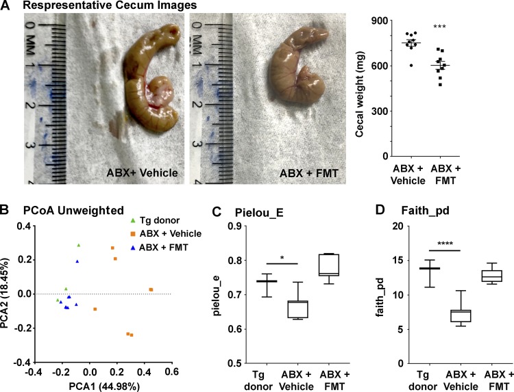
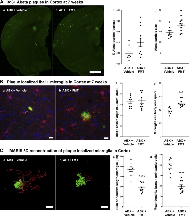
We demonstrated that an antibiotic cocktail (ABX)-perturbed gut microbiome is associated with reduced amyloid-β (Aβ) plaque pathology and astrogliosis in the male amyloid precursor protein (APP)SWE/presenilin 1 (PS1)ΔE9 transgenic model of Aβ amyloidosis. We now show that in an independent, aggressive APPSWE/PS1L166P (APPPS1-21) mouse model of Aβ amyloidosis, an ABX-perturbed gut microbiome is associated with a reduction in Aβ pathology and alterations in microglial morphology, thus establishing the generality of the phenomenon. Most importantly, these latter alterations occur only in brains of male mice, not in the brains of female mice. Furthermore, ABX treatment lead to alterations in levels of selected microglial expressed transcripts indicative of the “M0” homeostatic state in male but not in female mice. Finally, we found that transplants of fecal microbiota from age-matched APPPS1-21 male mice into ABX-treated APPPS1-21 male restores the gut microbiome and partially restores Aβ pathology and microglial morphology, thus demonstrating a causal role of the microbiome in the modulation of Aβ amyloidosis and microglial physiology in mouse models of Aβ amyloidosis.
Overview
The claims and statistics of this paper haven’t been checked yet.
Run the check — free, no account needed. It reads the open-access text and checks the sources it cites.
Integrity signals
Registry
Cites
Statistics
Figures
Verification
Claims the paper makes
The claims and statistics of this paper haven’t been checked yet.
Run the check — free, no account needed. It reads the open-access text and checks the sources it cites.
Claims other papers make about this one
Each is checked against this paper’s own text: does it say what the citing paper says it does?
The cited source doesn’t support the claim as written.
In the citing paper
“Moreover, sex-specific differences have been noted, with female AD mouse models showing more pronounced microbiota-driven amyloid deposition and microglial activation compared to males140.”
From Artificial intelligence-driven multi-omics approaches in Alzheimer's disease: Progress, challenges, and future directions (2025) · cites this paper as [140]
Checked by a reasoning model — may be wrong.
Experimental — accuracy not yet measuredThe cited source says what the claim attributes to it.
In the citing paper
“and transplants of fecal microbiota of genotype- and age-matched male mice partially restored Aβ pathology and microglial morphology9.”
From Integrated analysis of behavioral, epigenetic, and gut microbiome analyses in AppNL-G-F, AppNL-F, and wild type mice (2021) · cites this paper as [9]
The cited source says what the claim attributes to it.
In the citing paper
“These data are consistent with the association of microbiome perturbations induced by antibiotics with neuropathological measures in male mice overexpressing hAPP with the Swedish mutation and presenilin 1, and the ability of transplants of fecal microbiota of genotype- and age-matched male mice to partially restore neuropathology9.”
From Integrated analysis of behavioral, epigenetic, and gut microbiome analyses in AppNL-G-F, AppNL-F, and wild type mice (2021) · cites this paper as [9]
In the citing paper
“Importantly, the AD pathologies in antibiotic-treated male mice were partially restored after 3-week FMT from age-matched male APP/PS1-21 mice.197”
From Microbiota–gut–brain axis and its therapeutic applications in neurodegenerative diseases (2024) · cites this paper as [197]
Self-check
Percentages, sums and reported differences re-computed from the paper’s own numbers.
The self-check needs the paper’s full text.
Citations
Cites
DADA2: High–resolution sample inference from Illumina amplicon data
Callahan, B. J., McMurdie, P. J., Rosen, M. J., Han, A. W., Johnson, A. J. A., & Holmes, S. P. (2016) · Nature Methods · doi:10.1038/nmeth.3869 · Cited by 35,698
MAFFT: a novel method for rapid multiple sequence alignment based on fast Fourier transform
Katoh, K. (2002) · Nucleic Acids Research · doi:10.1093/nar/gkf436 · Cited by 17,027
FastTree 2 – Approximately Maximum-Likelihood Trees for Large Alignments
Price, M. N., Dehal, P. S., & Arkin, A. P. (2010) · PLoS ONE · doi:10.1371/journal.pone.0009490 · Cited by 14,894
Greengenes, a Chimera-Checked 16S rRNA Gene Database and Workbench Compatible with ARB
DeSantis, T. Z., Hugenholtz, P., Larsen, N., Rojas, M., Brodie, E. L., Keller, K., Huber, T., Dalevi, D., Hu, P., & Andersen, G. L. (2006) · Applied and Environmental Microbiology · doi:10.1128/aem.03006-05 · Cited by 10,891
Neuroinflammation in Alzheimer's disease
Heneka, M. T., Carson, M. J., Khoury, J. E., Landreth, G. E., Brosseron, F., Feinstein, D. L., Jacobs, A. H., Wyss-Coray, T., Vitorica, J., Ransohoff, R. M., et al. (2015) · The Lancet Neurology · doi:10.1016/s1474-4422(15)70016-5 · Cited by 6,226
Induction of Intestinal Th17 Cells by Segmented Filamentous Bacteria
Ivanov, I. I., Atarashi, K., Manel, N., Brodie, E. L., Shima, T., Karaoz, U., Wei, D., Goldfarb, K. C., Santee, C. A., Lynch, S. V., et al. (2009) · Cell · doi:10.1016/j.cell.2009.09.033 · Cited by 4,554
Gut microbiota regulate motor deficits and neuroinflammation in a model of Parkinson's disease
Sampson, T. R., Debelius, J. W., Thron, T., Janssen, S., Shastri, G. G., Ilhan, Z. E., Challis, C., Schretter, C. E., Rocha, S., Gradinaru, V., et al. (2016) · Cell · doi:10.1016/j.cell.2016.11.018 · Cited by 3,622
Host microbiota constantly control maturation and function of microglia in the CNS
Erny, D., Hrabě de Angelis, A. L., Jaitin, D., Wieghofer, P., Staszewski, O., David, E., Keren-Shaul, H., Mahlakoiv, T., Jakobshagen, K., Buch, T., et al. (2015) · Nature Neuroscience · doi:10.1038/nn.4030 · Cited by 3,569
Normal gut microbiota modulates brain development and behavior
Heijtz, R. D., Wang, S., Anuar, F., Qian, Y., Björkholm, B., Samuelsson, A., Hibberd, M. L., Forssberg, H., & Pettersson, S. (2011) · Proceedings of the National Academy of Sciences · doi:10.1073/pnas.1010529108 · Cited by 3,228
TREM2 Variants in Alzheimer's Disease
Guerreiro, R., Wojtas, A., Bras, J., Carrasquillo, M., Rogaeva, E., Majounie, E., Cruchaga, C., Sassi, C., Kauwe, J. S. K., Younkin, S., et al. (2013) · New England Journal of Medicine · doi:10.1056/nejmoa1211851 · Cited by 3,020
Cited by
Sorted by citations
The gut microbiome in neurological disorders
Cryan, J. F., O'Riordan, K. J., Sandhu, K., Peterson, V., & Dinan, T. G. (2020) · The Lancet Neurology · doi:10.1016/s1474-4422(19)30356-4 · Cited by 1,336
Wang, X., Sun, G., Feng, T., Zhang, J., Huang, X., Wang, T., Xie, Z., Chu, X., Yang, J., Wang, H., et al. (2019) · Cell Research · doi:10.1038/s41422-019-0216-x · Cited by 1,144
Microbiota–gut–brain axis and its therapeutic applications in neurodegenerative diseases
Loh, J. S., Mak, W. Q., Tan, L. K. S., Ng, C. X., Chan, H. H., Yeow, S. H., Foo, J. B., Ong, Y. S., How, C. W., & Khaw, K. Y. (2024) · Signal Transduction and Targeted Therapy · doi:10.1038/s41392-024-01743-1 · Cited by 1,193
A Comprehensive Review on the Role of the Gut Microbiome in Human Neurological Disorders
Sorboni, S. G., Moghaddam, H. S., Esfehani, R. J., & Soleimanpour, S. (2022) · Clinical Microbiology Reviews · doi:10.1128/cmr.00338-20 · Cited by 586
Chen, C., Liao, J., Xia, Y., Liu, X., Jones, R., Haran, J., McCormick, B., Sampson, T. R., Alam, A., & Ye, K. (2022) · Gut · doi:10.1136/gutjnl-2021-326269 · Cited by 491
Metadata
- Authors
- Dodiya, H. B., Kuntz, T., Shaik, S. M., Baufeld, C., Leibowitz, J., Zhang, X., Gottel, N., Zhang, X., Butovsky, O., Gilbert, J. A., & Sisodia, S. S.
- Affiliations
- University of Chicago (US)
- Brigham and Women's Hospital (US)
- Scripps Institution of Oceanography (US)
- Corresponding author
- Sisodia, Sangram S.
- ORCID
- Venue
- Journal of Experimental Medicine
- Publisher
- Rockefeller University Press
- Published
- 2019-05-16
- Volume
- 216
- Issue
- 7
- Pages
- 1542-1560
- ISSN
- 0022-1007 (ISSN-L)
- 1540-9538
- PMC
- PMC6605759
- OpenAlex
- W2945327298
- Cited by
- 288
- References
- 71
- Citations by year
- 2026: 19 · 2025: 50 · 2024: 54 · 2023: 45 · 2022: 40 · 2021: 39 · 2020: 33 · 2019: 7
- FWCI
- 20.40
- Type
- journal article
- Language
- en
- Open access
- hybrid
- doi.org
- Licence
- Locations
- The Journal of Experimental Medicine (publishedVersion, cc-by-nc-sa)
- PubMed Central (publishedVersion, cc-by-nc-sa)
- PubMed (publishedVersion)
- eScholarship (California Digital Library) (submittedVersion)
- eScholarship (California Digital Library) (submittedVersion, cc-by-nc-sa)
- Funders
- Cure Alzheimer’s Fund
- Open Philanthropy Project and Good Ventures Foundation
- Edward H. Levi Fund
- National Institute of Neurological Disorders and Stroke (1R01NS088137, R21NS104609, R21NS101673)
- National Institute on Aging (R01AG051812, R01AG054672)
- National Multiple Sclerosis Society
- Amyotrophic Lateral Sclerosis Association
- Topics
- Life Sciences › Neuroscience › Cellular and Molecular Neuroscience › Alzheimer's disease research and treatments
- Life Sciences › Neuroscience › Neurology › Neuroinflammation and Neurodegeneration Mechanisms
- Life Sciences › Biochemistry, Genetics and Molecular Biology › Molecular Biology › Gut microbiota and health
- Keywords
- gut microbiota, Alzheimer's disease, microglia, sex differences, antibiotic perturbation, fecal microbiota transplantation, astrocyte activation, neuroinflammation
- MeSH terms
- Gastrointestinal Microbiome, Fecal Microbiota Transplantation, Animals, Anti-Bacterial Agents, Brain Diseases, Disease Models, Animal, Female, Male, Sex Factors, Amyloid beta-Protein Precursor, Microglia, Amyloid Neuropathies, Mice, Plaque, Amyloid
About these checks
Registry, retraction, Cites and statistics results are computed, not judged, with one exception: a printed reference that no index lists is read once by a model before it counts as a citation error. Claims are extracted by a model and kept only when their quote appears verbatim in the text; each cited source is then checked, with a model’s help, for whether it says what the claim relies on. Those verdicts can be wrong — read the claim’s quote and the cited source before relying on one.
Statistics and arithmetic are recomputed, not judged.
Confidence bands are shown only for fields calibrated on our annotated test set. None is calibrated yet, so no band is shown.
What counts as a claim ›How we measure confidence ›How we check this ›
Are you an author, or do you see a problem with this page? Request a review
