

The DOI matches its Crossref record; 6 of the 6 references it cites were found in a registry or an index.
A loss-of-function IFNAR1 allele in Polynesia underlies severe viral diseases in homozygotes
Abstract
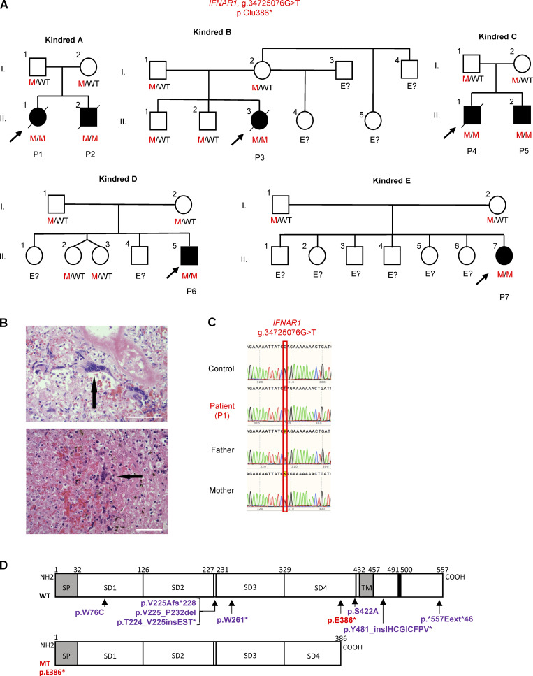
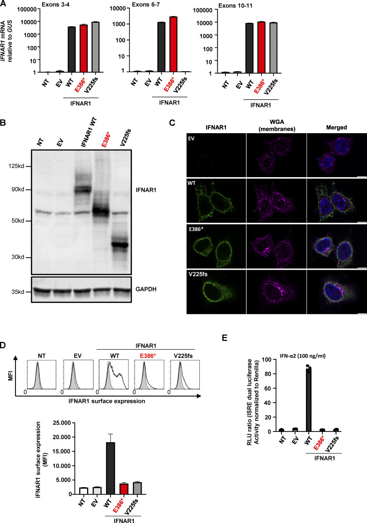
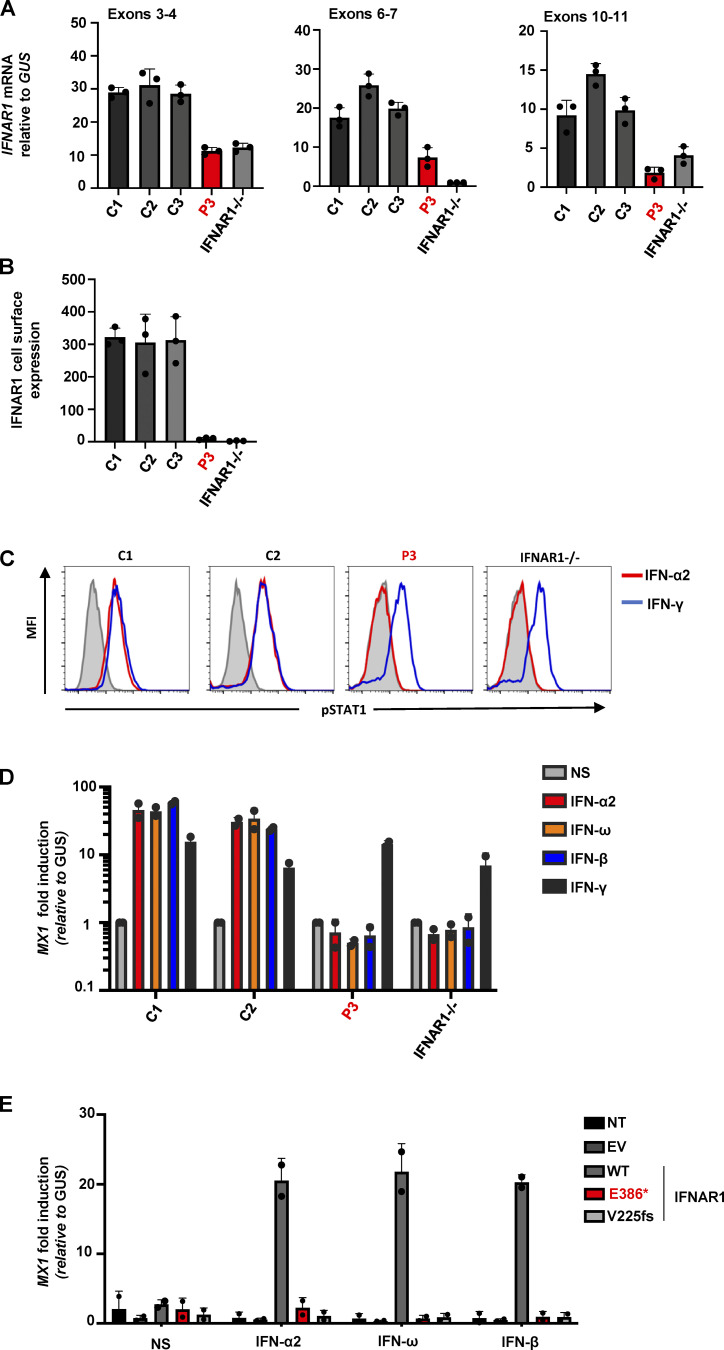
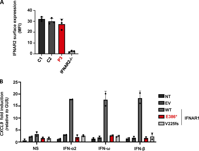
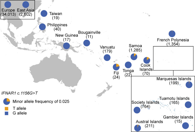
Globally, autosomal recessive IFNAR1 deficiency is a rare inborn error of immunity underlying susceptibility to live attenuated vaccine and wild-type viruses. We report seven children from five unrelated kindreds of western Polynesian ancestry who suffered from severe viral diseases. All the patients are homozygous for the same nonsense IFNAR1 variant (p.Glu386*). This allele encodes a truncated protein that is absent from the cell surface and is loss-of-function. The fibroblasts of the patients do not respond to type I IFNs (IFN-α2, IFN-ω, or IFN-β). Remarkably, this IFNAR1 variant has a minor allele frequency >1% in Samoa and is also observed in the Cook, Society, Marquesas, and Austral islands, as well as Fiji, whereas it is extremely rare or absent in the other populations tested, including those of the Pacific region. Inherited IFNAR1 deficiency should be considered in individuals of Polynesian ancestry with severe viral illnesses.
Overview
The claims and statistics of this paper haven’t been checked yet.
Run the check — free, no account needed. It reads the open-access text and checks the sources it cites.
Integrity signals
Registry
Cites
Statistics
Figures
Verification
Claims the paper makes
The claims and statistics of this paper haven’t been checked yet.
Run the check — free, no account needed. It reads the open-access text and checks the sources it cites.
Claims other papers make about this one
Each is checked against this paper’s own text: does it say what the citing paper says it does?
The cited source says what the claim attributes to it.
In the citing paper
“In particular, sequencing founder populations has already enabled the identification of certain Mendelian diseases (52, 53, 54).”
From Message in a bottleneck: Nested founder effects from French Polynesia to Rapa Nui and Hawaiʻi (2026) · cites this paper as [52]
Self-check
Percentages, sums and reported differences re-computed from the paper’s own numbers.
The self-check needs the paper’s full text.
Citations
Cites
Autoantibodies against type I IFNs in patients with life-threatening COVID-19
Bastard, P., Rosen, L. B., Zhang, Q., Michailidis, E., Hoffmann, H.-H., Zhang, Y., Dorgham, K., Philippot, Q., Rosain, J., Béziat, V., et al. (2020) · Science · doi:10.1126/science.abd4585 · Cited by 2,662
Self-citationInsights into human genetic variation and population history from 929 diverse genomes
Bergström, A., McCarthy, S. A., Hui, R., Almarri, M. A., Ayub, Q., Danecek, P., Chen, Y., Felkel, S., Hallast, P., Kamm, J., et al. (2020) · Science · doi:10.1126/science.aay5012 · Cited by 1,020
Deletion 17q12 is a recurrent copy number variant that confers high risk of autism and schizophrenia
Moreno-De-Luca, D., Mulle, J. G., Kaminsky, E. B., Sanders, S. J., Myers, S. M., Adam, M. P., Pakula, A. T., Eisenhauer, N. J., Uhas, K., Weik, L., et al. (2010) · The American Journal of Human Genetics · doi:10.1016/j.ajhg.2010.10.004 · Cited by 324
Paths and timings of the peopling of Polynesia inferred from genomic networks
Ioannidis, A. G., Blanco-Portillo, J., Sandoval, K., Hagelberg, E., Barberena-Jonas, C., Hill, A. V. S., Rodríguez-Rodríguez, J. E., Fox, K., Robson, K., Haoa-Cardinali, S., et al. (2021) · Nature · doi:10.1038/s41586-021-03902-8 · Cited by 124 · cited 5×
“However, the variant was present in the Austral Islands ( n = 211, AF = 0.00474), the Society Islands ( n = 764, AF = 0.000654), the Marquesas Islands ( n = 199, AF = 0.00251), the Cook Islands ( n = 70, AF = 0.0143), and Fiji ( n = 24, AF = 0.0208; Fig.”
BackgroundMethodInfluentialSelf-citationRethinking Polynesians origins: A West-Polynesia Triple-I Model
ADDISON, D. J., & MATISOO‐SMITH, E. (2010) · Archaeology in Oceania · doi:10.1002/j.1834-4453.2010.tb00072.x · Cited by 73
Research works printed but not linked: 50
- Abolhassani, 2022, Inherited IFNAR1 deficiency in a child with both critical COVID-19 pneumonia and multisystem inflammatory syndrome, J. Clin. Immunol., 1, 10.1007/s10875-022-01215-7 · DOI
- Amatuni, 2019, Newborn screening for severe combined immunodeficiency and T-cell lymphopenia in California, 2010-2017, Pediatrics, 143, 10.1542/peds.2018-2300 · DOI
- Bastard, 2021, Autoantibodies neutralizing type I IFNs are present in ∼4% of uninfected individuals over 70 years old and account for ∼20% of COVID-19 deaths, Sci. Immunol., 6, 10.1126/sciimmunol.abl4340 · DOI
- Bastard, 2021, Herpes simplex encephalitis in a patient with a distinctive form of inherited IFNAR1 deficiency, J. Clin. Invest., 131, 10.1172/jci139980 · DOI
- Bastard, 2021, Auto-antibodies to type I IFNs can underlie adverse reactions to yellow fever live attenuated vaccine, J. Exp. Med., 218, 10.1084/jem.20202486 · DOI
- Becroft, 1980, The lungs in fatal measles infection in childhood: Pathological, radiological and immunological correlations, Histopathology, 4, 401, 10.1111/j.1365-2559.1980.tb02935.x · DOI
- Biggs, 2017, Newborn screening for severe combined immunodeficiency: A primer for clinicians, CMAJ, 189, E1551, 10.1503/cmaj.170561 · DOI
- Bousfiha, 2020, Human inborn errors of immunity: 2019 update of the IUIS phenotypical classification, J. Clin. Immunol., 40, 66, 10.1007/s10875-020-00758-x · DOI
- Casanova, 2021, Mechanisms of viral inflammation and disease in humans, Science, 374, 1080, 10.1126/science.abj7965 · DOI
- Champredon, 2020, Curbing the 2019 Samoa measles outbreak, Lancet Infect. Dis., 20, 287, 10.1016/S1473-3099(20)30044-X · DOI
- Choin, 2021, Genomic insights into population history and biological adaptation in Oceania, Nature, 592, 583, 10.1038/s41586-021-03236-5 · DOI · Paper page
- Craig, 2020, Measles epidemic in Samoa and other Pacific islands, Lancet Infect. Dis., 20, 273, 10.1016/S1473-3099(20)30053-0 · DOI
- Davies, 1894, Epidemic measles at Samoa, Br. Med. J., 1, 1077, 10.1136/bmj.1.1742.1077 · DOI
- Duncan, 2015, Human IFNAR2 deficiency: Lessons for antiviral immunity, Sci. Transl Med., 7, 307ra154, 10.1126/scitranslmed.aac4227 · DOI
- Duncan, 2021, Genetic lesions of type I interferon signalling in human antiviral immunity, Trends Genet., 37, 46, 10.1016/j.tig.2020.08.017 · DOI
- Dupuis, 2003, Impaired response to interferon-alpha/beta and lethal viral disease in human STAT1 deficiency, Nat. Genet., 33, 388, 10.1038/ng1097 · DOI
- Eletto, 2016, Biallelic JAK1 mutations in immunodeficient patient with mycobacterial infection, Nat. Commun., 7, 13992, 10.1038/ncomms13992 · DOI
- Gothe, 2022, A novel case of homozygous interferon alpha/beta receptor alpha chain (IFNAR1) deficiency with hemophagocytic lymphohistiocytosis, Clin. Infect. Dis., 74, 136, 10.1093/cid/ciaa1790 · DOI
- Gothe, 2021, Monogenic susceptibility to live viral vaccines, Curr. Opin. Immunol., 72, 167, 10.1016/j.coi.2021.05.006 · DOI
- Griffith, 2020, Collider bias undermines our understanding of COVID-19 disease risk and severity, Nat. Commun., 11, 5749, 10.1038/s41467-020-19478-2 · DOI · Paper page
- Hambleton, 2013, STAT2 deficiency and susceptibility to viral illness in humans, Proc. Natl. Acad. Sci. USA, 110, 3053, 10.1073/pnas.1220098110 · DOI
- Harris, 2020, Evolutionary history of modern Samoans, Proc Natl. Acad. Sci. USA, 117, 9458, 10.1073/pnas.1913157117 · DOI · Paper page
- Hawley, 2014, Prevalence of adiposity and associated cardiometabolic risk factors in the Samoan genome-wide association study, Am. J. Hum. Biol., 26, 491, 10.1002/ajhb.22553 · DOI
- Hernandez, 2019, Inherited IFNAR1 deficiency in otherwise healthy patients with adverse reaction to measles and yellow fever live vaccines, J. Exp. Med., 216, 2057, 10.1084/jem.20182295 · DOI
- Hernandez, 2018, Life-threatening influenza pneumonitis in a child with inherited IRF9 deficiency, J. Exp. Med., 215, 2567, 10.1084/jem.20180628 · DOI
- Ioannidis, 2020, Native American gene flow into Polynesia predating Easter Island settlement, Nature, 583, 572, 10.1038/s41586-020-2487-2 · DOI · Paper page
- Karczewski, 2020, The mutational constraint spectrum quantified from variation in 141,456 humans, Nature, 581, 434, 10.1038/s41586-020-2308-7 · DOI · Paper page
- Khanmohammadi, 2022, A case of autosomal recessive interferon alpha/beta receptor alpha chain (IFNAR1) deficiency with severe COVID-19, J. Clin. Immunol., 42, 19, 10.1007/s10875-021-01166-5 · DOI
- Kreins, 2015, Human TYK2 deficiency: Mycobacterial and viral infections without hyper-IgE syndrome, J. Exp. Med., 212, 1641, 10.1084/jem.20140280 · DOI
- Kwan, 2014, Newborn screening for severe combined immunodeficiency in 11 screening programs in the United States, JAMA, 312, 729, 10.1001/jama.2014.9132 · DOI
- Laksono, 2016, Measles virus host invasion and pathogenesis, Viruses, 8, 210, 10.3390/v8080210 · DOI
- Loirat, 2010, Autism in three patients with cystic or hyperechogenic kidneys and chromosome 17q12 deletion, Nephrol. Dial. Transplant., 25, 3430, 10.1093/ndt/gfq380 · DOI
- Meyts, 2021, Viral infections in humans and mice with genetic deficiencies of the type I IFN response pathway, Eur. J. Immunol., 51, 1039, 10.1002/eji.202048793 · DOI
- Minegishi, 2006, Human tyrosine kinase 2 deficiency reveals its requisite roles in multiple cytokine signals involved in innate and acquired immunity, Immunity, 25, 745, 10.1016/j.immuni.2006.09.009 · DOI
- Minster, 2016, A thrifty variant in CREBRF strongly influences body mass index in Samoans, Nat. Genet., 48, 1049, 10.1038/ng.3620 · DOI
- Nagamani, 2010, Clinical spectrum associated with recurrent genomic rearrangements in chromosome 17q12, Eur. J. Hum. Genet., 18, 278, 10.1038/ejhg.2009.174 · DOI
- NHLBI Trans-Omics for Precision Medicine, 2021, TOPMed Whole Genome Sequencing Methods: Freeze 9
- Notarangelo, 2020, Human inborn errors of immunity: An expanding universe, Sci. Immunol., 5, 10.1126/sciimmunol.abb1662 · DOI
- Nozawa, 1994, An immunohistochemical study of Warthin-Finkeldey cells in measles, Pathol. Int., 44, 442, 10.1111/j.1440-1827.1994.tb01708.x · DOI
- Passarelli, 2020, IFNAR2 deficiency causing dysregulation of NK cell functions and presenting with hemophagocytic lymphohistiocytosis, Front. Genet., 11, 937, 10.3389/fgene.2020.00937 · DOI
- Petchey, 2001, Radiocarbon determinations from the Mulifanua Lapita site, Upolu, Western Samoa, Radiocarbon, 43, 63, 10.1017/s0033822200031635 · DOI
- Poyhonen, 2019, Life-threatening infections due to live-attenuated vaccines: Early manifestations of inborn errors of immunity, J. Clin. Immunol., 39, 376, 10.1007/s10875-019-00642-3 · DOI
- Quintana-Murci, 2019, Human immunology through the lens of evolutionary genetics, Cell, 177, 184, 10.1016/j.cell.2019.02.033 · DOI
- Quintana-Murci, 2007, Immunology in natura: Clinical, epidemiological and evolutionary genetics of infectious diseases, Nat. Immunol., 8, 1165, 10.1038/ni1535 · DOI
- Sirugo, 2019, The missing diversity in human genetic studies, Cell, 177, 1080, 10.1016/j.cell.2019.04.032 · DOI
- Staples, 2013, Utilizing graph theory to select the largest set of unrelated individuals for genetic analysis, Genet. Epidemiol., 37, 136, 10.1002/gepi.21684 · DOI
- Taliun, 2021, Sequencing of 53,831 diverse genomes from the NHLBI TOPMed Program, Nature, 590, 290, 10.1038/s41586-021-03205-y · DOI · Paper page
- Tangye, 2020, Human inborn errors of immunity: 2019 update on the Classification from the International Union of Immunological Societies Expert Committee, J. Clin. Immunol., 40, 24, 10.1007/s10875-019-00737-x · DOI
- Zhang, 2018, A proline deletion in IFNAR1 impairs IFN-signaling and underlies increased resistance to tuberculosis in humans, Nat. Commun., 9, 85, 10.1038/s41467-017-02611-z · DOI
- Zhang, 2020, Inborn errors of type I IFN immunity in patients with life-threatening COVID-19, Science, 370, 10.1126/science.abd4570 · DOI · Paper page
Cited by
Sorted by citations
Message in a bottleneck: Nested founder effects from French Polynesia to Rapa Nui and Hawaiʻi
Shanks, C., Huang, E. C., Thomassin, C., Emde, A.-K., Rodière, V., Moraga, M., Sandoval, K., Merriman, T., Pascart, T., Moreno-Estrada, A., et al. (2026) · Science · doi:10.1126/science.aec7662
Self-citation
Metadata
- Authors
- Bastard, P., Hsiao, K.-C., Zhang, Q., Choin, J., Best, E., Chen, J., Gervais, A., Bizien, L., Materna, M., Harmant, C., Roux, M., Hawley, N. L., Weeks, D. E., McGarvey, S. T., Sandoval, K., Barberena-Jonas, C., Quinto-Cortés, C. D., Hagelberg, E., Mentzer, A. J., Robson, K., Coulibaly, B., Seeleuthner, Y., Bigio, B., Li, Z., Uzé, G., Pellegrini, S., Lorenzo, L., Sbihi, Z., Latour, S., Besnard, M., Adam de Beaumais, T., Jacqz Aigrain, E., Béziat, V., Deka, R., Esera Tulifau, L., Viali, S., Reupena, M. S., Naseri, T., McNaughton, P., Sarkozy, V., Peake, J., Blincoe, A., Primhak, S., Stables, S., Gibson, K., Woon, S.-T., Drake, K. M., Hill, A. V. S., Chan, C.-Y., King, R., Ameratunga, R., Teiti, I., Aubry, M., Cao-Lormeau, V.-M., Tangye, S. G., Zhang, S.-Y., Jouanguy, E., Gray, P., Abel, L., Moreno-Estrada, A., Minster, R. L., Quintana-Murci, L., Wood, A. C., & Casanova, J.-L.
- Affiliations
- Laboratory of Human Genetics of Infectious Diseases, Necker Branch, INSERM U1163, Necker Hospital for Sick Children, Paris, France 1
- Starship Child Health, Auckland, New Zealand 5
- Institut Pasteur, Université de Paris, CNRS UMR2000, Human Evolutionary Genetics Unit, Paris, France 9
- St. Giles Laboratory of Human Genetics of Infectious Diseases, Rockefeller Branch, The Rockefeller University, New York, NY 2
- Department of Chronic Disease Epidemiology, Yale University School of Public Health, New Haven, CT 14
- Department of Human Genetics, School of Public Health, University of Pittsburgh, Pittsburgh, PA 16
- International Health Institute, Department of Epidemiology, School of Public Health, Brown University, Providence, RI 15
- National Laboratory of Genomics for Biodiversity (LANGEBIO) - UGA, CINVESTAV, Irapuato, Guanajuato, Mexico 19
- Department of Biology, University of Oslo, Oslo, Norway 20
- Wellcome Centre for Human Genetics, University of Oxford, Oxford, UK 21
- MRC Weatherall Institute of Molecular Medicine, University of Oxford, Oxford, UK 23
- Institute for Regenerative Medicine and Biotherapy, Université Montpellier, INSERM, CNRS, Montpellier, France 25
- Unit of Cytokine Signaling, Pasteur Institute, INSERM U1224, Paris, France 24
- Laboratory of Lymphocyte Activation and Susceptibility to EBV Infection, INSERM UMR 1163, Imagine Institute, Paris, France 26
- Paris Cité University, Imagine Institute, Paris, France 3
- Department of Neonatology, Centre Hospitalier de Polynésie Française, Papeete, French Polynesia 27
- Precision Cancer Medicine Team, Institut Gustave Roussy, Villejuif, France 28
- Paris Cité University, Paris, France 11
- Department of Environmental and Public Health Sciences, College of Medicine, University of Cincinnati, Cincinnati, OH 30
- Tupua Tamasese Meaole Hospital, Apia, Samoa 31
- School of Medicine, National University of Samoa, Apia, Samoa 32
- Lutia i Puava ae Mapu i Fagalele, Apia, Samoa 33
- Clinical Immunogenomics Research Consortium Australasia, Sydney, Australia 8
- Tumbatin Developmental Services, Sydney Children’s Hospital, Randwick, New South Wales, Australia 36
- Department of Forensic Pathology, Auckland City Hospital, Auckland, New Zealand 38
- Clinical Geneticist, South Island Hub, Genetic Health Service, Christchurch, New Zealand 39
- Department of Virology and Immunology, LabPLUS, Auckland City Hospital, Auckland, New Zealand 40
- Molecular Pathology, Canterbury Health Laboratories, Christchurch, New Zealand 42
- Chemical Pathology and Genetics, Canterbury Health Laboratories, Christchurch, New Zealand 44
- Laboratory of Research on Infectious Vector-borne Diseases, Institut Louis Malardé, Papeete, French Polynesia 46
- ORCID
- Bastard, Paul
- Hsiao, Kuang-Chih
- Zhang, Qian
- Choin, Jeremy
- Best, Emma
- Chen, Jie
- Gervais, Adrian
- Bizien, Lucy
- Materna, Marie
- Harmant, Christine
- Roux, Maguelonne
- Hawley, Nicola L.
- Weeks, Daniel E.
- McGarvey, Stephen T.
- Sandoval, Karla
- Barberena-Jonas, Carmina
- Quinto-Cortés, Consuelo D.
- Hagelberg, Erika
- Mentzer, Alexander J.
- Robson, Kathryn
- Coulibaly, Boubacar
- Seeleuthner, Yoann
- Bigio, Benedetta
- Li, Zhi
- Uzé, Gilles
- Pellegrini, Sandra
- Lorenzo, Lazaro
- Sbihi, Zineb
- Latour, Sylvain
- Besnard, Marianne
- Adam de Beaumais, Tiphaine
- Jacqz Aigrain, Evelyne
- Béziat, Vivien
- Deka, Ranjan
- Esera Tulifau, Litara
- Viali, Satupa‘itea
- Naseri, Take
- McNaughton, Peter
- Sarkozy, Vanessa
- Peake, Jane
- Blincoe, Annaliesse
- Primhak, Sarah
- Stables, Simon
- Gibson, Kate
- Woon, See-Tarn
- Drake, Kylie Marie
- Hill, Adrian V.S.
- Chan, Cheng-Yee
- King, Richard
- Ameratunga, Rohan
- Teiti, Iotefa
- Aubry, Maite
- Cao-Lormeau, Van-Mai
- Tangye, Stuart G.
- Zhang, Shen-Ying
- Jouanguy, Emmanuelle
- Gray, Paul
- Abel, Laurent
- Moreno-Estrada, Andrés
- Minster, Ryan L.
- Quintana-Murci, Lluis
- Wood, Andrew C.
- Casanova, Jean-Laurent
- Venue
- Journal of Experimental Medicine
- Publisher
- Rockefeller University Press
- Published
- 2022-04-20
- Volume
- 219
- Issue
- 6
- ISSN
- 0022-1007
- 1540-9538
- PMC
- PMC9026234
- Cited by
- 70
- References
- 6
- Language
- en
- Funders
- MATAEA (03557/MED/REC_29/05/2019)
- Institut Pasteur
- Collège de France
- Centre national de la recherche scientifique
- Fondation Allianz-Institut de France
- French Government’s Investissement d’Avenir program
- Milieu Intérieur (ANR-10-LABX-69-01)
- Fondation de France (00106080)
- Fondation pour la Recherche Médicale (Equipe FRM DEQ20180339214, EQU201903007798, EA20170638020)
- Howard Hughes Medical Institute
- Rockefeller University
- St. Giles Foundation
- National Institutes of Health (R01AI088364, R01AI163029, R01-HL093093, R01-HL133040)
- National Center for Advancing Translational Sciences
- NIH Clinical and Translational Science Award (UL1 TR001866)
- Emergent Ventures
- Mercatus Center at George Mason University
- Yale Center for Mendelian Genomics
- National Human Genome Research Institute (UM1HG006504, U24HG008956)
- Yale High-Performance Computing Center (S10OD018521)
- Fisher Center for Alzheimer’s Research Foundation
- Meyer Foundation
- French National Research Agency (ANR-10-IAHU-01)
- Integrative Biology of Emerging Infectious Diseases Laboratory of Excellence (ANR-10-LABX-62-IBEID)
- FRM and ANR GENCOVID project
- ANRS-COV05
- ANR GENVIR (ANR-20-CE93-003)
- ANR AABIFNCOV (ANR-20-CO11-0001)
- European Union’s Horizon 2020 research and innovation program (824110)
- Square Foundation
- Grandir - Fonds de solidarité pour l’enfance
- Fondation du Souffle
- SCOR Corporate Foundation for Science
- Institut National de la Santé et de la Recherche Médicale
- French Ministry of Higher Education, Research, and Innovation
- University of Paris
- Imagine Institute
- Fondation Bettencourt-Schueller
- Consejo Nacional de Ciencia y Technologia (FONCICYT/50/2016)
- Medical Research Council (MR/N028937/1)
- International Center for Genetic Engineering and Biotechnology (CRP/MEX20-01)
- National Health and Medical Research Council of Australia (1176665)
- John Brown Cook Foundation
- National Heart, Lung and Blood Institute
- Northwest Genomics Center (HHSN268201100037C)
- New York Genome Center (HHSN268201500016C)
- TOPMed Informatics Research Center (3R01-HL117626-02S1, HHSN268201800002I)
- TOPMed Data Coordinating Center (01-HL120393, U01-HL120393, HHSN268201800001I)
- Jeffrey Modell Foundation
About these checks
Registry, retraction, Cites and statistics results are computed, not judged, with one exception: a printed reference that no index lists is read once by a model before it counts as a citation error. Claims are extracted by a model and kept only when their quote appears verbatim in the text; each cited source is then checked, with a model’s help, for whether it says what the claim relies on. Those verdicts can be wrong — read the claim’s quote and the cited source before relying on one.
Statistics and arithmetic are recomputed, not judged.
Confidence bands are shown only for fields calibrated on our annotated test set. None is calibrated yet, so no band is shown.
What counts as a claim ›How we measure confidence ›How we check this ›
Are you an author, or do you see a problem with this page? Request a review
