

The DOI matches its Crossref record; 5 of the 5 references it cites were found in a registry or an index.
Targeting helminths: The expanding world of type 2 immune effector mechanisms
Abstract
In this new review, Rick Maizels and Bill Gause summarize how type 2 immune responses combat helminth parasites through novel mechanisms, coordinating multiple innate and adaptive cell and molecular players that can eliminate infection and repair-resultant tissue damage.
Overview
The claims and statistics of this paper haven’t been checked yet.
Run the check — free, no account needed. It reads the open-access text and checks the sources it cites.
Integrity signals
Registry
Cites
Statistics
Figures
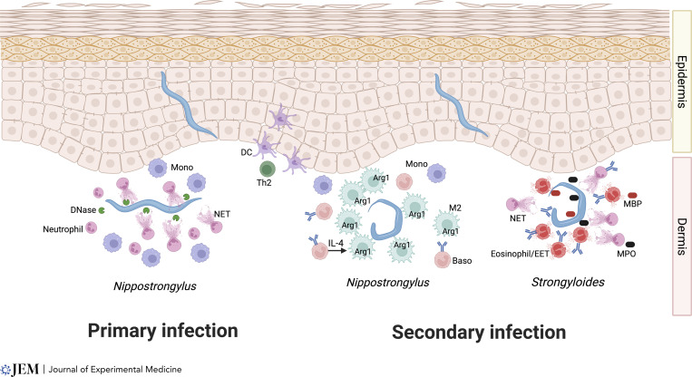
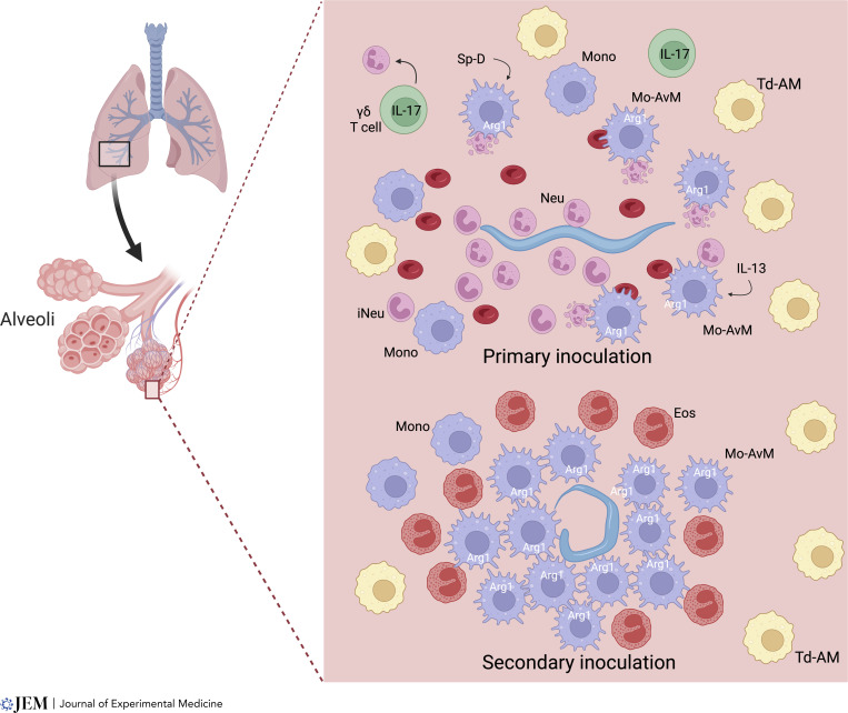
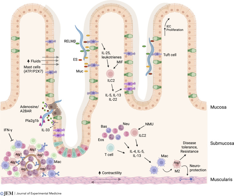
Verification
Claims the paper makes
The claims and statistics of this paper haven’t been checked yet.
Run the check — free, no account needed. It reads the open-access text and checks the sources it cites.
Claims other papers make about this one
Each is checked against this paper’s own text: does it say what the citing paper says it does?
In the citing paper
“However, in endemic environments with constant, low-level exposure to helminths, infections tend to be clinically asymptomatic due to immune tolerance”
From Parasites and allergies: a complex bidirectional relationship from evolutionary origins to modern therapeutics (2026) · cites this paper as Maizels and Gause 2023
Self-check
Percentages, sums and reported differences re-computed from the paper’s own numbers.
The self-check needs the paper’s full text.
Citations
Cites
Monocyte-derived alveolar macrophages drive lung fibrosis and persist in the lung over the life span
Misharin, A. V., Morales-Nebreda, L., Reyfman, P. A., Cuda, C. M., Walter, J. M., McQuattie-Pimentel, A. C., Chen, C.-I., Anekalla, K. R., Joshi, N., Williams, K. J. N., et al. (2017) · Journal of Experimental Medicine · doi:10.1084/jem.20162152 · Cited by 1,133
Type 2 immunity in tissue repair and fibrosis
Gieseck, R. L., Wilson, M. S., & Wynn, T. A. (2018) · Nature Reviews Immunology · doi:10.1038/nri.2017.90 · Cited by 1,055
Neuronal regulation of type 2 innate lymphoid cells via neuromedin U
Cardoso, V., Chesné, J., Ribeiro, H., García-Cassani, B., Carvalho, T., Bouchery, T., Shah, K., Barbosa-Morais, N. L., Harris, N., & Veiga-Fernandes, H. (2017) · Nature · doi:10.1038/nature23469 · Cited by 592
Macrophage function in tissue repair and remodeling requires IL-4 or IL-13 with apoptotic cells
Bosurgi, L., Cao, Y. G., Cabeza-Cabrerizo, M., Tucci, A., Hughes, L. D., Kong, Y., Weinstein, J. S., Licona-Limon, P., Schmid, E. T., Pelorosso, F., et al. (2017) · Science · doi:10.1126/science.aai8132 · Cited by 560
Anthony, R. M., Urban, J. F., Alem, F., Hamed, H. A., Rozo, C. T., Boucher, J.-L., Van Rooijen, N., & Gause, W. C. (2006) · Nature Medicine · doi:10.1038/nm1451 · Cited by 522
Self-citation
Research works printed but not linked: 114
- Aegerter, 2020, Influenza-induced monocyte-derived alveolar macrophages confer prolonged antibacterial protection, Nat. Immunol., 21, 145, 10.1038/s41590-019-0568-x · DOI
- Aegerter, 2022, Biology of lung macrophages in health and disease, Immunity, 55, 1564, 10.1016/j.immuni.2022.08.010 · DOI
- Ahrends, 2021, Enteric pathogens induce tissue tolerance and prevent neuronal loss from subsequent infections, Cell, 184, 5715, 10.1016/j.cell.2021.10.004 · DOI
- Ajendra, 2020, IL-17A both initiates, via IFNγ suppression, and limits the pulmonary type-2 immune response to nematode infection, Mucosal Immunol., 13, 958, 10.1038/s41385-020-0318-2 · DOI
- Anthony, 2007, Protective immune mechanisms in helminth infection, Nat. Rev. Immunol., 7, 975, 10.1038/nri2199 · DOI · Paper page
- Artis, 2004, RELMbeta/FIZZ2 is a goblet cell-specific immune-effector molecule in the gastrointestinal tract, Proc. Natl. Acad. Sci. USA, 101, 13596, 10.1073/pnas.0404034101 · DOI
- Bonne-Année, 2014, Extracellular traps are associated with human and mouse neutrophil and macrophage mediated killing of larval Strongyloides stercoralis, Microbes Infect., 16, 502, 10.1016/j.micinf.2014.02.012 · DOI
- Bouchery, 2015, ILC2s and T cells cooperate to ensure maintenance of M2 macrophages for lung immunity against hookworms, Nat. Commun., 6, 6970, 10.1038/ncomms7970 · DOI · Paper page
- Bouchery, 2020, Hookworms evade host immunity by secreting a deoxyribonuclease to degrade neutrophil extracellular traps, Cell Host Microbe, 27, 277, 10.1016/j.chom.2020.01.011 · DOI
- Chen, 2022, Helminth resistance is mediated by differential activation of recruited monocyte-derived alveolar macrophages and arginine depletion, Cell Rep., 38, 110215, 10.1016/j.celrep.2021.110215 · DOI
- Chen, 2012, An essential role for TH2-type responses in limiting acute tissue damage during experimental helminth infection, Nat. Med., 18, 260, 10.1038/nm.2628 · DOI
- Chen, 2018, B cells produce the tissue-protective protein RELMα during helminth infection, which inhibits IL-17 expression and limits emphysema, Cell Rep., 25, 2775, 10.1016/j.celrep.2018.11.038 · DOI
- Chen, 2014, Neutrophils prime a long-lived effector macrophage phenotype that mediates accelerated helminth expulsion, Nat. Immunol., 15, 938, 10.1038/ni.2984 · DOI
- Chen, 2016, Comparison of RELMα and RELMβ single- and double-gene-deficient mice reveals that RELMα expression dictates inflammation and worm expulsion in hookworm infection, Infect. Immun., 84, 1100, 10.1128/IAI.01479-15 · DOI
- Chu, 2021, The ChAT-acetylcholine pathway promotes group 2 innate lymphoid cell responses and anti-helminth immunity, Sci Immunol., 6, 10.1126/sciimmunol.abe3218 · DOI
- Classon, 2022, Intestinal helminth infection transforms the CD4+ T cell composition of the skin, Mucosal Immunol., 15, 257, 10.1038/s41385-021-00473-9 · DOI
- Cliffe, 2005, Accelerated intestinal epithelial cell turnover: A new mechanism of parasite expulsion, Science, 308, 1463, 10.1126/science.1108661 · DOI
- Coakley, 2020, The intestinal epithelium at the forefront of host-helminth interactions, Trends Parasitol, 36, 761, 10.1016/j.pt.2020.07.002 · DOI
- Connor, 2017, Th2 responses are primed by skin dendritic cells with distinct transcriptional profiles, J. Exp. Med., 214, 125, 10.1084/jem.20160470 · DOI
- Cunningham, 2021, Helminth imprinting of hematopoietic stem cells sustains anti-inflammatory trained innate immunity that attenuates autoimmune disease, J. Immunol., 206, 1618, 10.4049/jimmunol.2001225 · DOI
- de Los Reyes Jiménez, 2020, An anti-inflammatory eicosanoid switch mediates the suppression of type-2 inflammation by helminth larval products, Sci Transl Med., 12, 10.1126/scitranslmed.aay0605 · DOI
- Doolan, 2022, Hookworm infections: Reappraising the evidence for a role of neutrophils in light of NETosis, Parasite Immunol., 44, 10.1111/pim.12911 · DOI
- Douglas, 2021, Immune system investigation using parasitic helminths, Annu. Rev. Immunol., 39, 639, 10.1146/annurev-immunol-093019-122827 · DOI
- Drurey, 2022, Intestinal epithelial tuft cell induction is negated by a murine helminth and its secreted products, J. Exp. Med, 219, 10.1084/jem.20211140 · DOI
- Ehrens, 2021, Microfilariae trigger eosinophil extracellular DNA traps in a dectin-1-dependent manner, Cell Rep., 34, 108621, 10.1016/j.celrep.2020.108621 · DOI
- Ehrens, 2021, Eosinophils and neutrophils eliminate migrating Strongyloides ratti larvae at the site of infection in the context of extracellular DNA trap formation, Front. Immunol., 12, 715766, 10.3389/fimmu.2021.715766 · DOI
- El-Naccache, 2020, The NET effect of neutrophils during helminth infection, Cell Host Microbe, 27, 165, 10.1016/j.chom.2020.01.013 · DOI
- El-Naccache, 2022, Adenosine metabolized from extracellular ATP promotes type 2 immunity through triggering A2BAR signaling in intestinal epithelial cells, Cell Rep., 40, 111150, 10.1016/j.celrep.2022.111150 · DOI
- El-Naccache, 2021, Early events triggering the initiation of a type 2 immune response, Trends Immunol., 42, 151, 10.1016/j.it.2020.11.006 · DOI
- Entwistle, 2017, Epithelial-cell-derived phospholipase A2 group 1b is an endogenous anthelmintic, Cell Host Microbe, 22, 484, 10.1016/j.chom.2017.09.006 · DOI
- Esser-von Bieren, 2013, Antibodies trap tissue migrating helminth larvae and prevent tissue damage by driving IL-4Rα-independent alternative differentiation of macrophages, PLoS Pathog., 9, 10.1371/journal.ppat.1003771 · DOI
- Esser-von Bieren, 2015, Antibody-mediated trapping of helminth larvae requires CD11b and Fcγ receptor I, J. Immunol., 194, 1154, 10.4049/jimmunol.1401645 · DOI
- Filbey, 2014, Innate and adaptive type 2 immune cell responses in genetically controlled resistance to intestinal helminth infection, Immunol. Cell Biol., 92, 436, 10.1038/icb.2013.109 · DOI · Paper page
- Gause, 2020, Heterogeneity in the initiation, development and function of type 2 immunity, Nat. Rev. Immunol., 20, 603, 10.1038/s41577-020-0301-x · DOI
- Gause, 2013, Type 2 immunity and wound healing: Evolutionary refinement of adaptive immunity by helminths, Nat. Rev. Immunol., 13, 607, 10.1038/nri3476 · DOI
- Gazzinelli-Guimaraes, 2019, Allergen presensitization drives an eosinophil-dependent arrest in lung-specific helminth development, J. Clin. Invest., 129, 3686, 10.1172/JCI127963 · DOI
- Gerbe, 2016, Intestinal epithelial tuft cells initiate type 2 mucosal immunity to helminth parasites, Nature, 529, 226, 10.1038/nature16527 · DOI
- Grencis, 2015, Immunity to helminths: Resistance, regulation, and susceptibility to gastrointestinal nematodes, Annu. Rev. Immunol., 33, 201, 10.1146/annurev-immunol-032713-120218 · DOI
- Gundra, 2017, Vitamin A mediates conversion of monocyte-derived macrophages into tissue-resident macrophages during alternative activation, Nat. Immunol., 18, 642, 10.1038/ni.3734 · DOI
- Guo, 2015, Innate immunological function of TH2 cells in vivo, Nat. Immunol., 16, 1051, 10.1038/ni.3244 · DOI
- Hammad, 2022, Emerging paradigms in Type 2 immunity, Annu. Rev. Immunol., 40, 443, 10.1146/annurev-immunol-101320-030339 · DOI
- Hammad, 2015, Barrier epithelial cells and the control of Type 2 immunity, Immunity, 43, 29, 10.1016/j.immuni.2015.07.007 · DOI · Paper page
- Harris, 2017, Recent advances in type-2-cell-mediated immunity: Insights from helminth infection, Immunity, 47, 1024, 10.1016/j.immuni.2017.11.015 · DOI
- Hartung, 2022, Trained immunity in type 2 immune responses, Mucosal Immunol., 15, 1158, 10.1038/s41385-022-00557-0 · DOI
- Harvie, 2010, The lung is an important site for priming CD4 T-cell-mediated protective immunity against gastrointestinal helminth parasites, Infect. Immun., 78, 3753, 10.1128/IAI.00502-09 · DOI
- Hasnain, 2013, A new role for mucins in immunity: Insights from gastrointestinal nematode infection, Int. J. Biochem. Cell Biol., 45, 364, 10.1016/j.biocel.2012.10.011 · DOI
- Hepworth, 2012, Mast cells orchestrate type 2 immunity to helminths through regulation of tissue-derived cytokines, Proc. Natl. Acad. Sci. USA, 109, 6644, 10.1073/pnas.1112268109 · DOI · Paper page
- Herbert, 2004, Alternative macrophage activation is essential for survival during schistosomiasis and downmodulates T helper 1 responses and immunopathology, Immunity, 20, 623, 10.1016/S1074-7613(04)00107-4 · DOI
- Herbert, 2009, Intestinal epithelial cell secretion of RELM-β protects against gastrointestinal worm infection, J. Exp. Med., 206, 2947, 10.1084/jem.20091268 · DOI
- Heredia, 2013, Type 2 innate signals stimulate fibro/adipogenic progenitors to facilitate muscle regeneration, Cell, 153, 376, 10.1016/j.cell.2013.02.053 · DOI · Paper page
- Hewitson, 2015, Concerted activity of IgG1 antibodies and IL-4/IL-25-dependent effector cells trap helminth larvae in the tissues following vaccination with defined secreted antigens, providing sterile immunity to challenge infection, PLoS Pathog., 11, 10.1371/journal.ppat.1004676 · DOI
- Horák, 2015, Avian schistosomes and outbreaks of cercarial dermatitis, Clin. Microbiol. Rev., 28, 165, 10.1128/CMR.00043-14 · DOI
- Houlder, 2021, Schistosomes in the lung: Immunobiology and opportunity, Front. Immunol., 12, 635513, 10.3389/fimmu.2021.635513 · DOI
- Huang, 2018, S1P-dependent interorgan trafficking of group 2 innate lymphoid cells supports host defense, Science, 359, 114, 10.1126/science.aam5809 · DOI
- Hung, 2020, Cellular context of IL-33 expression dictates impact on anti-helminth immunity, Sci Immunol., 5, 10.1126/sciimmunol.abc6259 · DOI
- Ignacio, 2022, Small intestinal resident eosinophils maintain gut homeostasis following microbial colonization, Immunity, 55, 1250, 10.1016/j.immuni.2022.05.014 · DOI
- Inclan-Rico, 2022, “Every cell is an immune cell; contributions of non-hematopoietic cells to anti-helminth immunity”, Mucosal Immunol., 15, 1199, 10.1038/s41385-022-00518-7 · DOI
- Jacobson, 2021, The intestinal neuro-immune axis: Crosstalk between neurons, immune cells, and microbes, Mucosal Immunol., 14, 555, 10.1038/s41385-020-00368-1 · DOI
- Kabat, 2022, Resident TH2 cells orchestrate adipose tissue remodeling at a site adjacent to infection, Sci. Immunol., 7, 10.1126/sciimmunol.add3263 · DOI
- Karasuyama, 2018, Crucial role for basophils in acquired protective immunity to tick infestation, Front. Physiol., 9, 1769, 10.3389/fphys.2018.01769 · DOI
- Karo-Atar, 2022, Helminth-induced reprogramming of the stem cell compartment inhibits type 2 immunity, J Exp Med., 219, 10.1084/jem.20212311 · DOI
- Katona, 1988, The role of L3T4+ and Lyt-2+ T cells in the IgE response and immunity to Nippostrongylus brasiliensis, J. Immunol., 140, 3206, 10.4049/jimmunol.140.9.3206 · DOI
- Khan, 2001, Stat6 dependent goblet cell hyperplasia during intestinal nematode infection, Parasite Immunol., 23, 39, 10.1046/j.1365-3024.2001.00353.x · DOI
- Klose, 2017, The neuropeptide neuromedin U stimulates innate lymphoid cells and type 2 inflammation, Nature, 549, 282, 10.1038/nature23676 · DOI
- Knott, 2007, Impaired resistance in early secondary Nippostrongylus brasiliensis infections in mice with defective eosinophilopoeisis, Int. J. Parasitol., 37, 1367, 10.1016/j.ijpara.2007.04.006 · DOI
- Krljanac, 2019, RELMa-expressing macrophages protect against fatal lung damage and reduce parasite burden during helminth infection, Sci Immunol., 4, 10.1126/sciimmunol.aau3814 · DOI
- Kumamoto, 2013, CD301b(+) dermal dendritic cells drive T helper 2 cell-mediated immunity, Immunity, 39, 733, 10.1016/j.immuni.2013.08.029 · DOI
- Lacorcia, 2018, Maternal schistosomiasis: Immunomodulatory effects with lasting impact on allergy and vaccine responses, Front. Immunol., 9, 2960, 10.3389/fimmu.2018.02960 · DOI
- Lechner, 2021, Macrophage regulation & function in helminth infection, Semin. Immunol., 53, 101526, 10.1016/j.smim.2021.101526 · DOI
- Lindholm, 2022, BMP signaling in the intestinal epithelium drives a critical feedback loop to restrain IL-13-driven tuft cell hyperplasia, Sci. Immunol., 7, 10.1126/sciimmunol.abl6543 · DOI
- Liu, 2010, B cells have distinct roles in host protection against different nematode parasites, J. Immunol., 184, 5213, 10.4049/jimmunol.0902879 · DOI
- Lloyd, 2018, Type 2 immunity: Expanding our view, Sci. Immunol., 3, 10.1126/sciimmunol.aat1604 · DOI
- Marsland, 2008, Nippostrongylus brasiliensis infection leads to the development of emphysema associated with the induction of alternatively activated macrophages, Eur. J. Immunol., 38, 479, 10.1002/eji.200737827 · DOI
- Matheis, 2020, Adrenergic signaling in muscularis macrophages limits infection-induced neuronal loss, Cell, 180, 64, 10.1016/j.cell.2019.12.002 · DOI
- McDaniel, 2023, Initiation of Type 2 immunity at barrier surfaces, Mucosal Immunol., 10.1016/j.mucimm.2022.11.002 · DOI
- McGinty, 2020, Tuft-cell-derived leukotrienes drive rapid anti-helminth immunity in the small intestine but are dispensable for anti-protist immunity, Immunity, 52, 528, 10.1016/j.immuni.2020.02.005 · DOI
- McKay, 2017, Helminths and intestinal barrier function, Tissue Barriers, 5, 10.1080/21688370.2017.1283385 · DOI
- Medzhitov, 2012, Disease tolerance as a defense strategy, Science, 335, 936, 10.1126/science.1214935 · DOI · Paper page
- Moriyama, 2018, β2-adrenergic receptor-mediated negative regulation of group 2 innate lymphoid cell responses, Science, 359, 1056, 10.1126/science.aan4829 · DOI
- Obata-Ninomiya, 2013, The skin is an important bulwark of acquired immunity against intestinal helminths, J. Exp. Med., 210, 2583, 10.1084/jem.20130761 · DOI
- Ohnmacht, 2009, Basophil effector function and homeostasis during helminth infection, Blood, 113, 2816, 10.1182/blood-2008-05-154773 · DOI
- Oyesola, 2021, PGD2 and CRTH2 counteract Type 2 cytokine-elicited intestinal epithelial responses during helminth infection, J. Exp. Med., 218, 10.1084/jem.20202178 · DOI
- Oyesola, 2023, Exposure to lung-migrating helminth protects against murine SARS-CoV-2 infection through macrophage-dependent T cell activation, Sci. Immunol, 8, eadf8161, 10.1126/sciimmunol.adf8161 · DOI
- Palm, 2012, Allergic host defences, Nature, 484, 465, 10.1038/nature11047 · DOI · Paper page
- Peng, 2022, Monocytes maintain central nervous system homeostasis following helminth-induced inflammation, Proc. Natl. Acad. Sci. USA, 119, 10.1073/pnas.2201645119 · DOI
- Pesce, 2009, Arginase-1-expressing macrophages suppress Th2 cytokine-driven inflammation and fibrosis, PLoS Pathog., 5, 10.1371/journal.ppat.1000371 · DOI · Paper page
- Progatzky, 2021, Regulation of intestinal immunity and tissue repair by enteric glia, Nature, 599, 125, 10.1038/s41586-021-04006-z · DOI
- Reitz, 2018, Basophils are dispensable for the establishment of protective adaptive immunity against primary and challenge infection with the intestinal helminth parasite Strongyloides ratti, PLoS Negl. Trop. Dis., 12, 10.1371/journal.pntd.0006992 · DOI
- Reynolds, 2012, Immunity to the model intestinal helminth parasite Heligmosomoides polygyrus, Semin. Immunopathol., 34, 829, 10.1007/s00281-012-0347-3 · DOI
- Reynolds, 2014, MyD88 signaling inhibits protective immunity to the gastrointestinal helminth parasite Heligmosomoides polygyrus, J. Immunol., 193, 2984, 10.4049/jimmunol.1401056 · DOI
- Ricardo-Gonzalez, 2020, Tissue-specific pathways extrude activated ILC2s to disseminate type 2 immunity, J. Exp. Med., 217, 10.1084/jem.20191172 · DOI
- Roberts, 2021, Acetylcholine production by group 2 innate lymphoid cells promotes mucosal immunity to helminths, Sci. Immunol., 6, 10.1126/sciimmunol.abd0359 · DOI
- Rolot, 2018, Macrophage activation and functions during helminth infection: Recent advances from the laboratory mouse, J. Immunol. Res., 2018, 2790627, 10.1155/2018/2790627 · DOI
- Salgame, 2013, Effect of helminth-induced immunity on infections with microbial pathogens, Nat. Immunol., 14, 1118, 10.1038/ni.2736 · DOI
- Sanin, 2015, Sm16, a major component of Schistosoma mansoni cercarial excretory/secretory products, prevents macrophage classical activation and delays antigen processing, Parasit. Vectors, 8, 1, 10.1186/s13071-014-0608-1 · DOI
- Schneider, 2019, Regulation of immune responses by tuft cells, Nat. Rev. Immunol., 19, 584, 10.1038/s41577-019-0176-x · DOI
- Shimokawa, 2017, Mast cells are crucial for induction of group 2 innate lymphoid cells and clearance of helminth infections, Immunity, 46, 863, 10.1016/j.immuni.2017.04.017 · DOI
- Smith, 2016, Low-level regulatory T-cell activity is essential for functional type-2 effector immunity to expel gastrointestinal helminths, Mucosal Immunol., 9, 428, 10.1038/mi.2015.73 · DOI
- Steinfelder, 2017, Intestinal helminth infection induces highly functional resident memory CD4+ T cells in mice, Eur. J. Immunol., 47, 353, 10.1002/eji.201646575 · DOI
- Sutherland, 2014, Chitinase-like proteins promote IL-17-mediated neutrophilia in a tradeoff between nematode killing and host damage, Nat. Immunol., 15, 1116, 10.1038/ni.3023 · DOI
- Thawer, 2016, Surfactant protein-D Is essential for immunity to helminth infection, PLoS Pathog., 12, 10.1371/journal.ppat.1005461 · DOI
- Turner, 2013, IL-22 mediates goblet cell hyperplasia and worm expulsion in intestinal helminth infection, PLoS Pathog., 9, 10.1371/journal.ppat.1003698 · DOI
- Vannella, 2014, Incomplete deletion of IL-4Rα by LysM(Cre) reveals distinct subsets of M2 macrophages controlling inflammation and fibrosis in chronic schistosomiasis, PLoS Pathog., 10, 10.1371/journal.ppat.1004372 · DOI
- Vannella, 2016, Acidic chitinase primes the protective immune response to gastrointestinal nematodes, Nat. Immunol., 17, 538, 10.1038/ni.3417 · DOI
- Varyani, 2022, The IL-25-dependent tuft cell circuit driven by intestinal helminths requires macrophage migration inhibitory factor (MIF), Mucosal Immunol., 15, 1243, 10.1038/s41385-022-00496-w · DOI
- Wang, 2021, A basophil-neuronal axis promotes itch, Cell, 184, 422, 10.1016/j.cell.2020.12.033 · DOI
- Wculek, 2022, Metabolism of tissue macrophages in homeostasis and pathology, Cell. Mol. Immunol., 19, 384, 10.1038/s41423-021-00791-9 · DOI · Paper page
- Wilson, 2005, Suppression of allergic airway inflammation by helminth-induced regulatory T cells, J. Exp. Med., 202, 1199, 10.1084/jem.20042572 · DOI
- Winkel, 2018, Early induction of human regulatory dermal antigen presenting cells by skin-penetrating Schistosoma mansoni cercariae, Front. Immunol., 9, 2510, 10.3389/fimmu.2018.02510 · DOI
- Yordanova, 2022, The host peritoneal cavity harbors prominent memory Th2 and early recall responses to an intestinal nematode, Front. Immunol., 13, 842870, 10.3389/fimmu.2022.842870 · DOI
- Zaini, 2021, Context-dependent roles of B cells during intestinal helminth infection, PLoS Negl. Trop. Dis., 15, 10.1371/journal.pntd.0009340 · DOI
- Zaiss, 2006, Amphiregulin, a TH2 cytokine enhancing resistance to nematodes, Science, 314, 1746, 10.1126/science.1133715 · DOI
- Zhao, 2008, Th2 cytokine-induced alterations in intestinal smooth muscle function depend on alternatively activated macrophages, Gastroenterology, 135, 217, 10.1053/j.gastro.2008.03.077 · DOI
- Zhao, 2006, Contribution of 5-HT2A receptor in nematode infection-induced murine intestinal smooth muscle hypercontractility, Gastroenterology, 131, 568, 10.1053/j.gastro.2006.05.013 · DOI
Cited by
Sorted by citations
Understanding the Type 2 Immunity: An Evolving View Beyond Allergic Diseases
Jiménez‐Saiz, R., Iborra, S., Blanco, C., González‐Pérez, R., Nuñez‐Borque, E., Zeyneloglu, C., Babayev, H., Göksel, O., Göksel, T., Khaitov, M., et al. (2026) · Allergy · doi:10.1111/all.70512 · Cited by 2
Packi, K., Świerczewski, D., Klimczak, S., Ochab, D., Gałęziewska, J., Gromek, P., Kruczkowska, W., Płuciennik, E., Kałuzińska-Kołat, Ż., & Kołat, D. (2026) · Antonie van Leeuwenhoek · doi:10.1007/s10482-026-02425-4
Metadata
- Authors
- Maizels, R. M., & Gause, W. C.
- Venue
- Journal of Experimental Medicine
- Publisher
- Rockefeller University Press
- Published
- 2023-08-28
- Volume
- 220
- Issue
- 10
- ISSN
- 0022-1007
- 1540-9538
- PMC
- PMC10460967
- Cited by
- 45
- References
- 5
- Language
- en
- Funders
- Wellcome Trust (219530, 104111)
- Netherlands Lung Foundation (AWWA)
- National Institutes of Health (R01DK113790, R01AI131634)
About these checks
Registry, retraction, Cites and statistics results are computed, not judged, with one exception: a printed reference that no index lists is read once by a model before it counts as a citation error. Claims are extracted by a model and kept only when their quote appears verbatim in the text; each cited source is then checked, with a model’s help, for whether it says what the claim relies on. Those verdicts can be wrong — read the claim’s quote and the cited source before relying on one.
Statistics and arithmetic are recomputed, not judged.
Confidence bands are shown only for fields calibrated on our annotated test set. None is calibrated yet, so no band is shown.
What counts as a claim ›How we measure confidence ›How we check this ›
Are you an author, or do you see a problem with this page? Request a review
How to cite
Cite CoChat for the checks on this page, and the paper for its own findings.
- This page
- CoChat. (2026). Citation check: Targeting helminths: The expanding world of type 2 immune effector mechanisms. https://app.cochat.ai/papers/10.1084/jem.20221381
- The paper
- Maizels, R. M., & Gause, W. C. (2023). Targeting helminths: The expanding world of type 2 immune effector mechanisms. Journal of Experimental Medicine. https://doi.org/10.1084/jem.20221381
