

The DOI matches its Crossref record; 9 of the 9 references it cites were found in a registry or an index.
Gene variant effects across sodium channelopathies predict function and guide precision therapy
Abstract
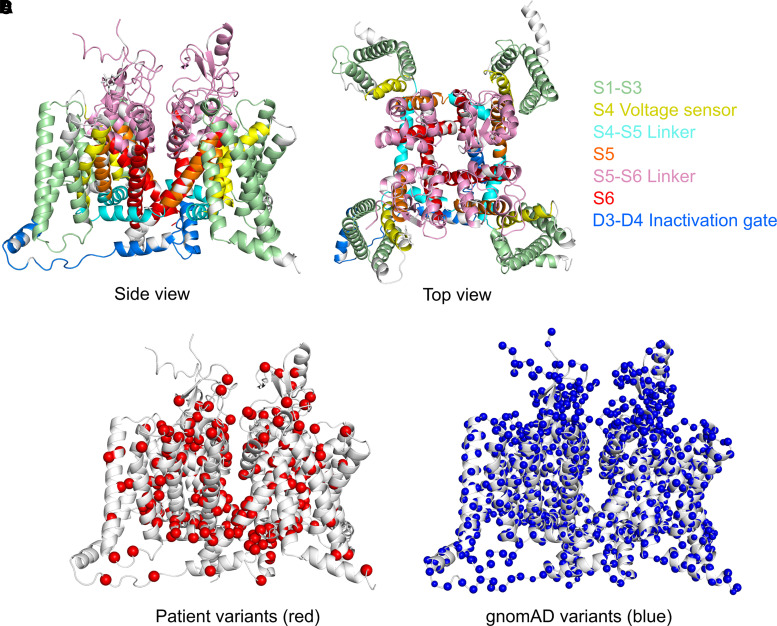
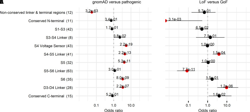
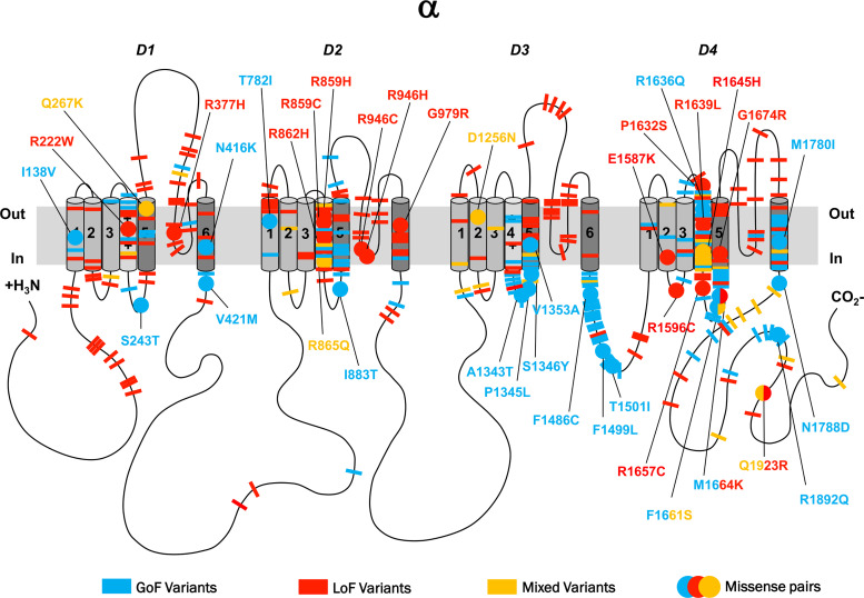
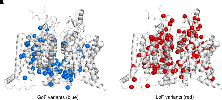
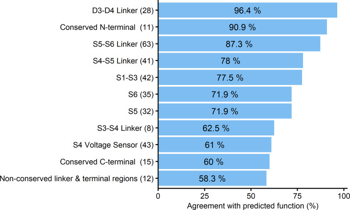
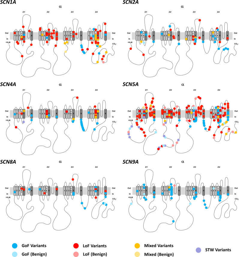
Pathogenic variants in the voltage-gated sodium channel gene family lead to early onset epilepsies, neurodevelopmental disorders, skeletal muscle channelopathies, peripheral neuropathies and cardiac arrhythmias. Disease-associated variants have diverse functional effects ranging from complete loss-of-function to marked gain-of-function. Therapeutic strategy is likely to depend on functional effect. Experimental studies offer important insights into channel function but are resource intensive and only performed in a minority of cases. Given the evolutionarily conserved nature of the sodium channel genes, we investigated whether similarities in biophysical properties between different voltage-gated sodium channels can predict function and inform precision treatment across sodium channelopathies. We performed a systematic literature search identifying functionally assessed variants in any of the nine voltage-gated sodium channel genes until 28 April 2021. We included missense variants that had been electrophysiologically characterized in mammalian cells in whole-cell patch-clamp recordings. We performed an alignment of linear protein sequences of all sodium channel genes and correlated variants by their overall functional effect on biophysical properties. Of 951 identified records, 437 sodium channel-variants met our inclusion criteria and were reviewed for functional properties. Of these, 141 variants were epilepsy-associated (SCN1/2/3/8A), 79 had a neuromuscular phenotype (SCN4/9/10/11A), 149 were associated with a cardiac phenotype (SCN5/10A) and 68 (16%) were considered benign. We detected 38 missense variant pairs with an identical disease-associated variant in a different sodium channel gene. Thirty-five out of 38 of those pairs resulted in similar functional consequences, indicating up to 92% biophysical agreement between corresponding sodium channel variants (odds ratio = 11.3; 95% confidence interval = 2.8 to 66.9; P < 0.001). Pathogenic missense variants were clustered in specific functional domains, whereas population variants were significantly more frequent across non-conserved domains (odds ratio = 18.6; 95% confidence interval = 10.9–34.4; P < 0.001). Pore-loop regions were frequently associated with loss-of-function variants, whereas inactivation sites were associated with gain-of-function (odds ratio = 42.1, 95% confidence interval = 14.5–122.4; P < 0.001), whilst variants occurring in voltage-sensing regions comprised a range of gain- and loss-of-function effects. Our findings suggest that biophysical characterisation of variants in one SCN-gene can predict channel function across different SCN-genes where experimental data are not available. The collected data represent the first gain- versus loss-of-function topological map of SCN proteins indicating shared patterns of biophysical effects aiding variant analysis and guiding precision therapy. We integrated our findings into a free online webtool to facilitate functional sodium channel gene variant interpretation (http://SCN-viewer.broadinstitute.org).
Overview
The claims and statistics of this paper haven’t been checked yet.
Run the check — free, no account needed. It reads the open-access text and checks the sources it cites.
Integrity signals
Registry
Cites
Statistics
Figures
Verification
Claims the paper makes
The claims and statistics of this paper haven’t been checked yet.
Run the check — free, no account needed. It reads the open-access text and checks the sources it cites.
Claims other papers make about this one
Each is checked against this paper’s own text: does it say what the citing paper says it does?
In the citing paper
“Cases of muscular dystrophies treated via splicing modulating ASOs have proven effective47, but overall, gene therapy in brain disease has shown mixed successes48.”
From Phenotypic complexities of rare heterozygous neurexin-1 deletions (2025) · cites this paper as [47]
Self-check
Percentages, sums and reported differences re-computed from the paper’s own numbers.
The self-check needs the paper’s full text.
Citations
Cites
Yu, F. H., Mantegazza, M., Westenbroek, R. E., Robbins, C. A., Kalume, F., Burton, K. A., Spain, W. J., McKnight, G. S., Scheuer, T., & Catterall, W. A. (2006) · Nature Neuroscience · doi:10.1038/nn1754 · Cited by 1,173
Overview of the voltage-gated sodium channel family
Yu (2003) · Genome biology · doi:10.1186/gb-2003-4-3-207 · Cited by 694
Han, Z., Chen, C., Christiansen, A., Ji, S., Lin, Q., Anumonwo, C., Liu, C., Leiser, S. C., Meena, Aznarez, I., et al. (2020) · Science Translational Medicine · doi:10.1126/scitranslmed.aaz6100 · Cited by 341
Progress in Understanding and Treating SCN2A-Mediated Disorders
Sanders, S. J., Campbell, A. J., Cottrell, J. R., Moller, R. S., Wagner, F. F., Auldridge, A. L., Bernier, R. A., Catterall, W. A., Chung, W. K., Empfield, J. R., et al. (2018) · Trends in Neurosciences · doi:10.1016/j.tins.2018.03.011 · Cited by 338
Self-citationVoltage sensor charge loss accounts for most cases of hypokalemic periodic paralysis
Matthews, E., Labrum, R., Sweeney, M. G., Sud, R., Haworth, A., Chinnery, P. F., Meola, G., Schorge, S., Kullmann, D. M., Davis, M. B., et al. (2009) · Neurology · doi:10.1212/01.wnl.0000342387.65477.46 · Cited by 201
Self-citationClinical spectrum of SCN2A mutations expanding to Ohtahara syndrome
Nakamura, K., Kato, M., Osaka, H., Yamashita, S., Nakagawa, E., Haginoya, K., Tohyama, J., Okuda, M., Wada, T., Shimakawa, S., et al. (2013) · Neurology · doi:10.1212/wnl.0b013e3182a43e57 · Cited by 201
Evolution of sodium channels predates the origin of nervous systems in animals
Liebeskind, B. J., Hillis, D. M., & Zakon, H. H. (2011) · Proceedings of the National Academy of Sciences · doi:10.1073/pnas.1106363108 · Cited by 149
Johannesen, K. M., Liu, Y., Koko, M., Gjerulfsen, C. E., Sonnenberg, L., Schubert, J., Fenger, C. D., Eltokhi, A., Rannap, M., Koch, N. A., et al. (2022) · Brain · doi:10.1093/brain/awab321 · Cited by 147
Self-citationPredicting functional effects of missense variants in voltage-gated sodium and calcium channels
Heyne, H. O., Baez-Nieto, D., Iqbal, S., Palmer, D. S., Brunklaus, A., May, P., Collaborative, E., Johannesen, K. M., Lauxmann, S., Lemke, J. R., et al. (2020) · Science Translational Medicine · doi:10.1126/scitranslmed.aay6848 · Cited by 140
Self-citation
Research works printed but not linked: 73
- Goldin, 2000, Nomenclature of voltage-gated sodium channels, Neuron, 28, 365, 10.1016/S0896-6273(00)00116-1 · DOI
- Hodgkin, 1952, A quantitative description of membrane current and its application to conduction and excitation in nerve, J Physiol, 117, 500, 10.1113/jphysiol.1952.sp004764 · DOI · Paper page
- Spillane, 2016, Genetic neurological channelopathies: molecular genetics and clinical phenotypes, J Neurol Neurosurg Psychiatry, 87, 37
- Brunklaus, 2014, Genotype phenotype associations across the voltage-gated sodium channel family, J Med Genet, 51, 650, 10.1136/jmedgenet-2014-102608 · DOI · Paper page
- Catterall, 2005, International union of pharmacology. XLVII. Nomenclature and structure-function relationships of voltage-gated sodium channels, Pharmacol Rev, 57, 397, 10.1124/pr.57.4.4 · DOI
- Yu, 2004, The VGL-chanome: a protein superfamily specialized for electrical signaling and ionic homeostasis, Sci STKE, 2004, re15, 10.1126/stke.2532004re15 · DOI
- Brunklaus, 2020, Precision medicine in sodium channelopathies - Moving beyond seizure control towards disease modification, Eur J Paediatr Neurol, 24, 7, 10.1016/j.ejpn.2020.01.008 · DOI
- Mantegazza, 2019, SCN1A/NaV1.1 channelopathies: Mechanisms in expression systems, animal models, and human iPSC models, Epilepsia, 60, S25, 10.1111/epi.14700 · DOI
- Cestèle, 2008, Self-limited hyperexcitability: Functional effect of a familial hemiplegic migraine mutation of the Nav1.1 (SCN1A) Na+ channel, J Neurosci, 28, 7273, 10.1523/JNEUROSCI.4453-07.2008 · DOI
- Cestèle, 2013, Nonfunctional NaV1.1 familial hemiplegic migraine mutant transformed into gain of function by partial rescue of folding defects, Proc Natl Acad Sci USA, 110, 17546, 10.1073/pnas.1309827110 · DOI
- Kahlig, 2008, Divergent sodium channel defects in familial hemiplegic migraine, Proc Natl Acad Sci USA, 105, 9799, 10.1073/pnas.0711717105 · DOI
- Barbieri, 2019, Late sodium current blocker GS967 inhibits persistent currents induced by familial hemiplegic migraine type 3 mutations of the SCN1A gene, J Headache Pain, 20, 107, 10.1186/s10194-019-1056-2 · DOI
- Berkovic, 2004, Benign familial neonatal-infantile seizures: characterization of a new sodium channelopathy, Ann Neurol, 55, 550, 10.1002/ana.20029 · DOI
- Shi, 2012, Clinical spectrum of SCN2A mutations, Brain Dev, 34, 541, 10.1016/j.braindev.2011.09.016 · DOI
- Bennett, 1995, Molecular mechanism for an inherited cardiac arrhythmia, Nature, 376, 683, 10.1038/376683a0 · DOI
- Fonseca, 2018, Cardiac channelopathies: The role of sodium channel mutations, Rev Port Cardiol, 37, 179, 10.1016/j.repc.2017.11.007 · DOI
- Jurkat-Rott, 2010, Sodium channelopathies of skeletal muscle result from gain or loss of function, Pflugers Arch, 460, 239, 10.1007/s00424-010-0814-4 · DOI
- Lee, 2009, Clinical diversity of SCN4A-mutation-associated skeletal muscle sodium channelopathy, J Clin Neurol, 5, 186, 10.3988/jcn.2009.5.4.186 · DOI
- Waxman, 2013, Painful Na-channelopathies: an expanding universe, Trends Mol Med, 19, 406, 10.1016/j.molmed.2013.04.003 · DOI
- Faber, 2012, Gain-of-function Nav1.8 mutations in painful neuropathy, Proc Natl Acad Sci USA, 109, 19444, 10.1073/pnas.1216080109 · DOI
- Huang, 2014, Gain-of-function mutations in sodium channel Na(v)1.9 in painful neuropathy, Brain, 137, 1627, 10.1093/brain/awu079 · DOI
- Elia, 2019, Myasthenic congenital myopathy from recessive mutations at a single residue in Na(V)1.4, Neurology, 92, e1405, 10.1212/WNL.0000000000007185 · DOI
- Habbout, 2016, A recessive Nav1.4 mutation underlies congenital myasthenic syndrome with periodic paralysis, Neurology, 86, 161, 10.1212/WNL.0000000000002264 · DOI
- Wolff, 2017, Genetic and phenotypic heterogeneity suggest therapeutic implications in SCN2A-related disorders, Brain, 140, 1316, 10.1093/brain/awx054 · DOI · Paper page
- Møller, 2016, Precision medicine: SCN8A encephalopathy treated with sodium channel blockers, Neurotherapeutics, 13, 190, 10.1007/s13311-015-0403-5 · DOI
- Brunklaus, 2012, Prognostic, clinical and demographic features in SCN1A mutation-positive Dravet syndrome, Brain, 135, 2329, 10.1093/brain/aws151 · DOI · Paper page
- Guerrini, 1998, Lamotrigine and seizure aggravation in severe myoclonic epilepsy, Epilepsia, 39, 508, 10.1111/j.1528-1157.1998.tb01413.x · DOI · Paper page
- Mazzanti, 2016, Gene-specific therapy with mexiletine reduces arrhythmic events in patients with long QT syndrome type 3, J Am Coll Cardiol, 67, 1053, 10.1016/j.jacc.2015.12.033 · DOI
- Kambouris, 2000, A revised view of cardiac sodium channel “blockade” in the long-QT syndrome, J Clin Invest, 105, 1133, 10.1172/JCI9212 · DOI
- Priori, 2013, HRS/EHRA/APHRS expert consensus statement on the diagnosis and management of patients with inherited primary arrhythmia syndromes: document endorsed by HRS, EHRA, and APHRS in May 2013 and by ACCF, AHA, PACES, and AEPC in June 2013, Heart Rhythm, 10, 1932, 10.1016/j.hrthm.2013.05.014 · DOI · Paper page
- Brunklaus, 2020, SCN1A variants from bench to bedside-improved clinical prediction from functional characterization, Hum Mutat, 41, 363, 10.1002/humu.23943 · DOI
- Pérez-Palma, 2020, Identification of pathogenic variant enriched regions across genes and gene families, Genome Res, 30, 62, 10.1101/gr.252601.119 · DOI
- Meisler, 2021, Sodium channelopathies in neurodevelopmental disorders, Nat Rev Neurosci, 22, 152, 10.1038/s41583-020-00418-4 · DOI
- Mantegazza, 2021, Sodium channelopathies of skeletal muscle and brain, Physiol Rev, 101, 1633, 10.1152/physrev.00025.2020 · DOI
- Brunklaus, 2020, Biological concepts in human sodium channel epilepsies and their relevance in clinical practice, Epilepsia, 61, 387, 10.1111/epi.16438 · DOI
- Rhodes, 2004, Noninactivating voltage-gated sodium channels in severe myoclonic epilepsy of infancy, Proc Natl Acad Sci USA, 101, 11147, 10.1073/pnas.0402482101 · DOI
- Thompson, 2012, Nontruncating SCN1A mutations associated with severe myoclonic epilepsy of infancy impair cell surface expression, J Biol Chem, 287, 42001, 10.1074/jbc.M112.421883 · DOI
- Fleischhauer, 1998, Effects of temperature and mexiletine on the F1473S Na+ channel mutation causing paramyotonia congenita, Pflugers Arch, 436, 757, 10.1007/s004240050699 · DOI
- Bechi, 2015, Rescuable folding defective NaV1.1 (SCN1A) mutants in epilepsy: properties, occurrence, and novel rescuing strategy with peptides targeted to the endoplasmic reticulum, Neurobiol Dis, 75, 100, 10.1016/j.nbd.2014.12.028 · DOI
- Fertleman, 2006, SCN9A mutations in paroxysmal extreme pain disorder: allelic variants underlie distinct channel defects and phenotypes, Neuron, 52, 767, 10.1016/j.neuron.2006.10.006 · DOI
- Dib-Hajj, 2008, Paroxysmal extreme pain disorder M1627K mutation in human Nav1.7 renders DRG neurons hyperexcitable, Mol Pain, 4, 37, 10.1186/1744-8069-4-37 · DOI
- Theile, 2011, Nav1.7 mutations associated with paroxysmal extreme pain disorder, but not erythromelalgia, enhance Navbeta4 peptide-mediated resurgent sodium currents, J Physiol, 589, 597, 10.1113/jphysiol.2010.200915 · DOI
- Winkel, 2015, The role of the sodium current complex in a nonreferred nationwide cohort of sudden infant death syndrome, Heart Rhythm, 12, 1241, 10.1016/j.hrthm.2015.03.013 · DOI
- Abdelsayed, 2017, Differential calcium sensitivity in NaV 1.5 mixed syndrome mutants, J Physiol, 595, 6165, 10.1113/JP274536 · DOI
- Rusconi, 2007, Modulatory proteins can rescue a trafficking defective epileptogenic Nav1.1 Na+ channel mutant, J Neurosci, 27, 11037, 10.1523/JNEUROSCI.3515-07.2007 · DOI
- Catterall, 2014, Structure and function of voltage-gated sodium channels at atomic resolution, Exp Physiol, 99, 35, 10.1113/expphysiol.2013.071969 · DOI
- Zuberi, 2011, Genotype-phenotype associations in SCN1A-related epilepsies, Neurology, 76, 594, 10.1212/WNL.0b013e31820c309b · DOI
- Tarradas, 2013, A novel missense mutation, I890T, in the pore region of cardiac sodium channel causes Brugada syndrome, PLoS One, 8, e53220, 10.1371/journal.pone.0053220 · DOI
- Valdivia, 2002, A novel SCN5A arrhythmia mutation, M1766L, with expression defect rescued by mexiletine, Cardiovasc Res, 55, 279, 10.1016/S0008-6363(02)00445-5 · DOI
- Bechi, 2015, Rescuable folding defective NaV1.1 (SCN1A) mutants in epilepsy: properties, occurrence, and novel rescuing strategy with peptides targeted to the endoplasmic reticulum, Neurobiol Dis, 75, 100, 10.1016/j.nbd.2014.12.028 · DOI
- Dhifallah, 2018, Gain of function for the SCN1A/hNav1.1-L1670W mutation responsible for familial hemiplegic migraine, Front Mol Neurosci, 11, 232, 10.3389/fnmol.2018.00232 · DOI
- Horga, 2013, Prevalence study of genetically defined skeletal muscle channelopathies in England, Neurology, 80, 1472, 10.1212/WNL.0b013e31828cf8d0 · DOI
- Loussouarn, 2015, Physiological and pathophysiological insights of Nav1.4 and Nav1.5 comparison, Front Pharmacol, 6, 314
- Antzelevitch, 2005, Brugada syndrome: report of the second consensus conference, Heart Rhythm, 2, 429, 10.1016/j.hrthm.2005.01.005 · DOI · Paper page
- Ku, 2013, A new paradigm emerges from the study of de novo mutations in the context of neurodevelopmental disease, Mol Psychiatry, 18, 141, 10.1038/mp.2012.58 · DOI
- Holland, 2018, Location: A surrogate for personalized treatment of sodium channelopathies, Ann Neurol, 84, 1, 10.1002/ana.25268 · DOI
- Yoshinaga, 2015, Phenotypic variability in childhood of skeletal muscle sodium channelopathies, Pediatr Neurol, 52, 504, 10.1016/j.pediatrneurol.2015.01.014 · DOI
- Cooper, 2005, Distribution and intensity of constraint in mammalian genomic sequence, Genome Res, 15, 901, 10.1101/gr.3577405 · DOI · Paper page
- Balestrini, 2021, Real-life survey of pitfalls and successes of precision medicine in genetic epilepsies, J Neurol Neurosurg Psychiatry, 92, 1044, 10.1136/jnnp-2020-325932 · DOI · Paper page
- National Research Council., 2011, Toward precision medicine: building a knowledge network for biomedical research and a new taxonomy of disease, 142
- Gardella, 2019, Phenotypic and genetic spectrum of SCN8A-related disorders, treatment options, and outcomes, Epilepsia, 60, S77
- Suetterlin, 2015, Long-term safety and efficacy of mexiletine for patients with skeletal muscle channelopathies, JAMA Neurol, 72, 1531, 10.1001/jamaneurol.2015.2338 · DOI
- Mann, 2012, R222Q SCN5A mutation is associated with reversible ventricular ectopy and dilated cardiomyopathy, J Am Coll Cardiol, 60, 1566, 10.1016/j.jacc.2012.05.050 · DOI
- Sadleir, 2017, Not all SCN1A epileptic encephalopathies are Dravet syndrome: Early profound Thr226Met phenotype, Neurology, 89, 1035, 10.1212/WNL.0000000000004331 · DOI · Paper page
- Berecki, 2019, SCN1A gain of function in early infantile encephalopathy, Ann Neurol, 85, 514, 10.1002/ana.25438 · DOI
- Anderson, 2014, Antiepileptic activity of preferential inhibitors of persistent sodium current, Epilepsia, 55, 1274, 10.1111/epi.12657 · DOI
- Männikkö, 2018, Spider toxin inhibits gating pore currents underlying periodic paralysis, Proc Natl Acad Sci USA, 115, 4495, 10.1073/pnas.1720185115 · DOI
- Weuring, 2020, NaV1.1 and NaV1.6 selective compounds reduce the behavior phenotype and epileptiform activity in a novel zebrafish model for Dravet Syndrome, PLoS One, 15, 10.1371/journal.pone.0219106 · DOI
- Xu, 2019, Structural basis of Nav1.7 inhibition by a gating-modifier spider toxin, Cell, 176, 702, 10.1016/j.cell.2018.12.018 · DOI
- Castañeda, 2018, A novel ATP1A2 mutation in a patient with hypokalaemic periodic paralysis and CNS symptoms, Brain, 141, 3308, 10.1093/brain/awy283 · DOI
- Xu, 2019, Structural basis of Nav1.7 inhibition by a gating-modifier spider toxin, Cell, 176, 702, 10.1016/j.cell.2018.12.018 · DOI
- Mason, 2019, Resurgent and gating pore currents induced by De Novo SCN2A epilepsy mutations, eNeuro, 6, 10.1523/ENEURO.0141-19.2019 · DOI
- Dunlop, 2008, High-throughput electrophysiology: an emerging paradigm for ion-channel screening and physiology, Nat Rev Drug Discov, 7, 358, 10.1038/nrd2552 · DOI
Cited by
The gain of function SCN1A disorder spectrum: novel epilepsy phenotypes and therapeutic implications
Brunklaus, A., Brünger, T., Feng, T., Fons, C., Lehikoinen, A., Panagiotakaki, E., Vintan, M.-A., Symonds, J., Andrew, J., Arzimanoglou, A., et al. (2022) · Brain · doi:10.1093/brain/awac210 · Cited by 167
Self-citationPhenotypic complexities of rare heterozygous neurexin-1 deletions
Fernando, M. B., Fan, Y., Zhang, Y., Tokolyi, A., Murphy, A. N., Kammourh, S., Deans, P. J. M., Ghorbani, S., Onatzevitch, R., Pero, A., et al. (2025) · Nature · doi:10.1038/s41586-025-08864-9 · Cited by 30
Metadata
- Authors
- Brunklaus, A., Feng, T., Brünger, T., Perez-Palma, E., Heyne, H., Matthews, E., Semsarian, C., Symonds, J. D., Zuberi, S. M., Lal, D., & Schorge, S.
- Venue
- Brain
- Publisher
- Oxford University Press (OUP)
- Published
- 2022-01-17
- Volume
- 145
- Issue
- 12
- Pages
- 4275-4286
- ISSN
- 0006-8950
- 1460-2156
- PMC
- PMC9897196
- Cited by
- 114
- References
- 9
- Language
- en
- Funders
- Dravet Syndrome Foundation (272016)
- Agencia Nacional de Investigación y Desarrollo (PAI77200124)
- FamilieSCN2A foundation 2020 Action Potential Grant
- Dravet syndrome UK (16GLW00)
- National Health and Medical Research Council (#1154992)
About these checks
Registry, retraction, Cites and statistics results are computed, not judged, with one exception: a printed reference that no index lists is read once by a model before it counts as a citation error. Claims are extracted by a model and kept only when their quote appears verbatim in the text; each cited source is then checked, with a model’s help, for whether it says what the claim relies on. Those verdicts can be wrong — read the claim’s quote and the cited source before relying on one.
Statistics and arithmetic are recomputed, not judged.
Confidence bands are shown only for fields calibrated on our annotated test set. None is calibrated yet, so no band is shown.
What counts as a claim ›How we measure confidence ›How we check this ›
Are you an author, or do you see a problem with this page? Request a review
How to cite
Cite CoChat for the checks on this page, and the paper for its own findings.
- This page
- CoChat. (2026). Citation check: Gene variant effects across sodium channelopathies predict function and guide precision therapy. https://app.cochat.ai/papers/10.1093/brain/awac006
- The paper
- Brunklaus, A., Feng, T., Brünger, T., Perez-Palma, E., Heyne, H., Matthews, E., Semsarian, C., Symonds, J. D., Zuberi, S. M., Lal, D., & Schorge, S. (2022). Gene variant effects across sodium channelopathies predict function and guide precision therapy. Brain. https://doi.org/10.1093/brain/awac006
Data sources
Bibliographic data: Semantic Scholar, OpenAlex, Crossref.
Contains information from the Semantic Scholar Academic Graph Datasets, made available under the ODC Attribution License (ODC-BY 1.0).
Semantic Scholar’s data is described in: Kinney, R., et al. (2023). The Semantic Scholar Open Data Platform. arXiv:2301.10140.
