

The DOI matches its Crossref record; 59 of the 59 references it cites were found in a registry or an index.
Cerebrospinal fluid analysis detects cerebral amyloid-β accumulation earlier than positron emission tomography
Open accessAbstract
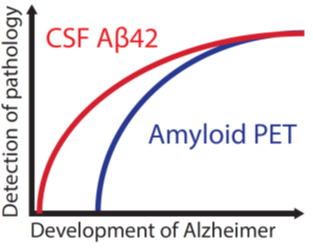
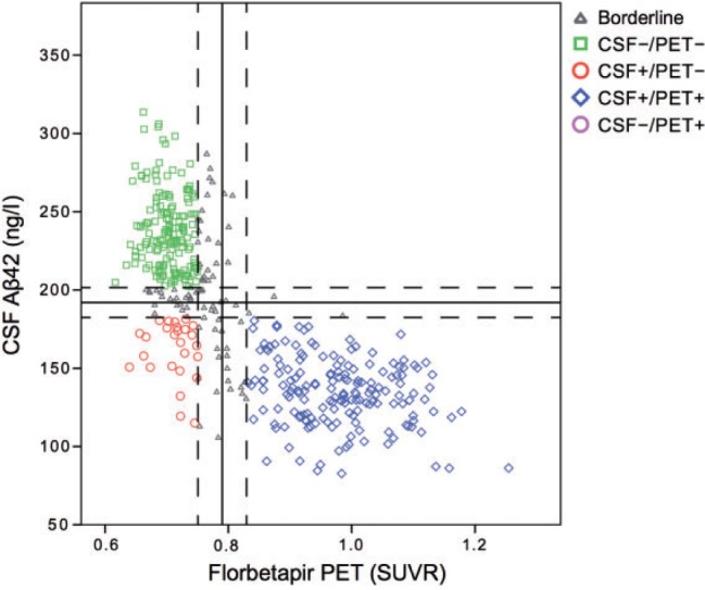
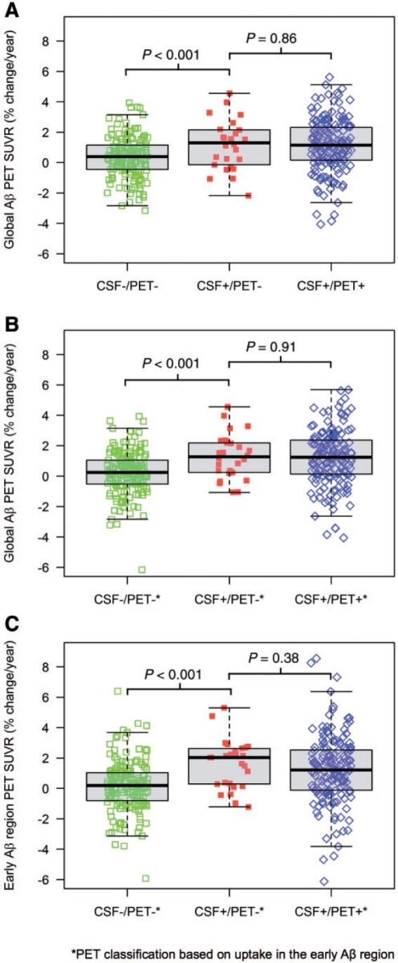
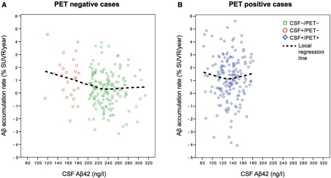
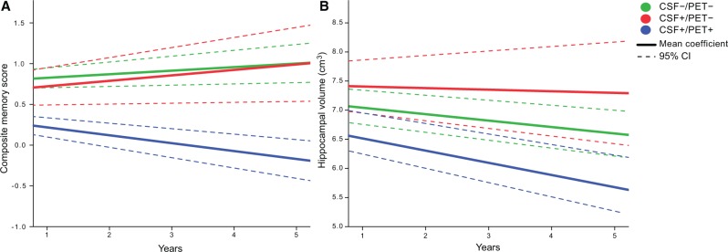
See Rabinovici (doi: 10.1093/brain/aww025 ) for a scientific commentary on this article. Cerebral accumulation of amyloid-β is thought to be the starting mechanism in Alzheimer’s disease. Amyloid-β can be detected by analysis of cerebrospinal fluid amyloid-β 42 or amyloid positron emission tomography, but it is unknown if any of the methods can identify an abnormal amyloid accumulation prior to the other. Our aim was to determine whether cerebrospinal fluid amyloid-β 42 change before amyloid PET during preclinical stages of Alzheimer’s disease. We included 437 non-demented subjects from the prospective, longitudinal Alzheimer’s Disease Neuroimaging Initiative (ADNI) study. All underwent 18 F-florbetapir positron emission tomography and cerebrospinal fluid amyloid-β 42 analysis at baseline and at least one additional positron emission tomography after a mean follow-up of 2.1 years (range 1.1–4.4 years). Group classifications were based on normal and abnormal cerebrospinal fluid and positron emission tomography results at baseline. We found that cases with isolated abnormal cerebrospinal fluid amyloid-β and normal positron emission tomography at baseline accumulated amyloid with a mean rate of 1.2%/year, which was similar to the rate in cases with both abnormal cerebrospinal fluid and positron emission tomography (1.2%/year, P = 0.86). The mean accumulation rate of those with isolated abnormal cerebrospinal fluid was more than three times that of those with both normal cerebrospinal fluid and positron emission tomography (0.35%/year, P = 0.018). The group differences were similar when analysing yearly change in standardized uptake value ratio of florbetapir instead of percentage change. Those with both abnormal cerebrospinal fluid and positron emission tomography deteriorated more in memory and hippocampal volume compared with the other groups ( P < 0.001), indicating that they were closer to Alzheimer’s disease dementia. The results were replicated after adjustments of different factors and when using different cut-offs for amyloid-β abnormality including a positron emission tomography classification based on the florbetapir uptake in regions where the initial amyloid-β accumulation occurs in Alzheimer’s disease. This is the first study to show that individuals who have abnormal cerebrospinal amyloid-β 42 but normal amyloid-β positron emission tomography have an increased cortical amyloid-β accumulation rate similar to those with both abnormal cerebrospinal fluid and positron emission tomography and higher rate than subjects where both modalities are normal. The results indicate that cerebrospinal fluid amyloid-β 42 becomes abnormal in the earliest stages of Alzheimer’s disease, before amyloid positron emission tomography and before neurodegeneration starts. See Rabinovici (doi: 10.1093/brain/aww025 ) for a scientific commentary on this article. The success of disease-modifying therapies in Alzheimer’s disease may depend on detecting the earliest signs of abnormal amyloid-β load. Palmqvist et al . show that CSF amyloid-β 42 becomes abnormal before amyloid PET and before neurodegeneration onset in preclinical Alzheimer’s disease. This may have implications for risk factor interventions and future therapies.
Overview
The claims and statistics of this paper haven’t been checked yet.
Run the check — free, no account needed. It reads the open-access text and checks the sources it cites.
Integrity signals
Registry
Cites
Statistics
Figures
Verification
Claims the paper makes
The claims and statistics of this paper haven’t been checked yet.
Run the check — free, no account needed. It reads the open-access text and checks the sources it cites.
Claims other papers make about this one
Each is checked against this paper’s own text: does it say what the citing paper says it does?
The cited source says what the claim attributes to it.
In the citing paper
“This may be due to a temporal offset between these two measures [121–123].”
From NIA‐AA Research Framework: Toward a biological definition of Alzheimer's disease (2018) · cites this paper as [122]
In the citing paper
“This indicates that individuals with AD may first be defined as being CSF+/PET−, and then progress to CSF+/PET+.”
From Earliest accumulation of β-amyloid occurs within the default-mode network and concurrently affects brain connectivity (2017) · cites this paper as [12]
In the citing paper
“those with abnormally low CSF levels of Aβ42, but with still normal Aβ levels detected by PET (CSF+/PET−), accumulated Aβ fibrils longitudinally at a similar rate as those who had both abnormal CSF levels of Aβ42 and abnormal levels of Aβ detected by PET (CSF+/PET+) and four times higher than those with normal biomarker results on both parameters (CSF−/PET−)12.”
From Earliest accumulation of β-amyloid occurs within the default-mode network and concurrently affects brain connectivity (2017) · cites this paper as [12]
In the citing paper
“In a recent study, we also showed that CSF+/PET− subjects accumulated Aβ at a similar rate as CSF+/PET+ subjects and about four times higher than CSF−/PET− subjects12.”
From Earliest accumulation of β-amyloid occurs within the default-mode network and concurrently affects brain connectivity (2017) · cites this paper as [12]
In the citing paper
“Mixture modeling statistics provide an unbiased threshold for abnormality (i.e., the diagnosis or status is not used in analysis) and it is a well-established method for defining Aβ PET cut-offs12, 17, 60, 61.”
From Earliest accumulation of β-amyloid occurs within the default-mode network and concurrently affects brain connectivity (2017) · cites this paper as [12]
In the citing paper
“reduced concentrations of Aβ42 in CSF and increased retention of Aβ tracers in the brain have been considered as early biomarkers of AD.255–258”
From Amyloid β-based therapy for Alzheimer’s disease: Challenges, successes and future (2023) · cites this paper as [258]
Self-check
Percentages, sums and reported differences re-computed from the paper’s own numbers.
The self-check needs the paper’s full text.
Citations
Cites
Folstein, M. F., Folstein, S. E., & McHugh, P. R. (1975) · Journal of Psychiatric Research · doi:10.1016/0022-3956(75)90026-6 · Cited by 88,282
Hirono, N., Mori, E., Ikejiri, Y., Imamura, T., Shimomura, T., Hashimoto, M., Yamashita, H., & Ikeda, M. (1997) · PubMed · Cited by 21,012
Kok, J. S., Berg, I. J., & Scherder, E. J. A. (2013) · Dementia and Geriatric Cognitive Disorders Extra · doi:10.1159/000353441 · Cited by 20,589
Older Adults' Reasons for Using Technology while Aging in Place
Peek, S. T. M., Luijkx, K. G., Rijnaard, M. D., Nieboer, M. E., van der Voort, C. S., Aarts, S., van Hoof, J., Vrijhoef, H. J., & Wouters, E. (2015) · Gerontology · doi:10.1159/000430949 · Cited by 19,852
Sensitivity and False Alarm Rate of a Fall Sensor in Long-Term Fall Detection in the Elderly
Kangas, M., Korpelainen, R., Vikman, I., Nyberg, L., & Jämsä, T. J. (2014) · Gerontology · doi:10.1159/000362720 · Cited by 19,007
McKhann, G. M., Knopman, D. S., Chertkow, H., Hyman, B. T., Jack, C. R., Kawas, C. H., Klunk, W. E., Koroshetz, W. J., Manly, J. J., Mayeux, R., et al. (2011) · Alzheimer's & Dementia · doi:10.1016/j.jalz.2011.03.005 · Cited by 18,657
Neuropathological stageing of Alzheimer-related changes.
Braak, H., & Braak, E. (1991) · Acta Neuropathologica · doi:10.1007/bf00308809 · Cited by 16,123 · cited 5×
“The results were robust using four different types of group classifications for CSF+/ and PET+/ and when classifying PET +/– based on amyloid-b deposition in regions that are affected in the initial phase of amyloid-b accumulation (Braak and Braak, 1991; Goedert, 2015).”
BackgroundMethodResultInfluentialAlbert, M. S., DeKosky, S. T., Dickson, D., Dubois, B., Feldman, H. H., Fox, N. C., Gamst, A., Holtzman, D. M., Jagust, W. J., Petersen, R. C., et al. (2011) · Alzheimer's & Dementia · doi:10.1016/j.jalz.2011.03.008 · Cited by 10,152 · cited 2×
“As the disease progresses, affected individuals show neurodegeneration and early clinical symptoms and may be diagnosed with mild cognitive impairment (MCI) (Albert et al., 2011).”
BackgroundThe Clinical Dementia Rating (CDR): Current version and scoring rules
Morris, J. C. (1993) · Neurology · doi:10.1212/wnl.43.11.2412-a · Cited by 9,882
Sperling, R. A., Aisen, P. S., Beckett, L. A., Bennett, D. A., Craft, S., Fagan, A. M., Iwatsubo, T., Jack, C. R., Kaye, J., Montine, T. J., et al. (2011) · Alzheimer's & Dementia · doi:10.1016/j.jalz.2011.03.003 · Cited by 7,155
Research works printed but not linked: 1
- Biogen. A Randomized, Blinded, Placebo-Controlled Single Ascending Dose Study of the Safety, Tolerability, and Pharmacokinetics of BIIB037 in Subjects with Mild to Moderate Alzheimer's Disease. In: AD/PD International Conference, Nice, France, 2015.
Cited by
Sorted by citations
Items 201–300 of 400
Is blood pTau a reliable indicator of the CSF status? A narrative review
Antonioni, A., Raho, E. M., & Lorenzo, F. D. (2023) · Neurological Sciences · doi:10.1007/s10072-023-07258-x · Cited by 20
Cerebrospinal Fluid Amyloid and Tau Biomarker Changes Across the Alzheimer Disease Clinical Spectrum
de Leeuw, D. M., Trieu, C., Vromen, E. M., Blujdea, E. R., Verberk, I. M. W., Duits, F. H., Teunissen, C. E., Pijnenburg, Y. A. L., van der Flier, W. M., van Harten, A. C., et al. (2025) · JAMA Network Open · doi:10.1001/jamanetworkopen.2025.19919 · Cited by 23
Milà‐Alomà, M., Shekari, M., Salvadó, G., Gispert, J. D., Arenaza‐Urquijo, E. M., Operto, G., Falcón, C., Vilor‐Tejedor, N., Grau‐Rivera, O., Sala‐Vila, A., et al. (2021) · Alzheimer s Research & Therapy · doi:10.1186/s13195-021-00863-y · Cited by 20
Hansen, É. D. O., Dias, N. S., Burgos, I. C. B., Costa, M. V., Teixeira‐Carvalho, A., Teixeira, A. L., Barbosa, I. G., dos Santos, L. A. V., Rosa, D. V., Ribeiro, A. J. F., et al. (2021) · Frontiers in Psychiatry · doi:10.3389/fpsyt.2021.716686 · Cited by 19
Canal-García, A., Gómez-Ruiz, E., Mijalkov, M., Chang, Y.-W., Volpe, G., Pereira, J. B., & Initiative, A. D. N. (2021) · Cerebral Cortex · doi:10.1093/cercor/bhab429 · Cited by 21
Protein fibril length in cerebrospinal fluid is increased in Alzheimer’s disease
Nirmalraj, P. N., Schneider, T. R., Lüder, L., & Felbecker, A. (2023) · Communications Biology · doi:10.1038/s42003-023-04606-7 · Cited by 17
Konijnenberg, E., den Braber, A., ten Kate, M., Tomassen, J., Mulder, S. D., Yaqub, M. M., Teunissen, C. E., Lammertsma, A. A., van Berckel, B. N. M., Scheltens, P., et al. (2019) · Neurobiology of Aging · doi:10.1016/j.neurobiolaging.2019.01.006 · Cited by 17
Meyer, P., McSweeney, M., Gonneaud, J., & Villeneuve, S. (2019) · Progress in molecular biology and translational science · doi:10.1016/bs.pmbts.2019.05.001 · Cited by 20
Laaker, C. J., Kovacs, K. G., Baenen, C. M., Hsu, M., Port, J. M., Vrba, S. M., Kumar, M., Hubacek, A. J., Herbáth, M., Priyathilaka, T. T., et al. (2025) · Science Advances · doi:10.1126/sciadv.adt9591 · Cited by 20
Engels, N., Riphagen, J. M., Egroo, M. V., Koops, E. A., Smegal, L. F., Becker, J. A., Prokopiou, P. C., Bueichekú, E., Kwong, K. K., Rentz, D. M., et al. (2024) · Annals of Neurology · doi:10.1002/ana.27022 · Cited by 19
Giacomucci, G., Mazzeo, S., Bagnoli, S., Casini, M., Padiglioni, S., Polito, C., Berti, V., Balestrini, J., Ferrari, C., Lombardi, G., et al. (2021) · Journal of Personalized Medicine · doi:10.3390/jpm11010047 · Cited by 16
Deciphering the factors that influence participation in studies requiring serial lumbar punctures
Day, G. S., Rappai, T., Sathyan, S., & Morris, J. C. (2020) · Alzheimer s & Dementia Diagnosis Assessment & Disease Monitoring · doi:10.1002/dad2.12003 · Cited by 16
Lin, R.-R., Xue, Y., Li, X., Chen, Y.-H., Tao, Q., & Wu, Z. (2021) · Frontiers in Aging Neuroscience · doi:10.3389/fnagi.2021.718959 · Cited by 15
Longitudinal changes in hippocampal texture from healthy aging to Alzheimer’s disease
Wearn, A. R., Rakêt, L. L., Collins, D. L., Spreng, R. N., & Initiative, A. D. N. (2023) · Brain Communications · doi:10.1093/braincomms/fcad195 · Cited by 15
Agreement of cerebrospinal fluid biomarkers and amyloid-PET in a multicenter study
Guillén, N., Contador, J., Buongiorno, M., Álvarez, I., Cullell, N., Alcolea, D. A., Lleó, A., Fortea, J., Piñol‐Ripoll, G., Carnes, A., et al. (2023) · European Archives of Psychiatry and Clinical Neuroscience · doi:10.1007/s00406-023-01701-y · Cited by 17
Continuous β-Amyloid CSF/PET Imbalance Model to Capture Alzheimer Disease Heterogeneity
Mastenbroek, S. E., Sala, A., García, D. V., Shekari, M., Salvadó, G., Lorenzini, L., Pieperhoff, L., Wink, A. M., Alves, I. L., Wolz, R., et al. (2024) · Neurology · doi:10.1212/wnl.0000000000209419 · Cited by 16
Self-citationWhich MCI Patients Should be Included in Prodromal Alzheimer Disease Clinical Trials?
Grill, J. D., Nuño, M. M., & Gillen, D. L. (2019) · Alzheimer Disease & Associated Disorders · doi:10.1097/wad.0000000000000303 · Cited by 15
Brain Amyloid Pathology and Cognitive Function
Visser, P. J., & Tijms, B. M. (2017) · JAMA · doi:10.1001/jama.2017.6895 · Cited by 15
Amyloid deposition in adults with drug‐resistant temporal lobe epilepsy
Fonseca, E., Lallana, S., Ortega, G., Cano, A. C., Sarria‐Estrada, S., Pareto, D., Quintana, M., Lorenzo‐Bosquet, C., López‐Maza, S., Gifreu, A., et al. (2024) · Epilepsia · doi:10.1111/epi.18142 · Cited by 15
Alzheimer disease biomarkers may aid in the prognosis of MCI cases initially reverted to normal
Vermunt, L., van Paasen, A. J. L., Teunissen, C. E., Scheltens, P., Visser, P. J., Tijms, B. M., & Initiative, A. D. N. (2019) · Neurology · doi:10.1212/wnl.0000000000007609 · Cited by 14
Deolankar, S. C., Patil, A. H., Koyangana, S. G., Subbannayya, Y., Prasad, T. S. K., & Modi, P. K. (2019) · OMICS A Journal of Integrative Biology · doi:10.1089/omi.2019.0085 · Cited by 14
Shen, X., Miao, D., Li, J., Tan, C., Cao, X., Tan, L., & Yu, J. (2019) · Aging · doi:10.18632/aging.101783 · Cited by 14
Benina, N., Buitrago, L., Simone, F. I. D., Radwan, R. R., Miller, M. C., Martin, K., Dickson, D. L., Ho, S. G., Moghekar, A., Albert, M., et al. (2026) · Brain · doi:10.1093/brain/awag001 · Cited by 15
Self-citationLibrizzi, D., Cabanel, N., Zavorotnyy, M., Riehl, E., Kircher, T. T. J., Luster, M., & Yousefi, B. H. (2021) · Molecules · doi:10.3390/molecules26051282 · Cited by 13
Patterns of Early Neocortical Amyloid-β Accumulation: A PET Population-Based Study
Lecy, E., Min, H.-K. P., Apgar, C. J., Maltais, D. D., Lundt, E. S., Albertson, S. M., Senjem, M. L., Schwarz, C. G., Botha, H., Graff‐Radford, J., et al. (2024) · Journal of Nuclear Medicine · doi:10.2967/jnumed.123.267150 · Cited by 18
Spatial extent as a sensitive amyloid‐PET metric in preclinical Alzheimer's disease
Farrell, M. E., Thibault, E. G., Becker, J. A., Price, J. C., Healy, B. C., Hanseeuw, B. J., Buckley, R. F., Jacobs, H. I. L., Schultz, A. P., Chen, C. D., et al. (2024) · Alzheimer s & Dementia · doi:10.1002/alz.14036 · Cited by 13
Amyloid solutions: lecanemab, gantenerumab, and donanemab in the treatment of Alzheimer's disease
Ameen, T. B., Ali, U., Salma, O., Samee, M. A., Abbas, S. M. I., Kashif, S. N., Arifi, M. A., Ali, M., Khowaja, M., Ali, S. M. S., et al. (2025) · The Egyptian Journal of Neurology Psychiatry and Neurosurgery · doi:10.1186/s41983-025-00968-3 · Cited by 20
Kulic, L., & Unschuld, P. G. (2016) · Current Opinion in Neurology · doi:10.1097/wco.0000000000000399 · Cited by 13
Jiang, C., Wang, Q., Xie, S., Chen, Z., Fu, L., Peng, Q., Liang, Y., Guo, H., Guo, T., & Initiative, A. D. N. (2022) · Brain Communications · doi:10.1093/braincomms/fcac084 · Cited by 14
Miller, M. R., Lee, Y. F., & Kastanenka, K. V. (2023) · Alzheimer s & Dementia · doi:10.1002/alz.13111 · Cited by 12
Mieling, M., Göttlich, M., Yousuf, M., Bunzeck, N., & Initative, T. A. D. N. (2023) · Brain Communications · doi:10.1093/braincomms/fcad262 · Cited by 13
Recent advances in pre-clinical diagnosis of Alzheimer’s disease
Zhou, J., Benoit, M., & Sharoar, M. G. (2021) · Metabolic Brain Disease · doi:10.1007/s11011-021-00733-4 · Cited by 14
Allen, A. C., Efrem, M., Umadevi, M., Guarino‐Hotz, M., Foley, A. R., Raskatov, J. A., Song, C., Lindley, S. A., Li, J., Chen, B., et al. (2021) · The Journal of Physical Chemistry B · doi:10.1021/acs.jpcb.1c06776 · Cited by 12
Jethwa, K. D., Dhillon, P. S., Meng, D., Auer, D. P., & Initiative, A. D. N. (2019) · American Journal of Neuroradiology · doi:10.3174/ajnr.a6313 · Cited by 12
How Microglia Manages Non-cell Autonomous Vicious Cycling of Aβ Toxicity in the Pathogenesis of AD
Seol, Y., Ki, S., Ryu, H. L., Chung, S., Lee, J.-H., & Ryu, H. (2020) · Frontiers in Molecular Neuroscience · doi:10.3389/fnmol.2020.593724 · Cited by 12
Teng, E., Manser, P. T., Shah, M. V., Pickthorn, K., Hu, N., Djakovic, S., Swendsen, H., Blendstrup, M., Faccin, G., Ostrowitzki, S., et al. (2022) · The Journal of Prevention of Alzheimer s Disease · doi:10.14283/jpad.2022.101 · Cited by 11
Machine learning prediction of future amyloid beta positivity in amyloid-negative individuals
Moradi, E., Prakash, M., Hall, A., Solomon, A., Strange, B. A., Tohka, J., & Initiative, A. D. N. (2024) · Alzheimer s Research & Therapy · doi:10.1186/s13195-024-01415-w · Cited by 13
Hajjar, I. M., Neal, R. M., Yang, Z., & Lah, J. J. (2024) · Alzheimer s & Dementia Diagnosis Assessment & Disease Monitoring · doi:10.1002/dad2.12581 · Cited by 10
Rathee, S., Sen, D., Pandey, V., & Jain, S. (2024) · Current Drug Targets · doi:10.2174/0113894501320096240627071400 · Cited by 16
Cacciaglia, R., Falcón, C., Sánchez‐Benavides, G., Brugulat‐Serrat, A., Alomà, M. M., Suárez‐Calvet, M., Molinuevo, J. L., Fauria, K., Minguillón, C., Kollmorgen, G., et al. (2025) · Alzheimer s & Dementia · doi:10.1002/alz.14415 · Cited by 14
Yu, H., Tan, L., Jiao, M.-J., Lv, Y.-J., Zhang, X.-H., Tan, C., Xu, W., & Initiative, A. D. N. (2025) · BMC Medicine · doi:10.1186/s12916-025-04092-0 · Cited by 15
Alzheimer Disease Biomarkers in Clinical Practice: A Blood-Based Diagnostic Revolution
Paczynski, M., & Day, G. S. (2022) · Journal of Primary Care & Community Health · doi:10.1177/21501319221141178 · Cited by 11
Weidman, D. A., ZAMRINI, E. Y., Sabbagh, M. N., Jacobson, S. A., Burke, A. D., Belden, C. M., Powell, J. J., Bhalla, N., Roontiva, A., Kuang, X., et al. (2017) · Neurocase · doi:10.1080/13554794.2017.1290806 · Cited by 11
Microstructural white matter injury contributes to cognitive decline: Besides amyloid and tau
Hu, H., Li, H., Gong, W., Huang, S., Fu, Y., Hu, H., Dong, Q., Cheng, W., Tan, L., Cui, M., et al. (2025) · The Journal of Prevention of Alzheimer s Disease · doi:10.1016/j.tjpad.2024.100037 · Cited by 11
Shi, D., Ye, C., Li, A., Sun, P., Lan, G., Zhang, L., Zhou, X. Y., Zhou, L., Liu, Z., Guo, T., et al. (2025) · Alzheimer s & Dementia · doi:10.1002/alz.70080 · Cited by 9
Ennis, G. E., Koscik, R. L., Ma, Y., Jonaitis, E. M., Hulle, C. A. V., Betthauser, T. J., Randall, A. M., Chin, N. A., Engelman, C. D., Anderson, R. M., et al. (2021) · Alzheimer s & Dementia Diagnosis Assessment & Disease Monitoring · doi:10.1002/dad2.12220 · Cited by 10
Biomarkers and Risk Assessment of Alzheimer’s Disease in Low- and Middle-Income Countries
Adewale, B. A., Coker, M. M., Ogunniyi, A. O., Kalaria, R. N., & Akinyemi, R. (2023) · Journal of Alzheimer s Disease · doi:10.3233/jad-221030 · Cited by 11
Brendel, M., Parvizi, T., Gnörich, J. S., Topfstedt, C. E., Büerger, K., Janowitz, D., Rauchmann, B., Perneczky, R., Kurz, C., Mehrens, D., et al. (2024) · Alzheimer s & Dementia Diagnosis Assessment & Disease Monitoring · doi:10.1002/dad2.70031 · Cited by 13
Mieling, M., Yousuf, M., Bunzeck, N., Initiative, A. D. N., Weiner, M. W., Aisen, P., Petersen, R. C., Jack, C. R., Jagust, W., Landau, S., et al. (2025) · GeroScience · doi:10.1007/s11357-025-01626-5 · Cited by 11
Cogswell, P. M., Wiste, H. J., Therneau, T. M., Griswold, M. E., Mattsson, N., Palmqvist, S., Binette, A. P., Stomrud, E., Bateman, R. J., Barthélemy, N. R., et al. (2025) · Alzheimer s & Dementia · doi:10.1002/alz.70625 · Cited by 9
Self-citationSchauer, S. P., Mylott, W. R., Jr., Yuan, M., Jenkins, R. G., Mathews, W. R., Honigberg, L. A., & Wildsmith, K. R. (2018) · Alzheimer s Research & Therapy · doi:10.1186/s13195-018-0445-0 · Cited by 9
Xia, H., Initiative, A. D. N., Wang, M., Li, J., Tan, C., Cao, X., Tan, L., & Yu, J. (2019) · Journal of Alzheimer s Disease · doi:10.3233/jad-180971 · Cited by 10
Quattrini, G., Marizzoni, M., Pizzini, F. B., Galazzo, I. B., Aiello, M., Didic, M., Soricelli, A., Albani, D., Romano, M., Blin, O., et al. (2021) · Journal of Alzheimer s Disease · doi:10.3233/jad-210531 · Cited by 10
Monfared, A. A. T., Houghton, K., Zhang, Q., Mauskopf, J., & Initiative, A. D. N. (2022) · Neurology and Therapy · doi:10.1007/s40120-022-00326-y · Cited by 9
Chun, M. Y., Jang, H., Kim, H. J., Kim, J. P., Gallacher, J., Allué, J. A., Sarasa, L., Castillo, S., Pascual‐Lucas, M., Na, D. L., et al. (2023) · Frontiers in Aging Neuroscience · doi:10.3389/fnagi.2023.1126799 · Cited by 9
Allegri, R., Méndez, P. C., Russo, M. J., Cohen, G., Calandri, I. L., Campos, J., Nahas, F., Surace, E. I., Vázquez, S., & Sevlever, G. E. (2018) · Neurología · doi:10.1016/j.nrl.2017.12.011 · Cited by 10
Ashford, M. T., Neuhaus, J. M., Jin, C., Camacho, M. R., Fockler, J., Truran, D., Mackin, R. S., Rabinovici, G. D., Weiner, M. W., & Nosheny, R. L. (2020) · Alzheimer s & Dementia Diagnosis Assessment & Disease Monitoring · doi:10.1002/dad2.12102 · Cited by 8
Biofluid biomarkers for Alzheimer’s disease
Wang, S., Xie, S., Zheng, Q., Zhang, Z., Wang, T., & Zhang, G. (2024) · Frontiers in Aging Neuroscience · doi:10.3389/fnagi.2024.1380237 · Cited by 9
Studart‐Neto, A., Barbosa, B. J. A. P., Coutinho, A. M., de Souza, L. C., Schilling, L. P., da Silva, M. N. M., Castilhos, R. M., Bertolucci, P. H. F., Borelli, W. V., Gomes, H. R., et al. (2024) · Dementia & Neuropsychologia · doi:10.1590/1980-5764-dn-2024-c001 · Cited by 11
Multimorbidity and fluid biomarkers of Alzheimer's disease: a systematic review
Valletta, M., Canevelli, M., Gasparini, F., Buscarnera, S., Salzillo, M., Triolo, F., Calderón‐Larrañaga, A., Marengoni, A., Vetrano, D. L., & Grande, G. (2025) · European Geriatric Medicine · doi:10.1007/s41999-025-01222-y · Cited by 8
MacGregor, E. A., Okonkwo, R. I., Detke, H. C., Polavieja, P., Fernandes, M. S., & Pavlovic, J. M. (2023) · Headache The Journal of Head and Face Pain · doi:10.1111/head.14652 · Cited by 9
Concatenating plasma p-tau to Alzheimer’s disease
Tijms, B. M., & Teunissen, C. E. (2020) · Brain · doi:10.1093/brain/awaa422 · Cited by 8
Lozupone, M., Dibello, V., Resta, E., Sardone, R., Castellana, F., Zupo, R., Lampignano, L., Bortone, I., Mollica, A., Berardino, G., et al. (2025) · Expert Review of Neurotherapeutics · doi:10.1080/14737175.2025.2500752 · Cited by 9
Amyloid biomarkers: pushing the limits of early detection
Rabinovici, G. D. (2016) · Brain · doi:10.1093/brain/aww025 · Cited by 7
Albright, J., Ashford, M. T., Jin, C., Neuhaus, J. M., Rabinovici, G. D., Truran, D., Maruff, P. T., Mackin, R. S., Nosheny, R. L., & Weiner, M. W. (2021) · Alzheimer s & Dementia Diagnosis Assessment & Disease Monitoring · doi:10.1002/dad2.12207 · Cited by 6
Teipel, S. J., Dyrba, M., Vergallo, A., Lista, S., Habert, M. O., Potier, M., Lamari, F., Dubois, B., Hampel, H. J., & Grothe, M. J. (2021) · Frontiers in Aging Neuroscience · doi:10.3389/fnagi.2021.748198 · Cited by 7
Abiose, O., Rutledge, J., Losada, P. M., Belloy, M. E., Wilson, E. N., He, Z., Trelle, A. N., Channappa, D., Romero, A., Park, J., et al. (2023) · Alzheimer s & Dementia · doi:10.1002/alz.13576 · Cited by 6
Subcortical amyloid relates to cortical morphology in cognitively normal individuals
Rahayel, S., Bocti, C., Sévigny-Dupont, P., Joannette, M., Lavallée, M. M., Nikelski, J., Chertkow, H. M., & Joubert, S. (2019) · European Journal of Nuclear Medicine and Molecular Imaging · doi:10.1007/s00259-019-04446-w · Cited by 7
Discordant Alzheimer's neurodegenerative biomarkers and their clinical outcomes
Guo, Y., Li, H., Tan, L., Chen, S., Yang, Y., Ma, Y., Zuo, C., Dong, Q., Tan, L., Yu, J., et al. (2020) · Annals of Clinical and Translational Neurology · doi:10.1002/acn3.51196 · Cited by 7
Prevalence Rates of Amyloid Positivity—Updates and Relevance
Young, C. B., & Mormino, E. C. (2022) · JAMA Neurology · doi:10.1001/jamaneurol.2021.5225 · Cited by 7
Debatisse, J., Leng, F., Ashraf, A., & Edison, P. (2025) · Cells · doi:10.3390/cells14030155 · Cited by 6
Gao, F., Initiative, A. D. N., Zhang, P., Gao, J., Song, J., & Chi, S. (2022) · Journal of Alzheimer s Disease · doi:10.3233/jad-220716 · Cited by 7
Clinical significance of plasma candidate biomarkers of Alzheimer’s Disease
Lewczuk, P., Łukaszewicz‐Zając, M., Kornhuber, J., & Mroczko, B. (2024) · Neurologia i Neurochirurgia Polska · doi:10.5603/pjnns.100675 · Cited by 7
CSF phosphorylated tau as an indicator of subsequent tau accumulation
Cogswell, P. M., Wiste, H. J., Mielke, M. M., Schwarz, C. G., Weigand, S. D., Lowe, V. J., Therneau, T. M., Knopman, D. S., Graff‐Radford, J., Vemuri, P., et al. (2022) · Neurobiology of Aging · doi:10.1016/j.neurobiolaging.2022.02.015 · Cited by 8
Do we still need positron emission tomography for early Alzheimer’s disease diagnosis?
Lewczuk, P., & Kornhuber, J. (2016) · Brain · doi:10.1093/brain/aww168 · Cited by 6
Sacchi, L., Carandini, T., Fumagalli, G., Pietroboni, A. M., Contarino, V. E., Siggillino, S., Arcaro, M., Fenoglio, C., Zito, F., Marotta, G., et al. (2021) · Journal of Alzheimer s Disease · doi:10.3233/jad-210593 · Cited by 6
Neural network training with highly incomplete medical datasets
Chang, Y.-W., Natali, L., Jamialahmadi, O., Romeo, S., Pereira, J. B., & Volpe, G. (2022) · Machine Learning Science and Technology · doi:10.1088/2632-2153/ac7b69 · Cited by 6
Dou, K., Initiative, A. D. N., Zhang, C. M., Tan, C., Xu, W., Li, J., Cao, X., Tan, L., & Yu, J. (2019) · Aging · doi:10.18632/aging.102125 · Cited by 6
Taking the A Train? Limited Consistency of Aβ42 and the Aβ42/40 Ratio in the AT(N) Classification
Gouilly, D., Tisserand, C., Nogueira, L., Saint‐Lary, L., Rousseau, V., Bénaiteau, M., Rafiq, M., Carlier, J., Milongo-Rigal, E., Pagès, J., et al. (2021) · Journal of Alzheimer s Disease · doi:10.3233/jad-210236 · Cited by 6
Ollen‐Bittle, N., Wang, W., Bean, K. M., Zhao, S., Buzatto, A. Z., Dong, Y., Li, L., & Whitehead, S. N. (2025) · Journal of Neurochemistry · doi:10.1111/jnc.70309 · Cited by 6
An overview in interactions and biosensors of multiple biomarkers of Alzheimer’s disease
Chen, X., Wei, Y., Wang, X., Liu, X., & Wang, X. (2025) · Chemical Engineering Journal · doi:10.1016/j.cej.2025.161121 · Cited by 5
Abbatantuono, C., Alfeo, F., Clemente, L., Lancioni, G. E., Caro, M. F. D., Livrea, P., & Taurisano, P. (2023) · Brain Sciences · doi:10.3390/brainsci13101443 · Cited by 7
Petersen, R. C., Graf, A., Brady, C., Santi, S. D., Florian, H., Landen, J. W., Pontecorvo, M., RANDOLPH, C. C., Sink, K. M., Carrillo, M. C., et al. (2023) · Alzheimer s & Dementia Translational Research & Clinical Interventions · doi:10.1002/trc2.12434 · Cited by 4
Challenges in Alzheimer’s Disease Diagnostic Work-Up: Amyloid Biomarker Incongruences
Lombardi, G., Pupi, A., Bessi, V., Polito, C., Padiglioni, S., Ferrari, C., Lucidi, G., Berti, V., Cristofaro, M. T. D., Piaceri, I., et al. (2020) · Journal of Alzheimer s Disease · doi:10.3233/jad-200119 · Cited by 5
Aggregation-induced emission materials for protein fibrils imaging
Patel, K., Shah, S. K. H., & Prabhakaran, P. (2021) · Progress in molecular biology and translational science · doi:10.1016/bs.pmbts.2021.06.011 · Cited by 5
Churkina, A. S., Shakhov, A. S., Kotlobay, A. A., & Alieva, I. B. (2022) · International Journal of Molecular Sciences · doi:10.3390/ijms232415533 · Cited by 5
Reply: Do we still need positron emission tomography for early Alzheimer’s disease diagnosis?
Palmqvist, S., Mattsson, N., & Hansson, O. (2016) · Brain · doi:10.1093/brain/aww169 · Cited by 5
Self-citationPaitel, E. R., Pettigrew, C. A., Callow, D. D., Moghekar, A., Miller, M. I., Faria, A. V., Oishi, K., Albert, M., & Soldan, A. (2025) · Human Brain Mapping · doi:10.1002/hbm.70398 · Cited by 4
Guo, F., Tan, M., Hu, H., Ou, Y., Zhang, M., Sheng, Z.-H., Chi, H., Tan, L., & Initiative, A. D. N. (2024) · Journal of Alzheimer s Disease · doi:10.3233/jad-240372 · Cited by 4
Management of Alzheimer's disease takes a leap forward
Frisoni, G. B., & Hansson, O. (2021) · The Lancet Neurology · doi:10.1016/s1474-4422(21)00198-8 · Cited by 4
Self-citationWang, T., Zhang, W., Maclin, J. M. A., Xu, H. J., Hong, B., Yan, F., Liu, Y., He, H., Liang, H., Li, C., et al. (2024) · Journal of Alzheimer s Disease · doi:10.3233/jad-231446 · Cited by 4
Biomarkers study in atypical dementia: proof of a diagnostic work-up
Lombardi, G., Polito, C., Berti, V., Ferrari, C., Lucidi, G., Bagnoli, S., Piaceri, I., Nacmias, B., Pupi, A., & Sorbi, S. (2018) · Neurological Sciences · doi:10.1007/s10072-018-3400-8 · Cited by 4
Neuroimaging Findings in Mild Cognitive Impairment
Massa, F., Bauckneht, M., Peira, E., Lapucci, C., Picco, A., Capitanio, S., Arnaldi, D., Roccatagliata, L., Chincarini, A., & Nobili, F. M. (2020) · Springer eBooks · doi:10.1007/978-3-030-53168-3_12 · Cited by 4
Dynamic measurements of β-amyloid accumulation
Sánchez‐Juan, P., & Seshadri, S. S. (2017) · Neurology · doi:10.1212/wnl.0000000000004344 · Cited by 3
Xu, C., Xu, E., Xiao, Y., Yang, D., Guorong Wu, A., & Chen, M. (2025) · International Journal of Biological Macromolecules · doi:10.1016/j.ijbiomac.2025.142887 · Cited by 3
Plasma Aβ42/40 predicts progression from Aβ-amyloid negative to positive PET scans
Feizpour, A., Doré, V., Bourgeat, P., Doecke, J. D., Cánovas, R., Laws, S. M., Porter, T., Huang, K., Fowler, C. J., Martins, R. N., et al. (2026) · The Journal of Prevention of Alzheimer s Disease · doi:10.1016/j.tjpad.2025.100455 · Cited by 4
Alzheimer's disease polygenic risk in early‐ and late‐onset Alzheimer's disease
Pentchev, J. V., Jackson, T., Khan, N., Rosewood, T. J., Huang, Y.-N., Nho, K. T., Saykin, A. J., Eloyan, A., Taurone, A., Thangarajah, M., et al. (2026) · Alzheimer s & Dementia · doi:10.1002/alz.71066 · Cited by 3
Nerattini, M., Rubino, F., Arnone, A., Polito, C., Mazzeo, S., Lombardi, G., Puccini, G., Nacmias, B., Cristofaro, M. T. D., Sorbi, S., et al. (2021) · Neurological Sciences · doi:10.1007/s10072-021-05704-2 · Cited by 4
Research progress on humoral biomarkers of Alzheimer’s disease: A review
Zhang, H., Fu, X., Yang, M., Song, X., Li, M., & Wang, X. (2024) · Medicine · doi:10.1097/md.0000000000038978 · Cited by 4
Biomarkers for Alzheimer's Disease – An Overview
Tyagi, U., Tripathy, D. B., Gupta, A., Bisht, K., & Nain, A. (2024) · Macromolecular Symposia · doi:10.1002/masy.202200218 · Cited by 3
Metadata
- Authors
- Palmqvist, S., Mattsson, N., Hansson, O., & Initiative, F. T. A. D. N.
- Affiliations
- Lund University (SE)
- Corresponding author
- Palmqvist, Sebastian
- Hansson, Oskar
- Venue
- Brain
- Publisher
- Oxford University Press (OUP)
- Published
- 2016-03-02
- Volume
- 139
- Issue
- 4
- Pages
- 1226-1236
- ISSN
- 0006-8950 (ISSN-L)
- 1460-2156
- PMC
- PMC4806222
- OpenAlex
- W2302528231
- Cited by
- 400
- References
- 59
- Citations by year
- 2026: 32 · 2025: 41 · 2024: 33 · 2023: 31 · 2022: 36 · 2021: 58 · 2020: 50 · 2019: 43 · 2018: 38 · 2017: 25 · 2016: 11
- FWCI
- 34.64
- Type
- journal article
- Language
- en
- Open access
- hybrid
- doi.org
- Licence
- cc-by-nc
- Locations
- Brain (publishedVersion, cc-by-nc)
- Europe PMC (PubMed Central) (publishedVersion, cc-by-nc)
- PubMed Central (publishedVersion, cc-by-nc)
- PubMed (publishedVersion)
- Lund University Publications (Lund University) (submittedVersion)
- HighWire Press Open Archive (submittedVersion)
- Funders
- Lunds Universitet
- National Institutes of Health (5u01ag024904-10)
- Canadian Institutes of Health Research
- National Institute on Aging (U01-AG-024904)
- Topics
- Health Sciences › Medicine › Psychiatry and Mental health › Dementia and Cognitive Impairment Research
- Life Sciences › Neuroscience › Cellular and Molecular Neuroscience › Alzheimer's disease research and treatments
- Health Sciences › Medicine › Clinical Neurology › Cerebrospinal fluid and hydrocephalus
- Keywords
- cerebrospinal fluid amyloid-beta 42, amyloid PET, florbetapir, preclinical Alzheimer's disease, standardized uptake value ratio, memory decline, hippocampal volume, Alzheimer's dementia, biomarker comparison, ADNI cohort, longitudinal follow-up
- MeSH terms
- Aged, Aged, 80 and over, Alzheimer Disease, Cerebral Cortex, Cross-Sectional Studies, Female, Humans, Male, Middle Aged, Peptide Fragments, Prospective Studies, Amyloid beta-Peptides, Early Diagnosis, Positron-Emission Tomography
About these checks
Registry, retraction, Cites and statistics results are computed, not judged, with one exception: a printed reference that no index lists is read once by a model before it counts as a citation error. Claims are extracted by a model and kept only when their quote appears verbatim in the text; each cited source is then checked, with a model’s help, for whether it says what the claim relies on. Those verdicts can be wrong — read the claim’s quote and the cited source before relying on one.
Statistics and arithmetic are recomputed, not judged.
Confidence bands are shown only for fields calibrated on our annotated test set. None is calibrated yet, so no band is shown.
What counts as a claim ›How we measure confidence ›How we check this ›
Are you an author, or do you see a problem with this page? Request a review
