

Visual dysfunction in Parkinson’s disease
Open accessThe DOI matches its Crossref record; 233 of the 233 references it cites were found in a registry or an index.
Abstract
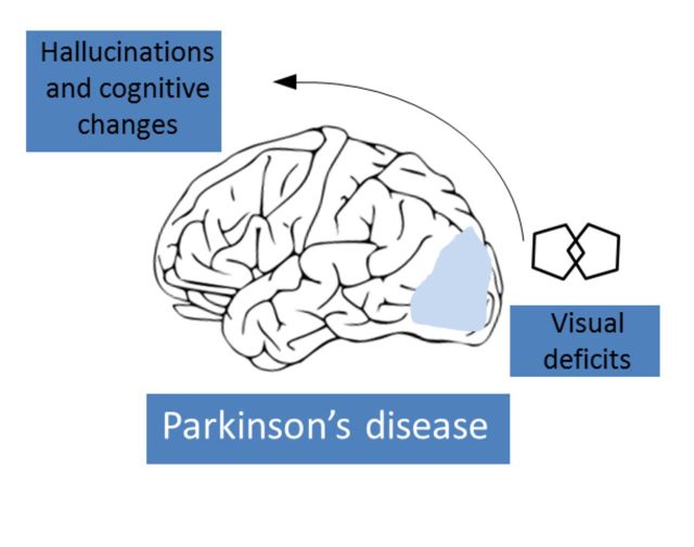
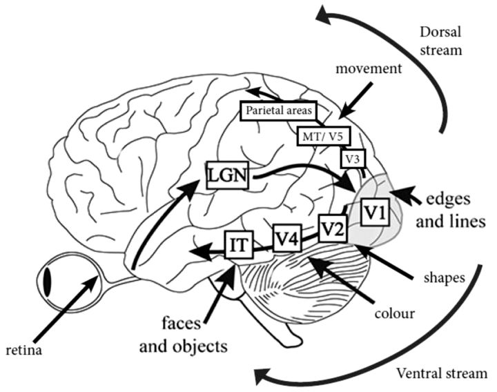
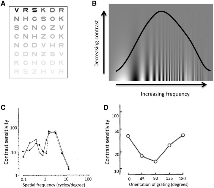
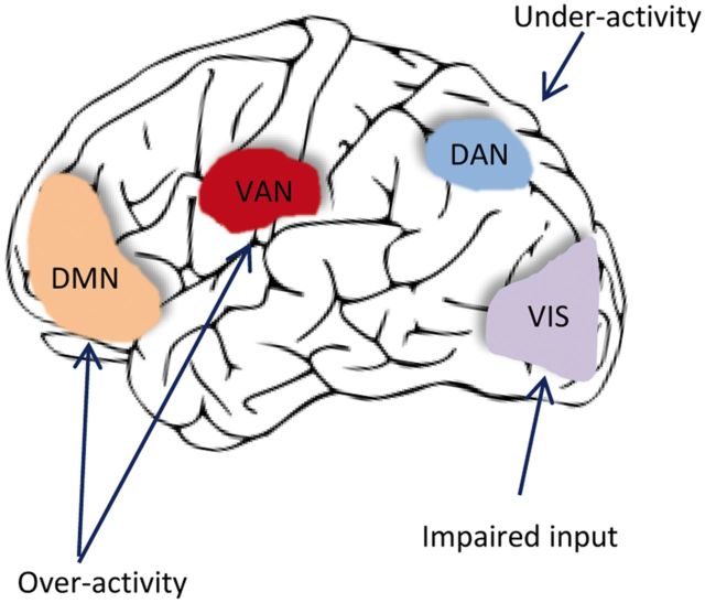
Patients with Parkinson's disease have a number of specific visual disturbances. These include changes in colour vision and contrast sensitivity and difficulties with complex visual tasks such as mental rotation and emotion recognition. We review changes in visual function at each stage of visual processing from retinal deficits, including contrast sensitivity and colour vision deficits to higher cortical processing impairments such as object and motion processing and neglect. We consider changes in visual function in patients with common Parkinson's disease-associated genetic mutations including GBA and LRRK2 . We discuss the association between visual deficits and clinical features of Parkinson's disease such as rapid eye movement sleep behavioural disorder and the postural instability and gait disorder phenotype. We review the link between abnormal visual function and visual hallucinations, considering current models for mechanisms of visual hallucinations. Finally, we discuss the role of visuo-perceptual testing as a biomarker of disease and predictor of dementia in Parkinson's disease.
Overview
The claims and statistics of this paper haven’t been checked yet.
Free: it reads the open-access text and checks the first 5 claims that cite a source we can check. Create a free account to check every claim.
Integrity signals
Registry
Cites
Statistics
Figures
Verification
Claims the paper makes
The claims and statistics of this paper haven’t been checked yet.
Free: it reads the open-access text and checks the first 5 claims that cite a source we can check. Create a free account to check every claim.
Self-check
Percentages, sums and reported differences re-computed from the paper’s own numbers.
The self-check needs the paper’s full text.
Citations
Cites
Items 201–233 of 233
Nagamachi, S., Wakamatsu, H., Kiyohara, S., Fujita, S., Futami, S., Tamura, S., Nakazato, M., Yamashita, S., Arita, H., Nishii, R., et al. (2008) · Annals of Nuclear Medicine · doi:10.1007/s12149-008-0156-x · Cited by 41 · cited 1×
“…have been reported using PET in occipital cortex (Eberling et al., 1994; Bohnen et al., 1999) and occipital and parietal hypoperfusion in Parkinson’s disease can be detected by measuring regional cerebral blood flow (Abe et al., 2003), that worsens with disease progression (Nagamachi et al., 2008).”
BackgroundManni, R., Sinforiani, E., Pacchetti, C., Zucchella, C., Cremascoli, R., & Terzaghi, M. (2013) · International Journal of Psychophysiology · doi:10.1016/j.ijpsycho.2013.04.003 · Cited by 40 · cited 1×
“Visuo-perceptual deficits have been reported in patients with RBD (who have not yet developed Parkinson’s disease), with impairments in colour vision and visuospatial construction (Boeve et al., 1998; Ferini-Strambi et al., 2004; Postuma et al., 2009; Manni et al., 2013).”
BackgroundTask-related laterality effects in the lateral occipital complex
Large, M., Aldcroft, A., & Vilis, T. (2006) · Brain Research · doi:10.1016/j.brainres.2006.10.023 · Cited by 40 · cited 1×
“In the healthy brain, object processing occurs in V2, V4 and the lateral occipital complex, especially in the right hemisphere (Grill-Spector et al., 1999; Kourtzi and Kanwisher, 2000; Large et al., 2007).”
BackgroundSun, L., Zhang, H., Gu, Z., Cao, M., Li, D., & Chan, P. (2014) · European journal of medical research · doi:10.1186/2047-783x-19-29 · Cited by 40 · cited 1×
“Depth perception is also affected in patients with Parkinson’s disease and is associated with worse motor dexterity and decreased colour perception (Sun et al., 2014).”
BackgroundProgress of visual dysfunction in Parkinson's disease
Müller, T., Woitalla, D., Peters, S. A., Kohla, K., & Przuntek, H. (2002) · Acta Neurologica Scandinavica · doi:10.1034/j.1600-0404.2002.1o154.x · Cited by 39 · cited 1×
“Colour vision deficits progress over time in both treated and untreated patients (Diederich et al., 2002; Muller et al., 2002).”
BackgroundRelation of clinical subtypes in Parkinson's disease with survival
de Lau, L. M. L., Verbaan, D., van Rooden, S. M., Marinus, J., & van Hilten, J. J. (2013) · Movement Disorders · doi:10.1002/mds.25652 · Cited by 39 · cited 1×
“…an increasing acknowledgement of the clinical heterogeneity of idiopathic Parkinson’s disease (Selikhova et al., 2009; Halliday et al., 2011; Sieber et al., 2014) and differences in survival rates have been reported between clinical subtypes defined in longitudinal analysis (de Lau et al., 2014).”
BackgroundRelation of Parkinson's Disease Subtypes to Visual Activities of Daily Living
Seichepine, D. R., Neargarder, S. A., Miller, I. N., Riedel, T. M., Gilmore, G. C., & Cronin‐Golomb, A. (2011) · Journal of the International Neuropsychological Society · doi:10.1017/s1355617711000853 · Cited by 38 · cited 2×
“In questionnaire studies, patients without tremor report worse visual function (Seichepine et al., 2011).”
BackgroundPROCESSING OF SPATIAL CONTRAST IN PERIPHERAL VISION IN PARKINSON'S DISEASE
Harris, J. P., Calvert, J., & PHILLISON, O. T. (1992) · Brain · doi:10.1093/brain/115.5.1447 · Cited by 38 · cited 1×
“Conversely, although patients with Parkinson’s disease are more distracted by targets shown in the periphery, they have greater difficulty discriminating details of peripheral images and perceive these images less strongly than healthy volunteers (Harris et al., 1992; Sampaio et al., 2011).”
BackgroundIrrelevant peripheral visual stimuli impair manual reaction times in Parkinson's disease
Mcdowell, S.-A., & Harris, J. (1997) · Vision Research · doi:10.1016/s0042-6989(97)00188-0 · Cited by 37 · cited 7×
“Contrast sensitivity loss is partly reversible with L-DOPA treatment, in both early (Bulens et al., 1987) and mid-stage Parkinson’s disease (Bodis-Wollner et al., 1987) but it eventually deteriorates again as the disease advances (Diederich et al., 2002).”
BackgroundInfluentialImpact of Load-Related Neural Processes on Feature Binding in Visuospatial Working Memory
Kochan, N. A., Valenzuela, M., Slavin, M. J., McCraw, S., Sachdev, P. S., & Breakspear, M. J. (2011) · PLoS ONE · doi:10.1371/journal.pone.0023960 · Cited by 35 · cited 2×
“, 2012), regions involved in visual perception (Cavanna and Trimble, 2006) and in visual memory (Kochan et al., 2011).”
BackgroundThe Neuroscience of Visual Hallucinations
Collerton, D., Mosimann, U. P., & Perry, E. (2014) · Wiley eBooks · doi:10.1002/9781118892794 · Cited by 34 · cited 1×
“They are usually non-frightening and occasionally enjoyed, but become increasingly distressing over time (for reviews see Fenelon et al., 2000; Diederich et al., 2009; Collerton et al., 2015).”
BackgroundA model of the Parkinsonian visual system: support for the dark adaptation hypothesis
Wink, B., & Harris, J. (2000) · Vision Research · doi:10.1016/s0042-6989(00)00036-5 · Cited by 34 · cited 2×
“, 2010), a process thought to be mediated in health by retinal dopaminergic neuromodulation (Wink and Harris, 2000).”
BackgroundQuality of Life and Mild Cognitive Impairment in Early Parkinson’s Disease: Does Subtype Matter?
Lawson, R. A., Yarnall, A. J., Duncan, G. W., Khoo, T. K., Breen, D. P., Barker, R. A., Collerton, D., Taylor, J., & Burn, D. J. (2015) · Journal of Parkinson s Disease · doi:10.3233/jpd-140390 · Cited by 33 · cited 3×
“Whether this reflects insensitivity of the Mini-Mental State Examination, or an absence of relationship between dementia and retinal thickness is not yet known.”
MethodLaudate, T. M., Neargarder, S. A., & Cronin‐Golomb, A. (2013) · Behavioral Neuroscience · doi:10.1037/a0031618 · Cited by 32 · cited 2×
“In the healthy brain, object processing occurs in V2, V4 and the lateral occipital complex, especially in the right hemisphere (Grill-Spector et al., 1999; Kourtzi and Kanwisher, 2000; Large et al., 2007).”
BackgroundStereopsis and extrastriate cortical atrophy in Parkinson’s disease
Koh, S., Suh, S., Kim, S. H., & Kim, J. H. (2013) · Neuroreport · doi:10.1097/wnr.0b013e32835edbc5 · Cited by 32 · cited 1×
“Untreated Parkinson’s patients with impaired depth perception have been shown, in neuroimaging studies, to have reduced grey matter volume in visual association areas (Koh et al., 2013).”
BackgroundClock-Drawing in Neurological Disorders
Pan, G. J. D., Stern, Y., Sano, M. C., & Mayeux, R. P. (1989) · Behavioural Neurology · doi:10.1155/1989/470135 · Cited by 31
Sleep aspects on video‐polysomnography in LRRK2 mutation carriers
Ehrminger, M., Leu‐Semenescu, S., Cormier‐Dequaire, F., Corvol, J., Vidailhet, M., Debellemanière, E., Brice, A., & Arnulf, I. (2015) · Movement Disorders · doi:10.1002/mds.26412 · Cited by 29
Neural Substrates of Cognitive Subtypes in Parkinson's Disease: A 3-Year Longitudinal Study
Shoji, Y., Nishio, Y., Baba, T., Uchiyama, M., Yokoi, K., Ishioka, T., Hosokai, Y., Hirayama, K., Fukuda, H., Aoki, M., et al. (2014) · PLoS ONE · doi:10.1371/journal.pone.0110547 · Cited by 29 · cited 1×
“Similarly, patients with new multi-domain dementia at 3-year follow-up showed impaired performance in an overlapping figures test at baseline (Shoji et al., 2014).”
BackgroundComplex paroxysmal nocturnal behaviors in Parkinson's disease
Manni, R., Terzaghi, M., Repetto, A., Zangaglia, R., & Pacchetti, C. (2010) · Movement Disorders · doi:10.1002/mds.22990 · Cited by 28 · cited 1×
“Although RBD is a rare prodromal sign in patients presenting with motor symptoms, up to a third of patients develop RBD in the course of their illness (Gagnon et al., 2002; Manni et al., 2010).”
BackgroundZhou, C., Zhang, J., Wang, M., & Peng, G. (2014) · Journal of Clinical Neuroscience · doi:10.1016/j.jocn.2013.10.041 · Cited by 25 · cited 11×
“Contrast sensitivity loss is partly reversible with L-DOPA treatment, in both early (Bulens et al., 1987) and mid-stage Parkinson’s disease (Bodis-Wollner et al., 1987) but it eventually deteriorates again as the disease advances (Diederich et al., 2002).”
BackgroundInfluentialSpecific impairment of visual spatial covert attention mechanisms in Parkinson's disease
Sampaio, J., Campos, E. B., André, R., Almeida, I. A. T., Faria, P., Januário, C., Freire, A., & Castelo-Branco, M. S. (2010) · Neuropsychologia · doi:10.1016/j.neuropsychologia.2010.11.002 · Cited by 23 · cited 1×
“Conversely, although patients with Parkinson’s disease are more distracted by targets shown in the periphery, they have greater difficulty discriminating details of peripheral images and perceive these images less strongly than healthy volunteers (Harris et al., 1992; Sampaio et al., 2011).”
BackgroundClock Test Deficits Related to the Global Cognitive State in Alzheimer’s and Parkinson’s Disease
Saur, R., Maier, C., Milian, M., Riedel, E., Berg, D., Liepelt‐Scarfone, I., & Leyhe, T. (2012) · Dementia and Geriatric Cognitive Disorders · doi:10.1159/000336598 · Cited by 21 · cited 2×
“In clock drawing tests, patients with Parkinson’s disease (and no dementia) make more errors than healthy controls (Dal et al., 1989; Stella et al., 2007; Saur et al., 2012) and errors in drawing houses are also reported in Parkinson’s disease (Kulkarni et al., 2013).”
BackgroundClock-drawing in neurological disorders.
Pan, G. J. D., Stern, Y., Sano, M. C., et al. (1989) · PubMed · doi:10.3233/ben-1989-2104 · Cited by 18 · cited 2×
“In clock drawing tests, patients with Parkinson’s disease (and no dementia) make more errors than healthy controls (Dal et al., 1989; Stella et al., 2007; Saur et al., 2012) and errors in drawing houses are also reported in Parkinson’s disease (Kulkarni et al., 2013).”
BackgroundVisual Problems in Parkinson’s Disease: A Questionnaire Survey
Mcdowell, S.-A., & Harris, J. P. (1997) · Behavioural Neurology · doi:10.1155/1997/230760 · Cited by 18
Colour vision abnormalities and movement time in Parkinson's disease
Müller, T. H., Kuhn, W., Büttner, T., & Przuntek, H. (1999) · European Journal of Neurology · doi:10.1046/j.1468-1331.1999.660711.x · Cited by 17 · cited 2×
“, 2011), with motor speed (in untreated patients) (Muller et al., 1999) and with disease duration (Price et al.”
BackgroundVisual problems in Parkinson's disease: A questionnaire survey.
Mcdowell, S.-A., & Harris, J. P. (1997) · PubMed · doi:10.3233/ben-1997-102-305 · Cited by 16 · cited 4×
“The combination of being more sensitive to irrelevant peripheral objects (McDowell and Harris, 1997a), and less able to discriminate details in the periphery could explain this phenomenon.”
BackgroundInfluentialThe “floating door sign” in Parkinson's disease
Kulkarni, O., LaFaver, K., & Tarsy, D. (2013) · Parkinsonism & Related Disorders · doi:10.1016/j.parkreldis.2013.04.013 · Cited by 15 · cited 2×
“, 2012) and errors in drawing houses are also reported in Parkinson’s disease (Kulkarni et al., 2013).”
BackgroundOff‐period screaming accompanying motor fluctuations in Parkinson's disease
Steiger, M. J., Quinn, N. P., Toone, B., & MARSDEN, C. D. (1991) · Movement Disorders · doi:10.1002/mds.870060120 · Cited by 13 · cited 1×
“Off period visual hallucinations are a recognized, if rare, phenomenon in L-DOPA-treated patients with motor fluctuations (Steiger et al., 1991).”
BackgroundImpaired pentagon drawing is an early predictor of cognitive decline in Parkinson's disease
Kaul, S., & Elble, R. J. (2014) · Movement Disorders · doi:10.1002/mds.25807 · Cited by 10 · cited 2×
“In nursing home residents with Parkinson’s disease, the presence of visual problems is a predictor of mortality (with age, cognitive impairment and pressure ulcers also predictive) (Fernandez and Lapane, 2002).”
BackgroundSixel‐Döring, F., Lohmann, K., Klein, C., Trenkwalder, C., & Mollenhauer, B. (2015) · Movement Disorders · doi:10.1002/mds.26193 · Cited by 6
Khadjevand, F., Shahzadi, S., & Abbassian, A. (2009) · Vision Research · doi:10.1016/j.visres.2009.09.026 · Cited by 4
Morrison, J., & David, A. S. (2005) · Behavioral and Brain Sciences · doi:10.1017/s0140525x05350138 · Cited by 3 · cited 1×
“It also implies that properly perceived objects should displace hallucinatory objects, causing the hallucination to disappear, which does not occur [see commentary by Morrison and David (2005) on Collerton et al., 2005)].”
BackgroundColor Discrimination in Patients with Gaucher Disease and Parkinson Disease
Simon-Tov, S., Dinur, T., Giladi, N., Bar‐Shira, A., Zelis, M., Zimran, A., & Elstein, D. (2015) · Journal of Parkinson s Disease · doi:10.3233/jpd-150585 · Cited by 2
Research works printed but not linked: 3
- Ogden, 1990, Deficits on visuospatiaol tests involving forward planning in high-functioning Parkinsonians, Neuropsychiatry Neuropsychol Behav Neurol, 3, 125
- 10.1037/0033-295X.89.4.407 · doi:10.1037/0033-295x.89.4.407
- 10.1097/00006324-199404000-00002 · doi:10.1097/00006324-199404000-00002
Cited by
Abnormal intrinsic brain functional network dynamics in Parkinson’s disease
Kim, J., Criaud, M., Cho, S. S., Díez-Cirarda, M., Mihaescu, A., Coakeley, S., Ghadery, C., Valli, M., Jacobs, M. F., Houle, S., et al. (2017) · Brain · doi:10.1093/brain/awx233 · Cited by 369
“Visual function is also a major complex sensory domain affected by Parkinson’s disease (Mosimann et al., 2004; Weil et al., 2016).”
BackgroundDiederich, N. J., Goetz, C. G., & Stebbins, G. T. (2005) · Movement Disorders · doi:10.1002/mds.20308 · Cited by 290
Ko, F., Muthy, Z. A., Gallacher, J., Sudlow, C., Rees, G., Yang, Q., Keane, P. A., Petzold, A., Khaw, P. T., Reisman, C., et al. (2018) · JAMA Neurology · doi:10.1001/jamaneurol.2018.1578 · Cited by 241
Levodopa unresponsive symptoms in Parkinson disease: L-Dopa Unresponsive Symptoms in PD
Sethi, K. (2008) · Movement Disorders · doi:10.1002/mds.22049 · Cited by 175
Phosphorylated α‐synuclein in the retina is a biomarker of Parkinson's disease pathology severity
Ortuño‐Lizarán, I., Beach, T. G., Serrano, G. E., Walker, D. G., Adler, C. H., & Cuenca, N. (2018) · Movement Disorders · doi:10.1002/mds.27392 · Cited by 167
“Visual dysfunction and retinal changes in PD have been widely reported.(5,6) Patients suffering from PD have functional visual alterations such as reduced electroretinogram (ERG) responses and prolonged latency in visual evoked potentials.”
Background
Metadata
- Authors
- Weil, R. S., Schrag, A. E., Warren, J. D., Crutch, S. J., Lees, A. J., & Morris, H. R.
- Affiliations
- Royal Free London NHS Foundation Trust (GB)
- National Hospital for Neurology and Neurosurgery (GB)
- UCL Queen Square Institute of Neurology (GB)
- Corresponding author
- Weil, Rimona S.
- ORCID
- Venue
- Brain
- Publisher
- Oxford University Press (OUP)
- Published
- 2016-07-13
- Volume
- 139
- Issue
- 11
- Pages
- 2827-2843
- ISSN
- 0006-8950 (ISSN-L)
- 1460-2156
- PMC
- PMC5091042
- OpenAlex
- W2474900144
- Cited by
- 481
- References
- 232
- Citations by year
- 2026: 41 · 2025: 55 · 2024: 54 · 2023: 48 · 2022: 52 · 2021: 55 · 2020: 66 · 2019: 37 · 2018: 34 · 2017: 28 · 2016: 2
- FWCI
- 4.67
- Type
- journal article
- Language
- en
- Open access
- green
- www.ncbi.nlm.nih.gov
- Licence
- cc-by
- Locations
- Brain (publishedVersion)
- PubMed Central (publishedVersion, cc-by)
- PubMed (publishedVersion)
- UCL Discovery (University College London) (submittedVersion)
- HighWire Press Open Archive (submittedVersion)
- Funders
- National Institute for Health and Care Research
- Parkinson's UK (K-1501, J‐0804)
- Academy of Medical Sciences (AMS-SGCL13-Weil)
- Engineering and Physical Sciences Research Council (EP/M006093/1)
- Economic and Social Research Council (ES/L001810/1)
- Topics
- Health Sciences › Medicine › Clinical Neurology › Parkinson's Disease Mechanisms and Treatments
- Life Sciences › Neuroscience › Cognitive Neuroscience › Visual perception and processing mechanisms
- Life Sciences › Neuroscience › Cognitive Neuroscience › Hallucinations in medical conditions
- Keywords
- Parkinson's disease, visual dysfunction, contrast sensitivity, mental rotation, emotion recognition, visual neglect, motion processing, object processing, GBA mutations, LRRK2 mutations, REM sleep behavior disorder, postural instability, gait disturbance, visual hallucinations, biomarkers, dementia prediction
- MeSH terms
- Leucine-Rich Repeat Serine-Threonine Protein Kinase-2, beta-Glucosidase, Humans, Parkinson Disease, Vision Disorders, Visual Pathways, Visual Perception
About these checks
Registry, retraction, Cites and statistics results are computed, not judged, with one exception: a printed reference that no index lists is read once by a model before it counts as a citation error. Claims are extracted by a model and kept only when their quote appears verbatim in the text; each cited source is then checked, with a model’s help, for whether it says what the claim relies on. Those verdicts can be wrong — read the claim’s quote and the cited source before relying on one.
Statistics and arithmetic are recomputed, not judged.
Confidence bands are shown only for fields calibrated on our annotated test set. None is calibrated yet, so no band is shown.
What counts as a claim ›How we measure confidence ›How we check this ›
Are you an author, or do you see a problem with this page? Request a review
Data sources
Bibliographic data: Semantic Scholar, OpenAlex, Crossref.
Contains information from the Semantic Scholar Academic Graph Datasets, made available under the ODC Attribution License (ODC-BY 1.0).
Semantic Scholar’s data is described in: Kinney, R., et al. (2023). The Semantic Scholar Open Data Platform. arXiv:2301.10140.
