

Visual dysfunction in Parkinson’s disease
Open accessThe DOI matches its Crossref record; 233 of the 233 references it cites were found in a registry or an index.
Abstract
Patients with Parkinson's disease have a number of specific visual disturbances. These include changes in colour vision and contrast sensitivity and difficulties with complex visual tasks such as mental rotation and emotion recognition. We review changes in visual function at each stage of visual processing from retinal deficits, including contrast sensitivity and colour vision deficits to higher cortical processing impairments such as object and motion processing and neglect. We consider changes in visual function in patients with common Parkinson's disease-associated genetic mutations including GBA and LRRK2 . We discuss the association between visual deficits and clinical features of Parkinson's disease such as rapid eye movement sleep behavioural disorder and the postural instability and gait disorder phenotype. We review the link between abnormal visual function and visual hallucinations, considering current models for mechanisms of visual hallucinations. Finally, we discuss the role of visuo-perceptual testing as a biomarker of disease and predictor of dementia in Parkinson's disease.
Overview
The claims and statistics of this paper haven’t been checked yet.
Free: it reads the open-access text and checks the first 5 claims that cite a source we can check. Create a free account to check every claim.
Integrity signals
Registry
Cites
Statistics
Figures
Verification
Claims the paper makes
The claims and statistics of this paper haven’t been checked yet.
Free: it reads the open-access text and checks the first 5 claims that cite a source we can check. Create a free account to check every claim.
Self-check
Percentages, sums and reported differences re-computed from the paper’s own numbers.
The self-check needs the paper’s full text.
Citations
Cites
The precuneus: a review of its functional anatomy and behavioural correlates
Cavanna, A. E., & Trimble, M. R. (2006) · Brain · doi:10.1093/brain/awl004 · Cited by 5,128 · cited 2×
“Finally, in a structure from motion task, where volunteers detect an object that is formed from the contrast of moving dots, patients with Parkinson’s disease show higher thresholds for correctly identifying objects (Uc et al., 2005).”
BackgroundApplication of fourier analysis to the visibility of gratings
Campbell, F. W., & Robson, J. G. (1968) · The Journal of Physiology · doi:10.1113/jphysiol.1968.sp008574 · Cited by 3,019 · cited 3×
“This is more suggestive of a cortical rather than retinal or geniculate site of disruption as cortical neurons show stronger selectivity for different spatial frequencies than neurons in the retina or thalamus (Campbell and Robson, 1968; De Valois et al., 1982; Regan, 1982).”
BackgroundStages in the development of Parkinson’s disease-related pathology
Braak, H., Ghebremedhin, E., Rüb, U., Bratzke, H., & Del Tredici, K. (2004) · Cell and Tissue Research · doi:10.1007/s00441-004-0956-9 · Cited by 2,689 · cited 1×
“One theory of disease progression in Parkinson’s disease has suggested that the cerebral cortex is not damaged until the later stages of the disease (Braak et al., 2004).”
BackgroundThe Sydney multicenter study of Parkinson's disease: The inevitability of dementia at 20 years
Hely, M. A., Reid, W. G. J., Adena, M. A., Halliday, G. M., & Morris, J. G. L. (2008) · Movement Disorders · doi:10.1002/mds.21956 · Cited by 2,246 · cited 1×
“Visual hallucinations are also common in Parkinson’s disease, with a reported prevalence as high as 74% after 20 years of disease (Fenelon et al., 2000; Hely et al., 2008).”
BackgroundA direct demonstration of functional specialization in human visual cortex
Zeki, S., Watson, J., Lueck, C., Friston, K., Kennard, C., & Frackowiak, R. (1991) · The Journal of Neuroscience · doi:10.1523/jneurosci.11-03-00641.1991 · Cited by 1,716 · cited 2×
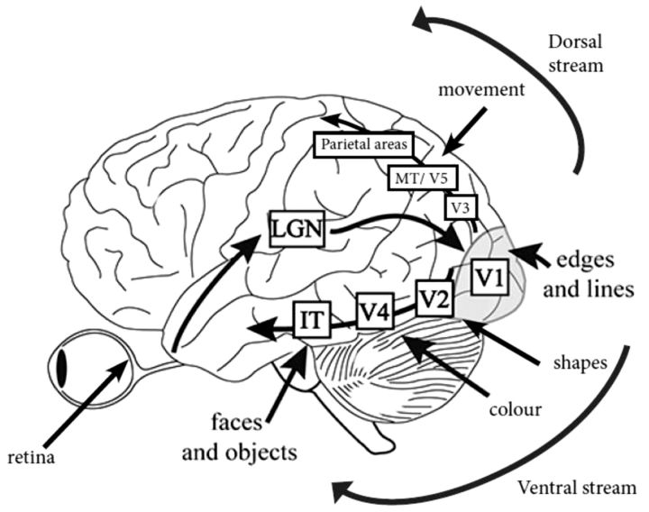
“IT = infero-temporal region; MT/V5 = motion processing region.”
BackgroundPharmacological Treatment of Parkinson Disease: A Review
Connolly, B. S., & Lang, A. E. (2014) · JAMA · doi:10.1001/jama.2014.3654 · Cited by 1,498 · cited 2×
“General advice is to consider visuo-perceptual ability and if there is any doubt, to refer the patient for a formal driving assessment via the national driving authority (Crizzle et al., 2012).”
BackgroundSpatial frequency selectivity of cells in macaque visual cortex
De Valois, R. L., Albrecht, D. G., & Thorell, L. G. (1982) · Vision Research · doi:10.1016/0042-6989(82)90113-4 · Cited by 1,489 · cited 2×
“This is more suggestive of a cortical rather than retinal or geniculate site of disruption as cortical neurons show stronger selectivity for different spatial frequencies than neurons in the retina or thalamus (Campbell and Robson, 1968; De Valois et al., 1982; Regan, 1982).”
Functional analysis of human MT and related visual cortical areas using magnetic resonance imaging
Tootell, R., Reppas, J., Kwong, K., Malach, R., Born, R., Brady, T., Rosen, B., & Belliveau, J. (1995) · The Journal of Neuroscience · doi:10.1523/jneurosci.15-04-03215.1995 · Cited by 1,415 · cited 2×
“IT = infero-temporal region; MT/V5 = motion processing region.”
BackgroundMazzulli, J. R., Xu, Y.-H., Sun, Y., Knight, A. L., McLean, P. J., Caldwell, G. A., Sidransky, E., Grabowski, G. A., & Krainc, D. (2011) · Cell · doi:10.1016/j.cell.2011.06.001 · Cited by 1,341 · cited 1×
“Both the GBA and SNCA genes are linked with the lysosomal pathway (Mazzulli et al., 2011), suggesting the visual deficits associated with these genes may share a pathophysiological mechanism, with lysosomal dysfunction associated with cortical visual dysfunction.”
BackgroundTHE DESIGN OF A NEW LETTER CHART FOR MEASURING CONTRAST SENSITIVITY
Pelli, D. G., Robson, J. G., & Wilkins, A. J. (1988) · Cited by 1,308
Grill-Spector, K., Kushnir, T., Edelman, S., Avidan, G., Itzchak, Y., & Malach, R. (1999) · Neuron · doi:10.1016/s0896-6273(00)80832-6 · Cited by 1,166 · cited 1×
“In the healthy brain, object processing occurs in V2, V4 and the lateral occipital complex, especially in the right hemisphere (Grill-Spector et al., 1999; Kourtzi and Kanwisher, 2000; Large et al., 2007).”
BackgroundDistinct spatial frequency sensitivities for processing faces and emotional expressions
Vuilleumier, P., Armony, J. L., Driver, J., & Dolan, R. J. (2003) · Nature Neuroscience · doi:10.1038/nn1057 · Cited by 1,117 · cited 1×
“In contrast, subcortical pathways, including the amygdala, are activated by fearful faces, and are driven by coarse, low spatial frequency information (Vuilleumier et al., 2003).”
BackgroundHallucinations in Parkinson's disease: Prevalence, phenomenology and risk factors
Fenelon, G. (2000) · Brain · doi:10.1093/brain/123.4.733 · Cited by 1,037 · cited 5×
“Mechanistic considerations of minor hallucinations ‘Passage hallucinations’ are illusions of objects, commonly insects and small animals, moving across the peripheries of vision (Fenelon et al., 2000).”
BackgroundInfluentialVisuospatial Judgment: A Clinical Test
Benton, A. L. (1978) · Archives of Neurology · doi:10.1001/archneur.1978.00500300038006 · Cited by 1,012 · cited 2×
“In tests comparing the ability to distinguish between lines of differing directions (Benton et al., 1978), several studies showed impaired performance in patients with Parkinson’s disease (Montse et al., 2001; Uc et al., 2005; Gullett et al., 2013) that correlated with duration and severity of…”
ResultDreaming and the brain: toward a cognitive neuroscience of conscious states
Hobson, J. A., Pace-Schott, E. F., & Stickgold, R. (2000) · Behavioral and Brain Sciences · doi:10.1017/s0140525x00003976 · Cited by 985 · cited 2×
“The presence of RBD in Parkinson’s disease is also predictive of progressive cognitive dysfunction (Sinforiani et al., 2008; Postuma et al., 2012; Nomura et al., 2013).”
BackgroundMethodThe distinct cognitive syndromes of Parkinson's disease: 5 year follow-up of the CamPaIGN cohort
Williams-Gray, C. H., Evans, J. R., Goris, A., Foltynie, T., Ban, M., Robbins, T. W., Brayne, C., Kolachana, B. S., Weinberger, D. R., Sawcer, S. J., et al. (2009) · Brain · doi:10.1093/brain/awp245 · Cited by 965 · cited 5×
“The underlying mechanism for these findings may relate to differences in the cortical expression of 4- versus 3repeat isoforms of tau (Williams-Gray et al., 2009).”
BackgroundInfluentialKehagia, A. A., Barker, R. A., & Robbins, T. W. (2010) · The Lancet Neurology · doi:10.1016/s1474-4422(10)70212-x · Cited by 881 · cited 1×
“(Williams-Gray et al., 2009; Kehagia et al., 2010).”
Schenck, C. H., Boeve, B. F., & Mahowald, M. W. (2013) · Sleep Medicine · doi:10.1016/j.sleep.2012.10.009 · Cited by 814 · cited 2×
“Idiopathic RBD, characterized by loss of normal atonia during REM sleep, is considered a risk factor for Parkinson’s disease and other synucleinopathies: over 80% of idiopathic RBD patients may eventually develop a neurodegenerative disorder linked to -synuclein accumulation in the brain (Iranzo et al., 2013; Schenck…”
BackgroundIranzo, A., Tolosa, E., Gelpi, E., Molinuevo, J. L., Valldeoriola, F., Serradell, M., Sanchez-Valle, R., Vilaseca, I., Lomeña, F., Vilas, D., et al. (2013) · The Lancet Neurology · doi:10.1016/s1474-4422(13)70056-5 · Cited by 723 · cited 2×
“Idiopathic RBD, characterized by loss of normal atonia during REM sleep, is considered a risk factor for Parkinson’s disease and other synucleinopathies: over 80% of idiopathic RBD patients may eventually develop a neurodegenerative disorder linked to -synuclein accumulation in the brain (Iranzo et al., 2013; Schenck…”
BackgroundGlucocerebrosidase mutations in clinical and pathologically proven Parkinson’s disease
Neumann, J., Bras, J., Deas, E., O'Sullivan, S. S., Parkkinen, L., Lachmann, R. H., Li, A., Holton, J., Guerreiro, R., Paudel, R., et al. (2009) · Brain · doi:10.1093/brain/awp044 · Cited by 696 · cited 4×
“Patients with Parkinson’s disease that carry GBA mutations also show a higher frequency of clinical features that are often associated with visual dysfunction in Parkinson’s disease: they have a higher risk of cognitive deficits (Neumann et al., 2009; Brockmann et al., 2011; Winder-Rhodes et al., 2013) and rapid eye m…”
BackgroundInfluentialSelf-citationThe CamPaIGN study of Parkinson's disease: 10-year outlook in an incident population-based cohort
Williams-Gray, C. H., Mason, S. L., Evans, J. R., Foltynie, T., Brayne, C., Robbins, T. W., & Barker, R. A. (2013) · Journal of Neurology, Neurosurgery & Psychiatry · doi:10.1136/jnnp-2013-305277 · Cited by 681 · cited 5×
“Whether this reflects insensitivity of the Mini-Mental State Examination, or an absence of relationship between dementia and retinal thickness is not yet known.”
BackgroundMethodInfluentialCortical Regions Involved in Perceiving Object Shape
Kourtzi, Z., & Kanwisher, N. (2000) · The Journal of Neuroscience · doi:10.1523/jneurosci.20-09-03310.2000 · Cited by 673 · cited 1×
“In the healthy brain, object processing occurs in V2, V4 and the lateral occipital complex, especially in the right hemisphere (Grill-Spector et al., 1999; Kourtzi and Kanwisher, 2000; Large et al., 2007).”
BackgroundFereshtehnejad, S.-M., Romenets, S. R., Anang, J. B. M., Latreille, V., Gagnon, J.-F., & Postuma, R. B. (2015) · JAMA Neurology · doi:10.1001/jamaneurol.2015.0703 · Cited by 615 · cited 2×
“Patients with increased tremor perform better on tasks of line orientation, mental reconstruction and rotation tasks (Levin et al., 1991) and in longitudinal follow-up, presence of tremor is associated with a more benign course and fewer colour vision abnormalities (Fereshtehnejad et al., 2015).”
BackgroundVisual hallucinations in Lewy body disease relate to Lewy bodies in the temporal lobe
Harding, A. J., Broe, G. A., & Halliday, G. M. (2002) · Brain · doi:10.1093/brain/awf033 · Cited by 609 · cited 2×
“…function compared to those without visual hallucinations (Grossi et al., 2005; Barnes and Boubert, 2008; Shin et al., 2012) and higher cortical Lewy body density is seen in the middle frontal gyrus in patients with visual hallucinations (Harding et al., 2002; Papapetropoulos et al., 2006).”
BackgroundREM sleep behavior disorder and REM sleep without atonia in Parkinson’s disease
Gagnon, J. F., Bédard, M. A., Fantini, M. L., Petit, D., Panisset, M., Rompré, S., Carrier, J., & Montplaisir, J. (2002) · Neurology · doi:10.1212/wnl.59.4.585 · Cited by 598 · cited 1×
“Although RBD is a rare prodromal sign in patients presenting with motor symptoms, up to a third of patients develop RBD in the course of their illness (Gagnon et al., 2002; Manni et al., 2010).”
BackgroundAnatomical demonstration of orientation columns in macaque monkey
Hubel, D. H., Wiesel, T. N., & Stryker, M. P. (1978) · Journal of Comparative Neurology · doi:10.1002/cne.901770302 · Cited by 470 · cited 2×
“As receptive fields of neurons in primary visual cortex are tuned to specific orientations (Hubel et al., 1978), these findings implicate primary visual cortex as the locus for deficits in contrast sensitivity in Parkinson’s disease, rather than just the retina or the thalamus, which are not linked to orientation spec…”
BackgroundA clinico-pathological study of subtypes in Parkinson's disease
Selikhova, M., Williams, D. R., Kempster, P. A., Holton, J. L., Revesz, T., & Lees, A. J. (2009) · Brain · doi:10.1093/brain/awp234 · Cited by 461 · cited 2×
“There is an increasing acknowledgement of the clinical heterogeneity of idiopathic Parkinson’s disease (Selikhova et al., 2009; Halliday et al., 2011; Sieber et al., 2014) and differences in survival rates have been reported between clinical subtypes defined in longitudinal analysis (de Lau et al.”
BackgroundSelf-citationRisk factors for nursing home placement in advanced Parkinson's disease
Goetz, C. G., & Stebbins, G. T. (1993) · Neurology · doi:10.1212/wnl.43.11.2227 · Cited by 455 · cited 2×
“Visual hallucinations are strong predictors for nursing home placement (Goetz and Stebbins, 1993) and have been suggested as a harbinger for the onset of dementia in Parkinson’s disease (Santangelo et al., 2007).”
BackgroundCollerton, D., Perry, E., & McKeith, I. (2005) · Behavioral and Brain Sciences · doi:10.1017/s0140525x05000130 · Cited by 437 · cited 2×
“It also implies that properly perceived objects should displace hallucinatory objects, causing the hallucination to disappear, which does not occur [see commentary by Morrison and David (2005) on Collerton et al., 2005)].”
BackgroundNeuropathology underlying clinical variability in patients with synucleinopathies
Halliday, G. M., Holton, J. L., Revesz, T., & Dickson, D. W. (2011) · Acta Neuropathologica · doi:10.1007/s00401-011-0852-9 · Cited by 430 · cited 2×
“There is an increasing acknowledgement of the clinical heterogeneity of idiopathic Parkinson’s disease (Selikhova et al., 2009; Halliday et al., 2011; Sieber et al., 2014) and differences in survival rates have been reported between clinical subtypes defined in longitudinal analysis (de Lau et al.”
BackgroundBoeve, B. F., Silber, M. H., Ferman, T. J., Kokmen, E., Smith, G. E., Ivnik, R. J., Parisi, J. E., Olson, E. J., & Petersen, R. C. (1998) · Neurology · doi:10.1212/wnl.51.2.363 · Cited by 395
Predictors of dementia in Parkinson disease: A prospective cohort study
Anang, J. B. M., Gagnon, J.-F., Bertrand, J.-A., Romenets, S. R., Latreille, V., Panisset, M., Montplaisir, J., & Postuma, R. B. (2014) · Neurology · doi:10.1212/wnl.0000000000000842 · Cited by 379
Prospective study of hallucinations and delusions in Parkinson's disease
Holroyd, S. (2001) · Journal of Neurology, Neurosurgery & Psychiatry · doi:10.1136/jnnp.70.6.734 · Cited by 345 · cited 4×
“Visual hallucinations often occur in poor lighting and are associated with reduced visual acuity (Holroyd et al., 2001; Matsui et al., 2006; Archibald et al., 2011) but can also be seen in patients with normal visual acuity (Diederich et al., 1998; Biousse et al., 2004).”
BackgroundInfluentialRetinal nerve fiber layer thinning in Parkinson disease
Inzelberg, R., Ramirez, J. A., Nisipeanu, P., & Ophir, A. (2004) · Vision Research · doi:10.1016/j.visres.2004.06.009 · Cited by 343 · cited 2×
“The nerve fibre layer is thinned in Parkinson’s disease (Inzelberg et al., 2004; Altintas et al., 2008) and at the macula, thinning is found in the inner retinal layer (Hajee et al.”
BackgroundVisual control of locomotion in Parkinson's disease
Azulay, J.-P., Mesure, S., Amblard, B., Blin, O., Sangla, I., & Pouget, J. (1999) · Brain · doi:10.1093/brain/122.1.111 · Cited by 340 · cited 2×
“Patients with Parkinson’s disease carrying mutations in the gene for synuclein, SNCA, also show higher rates of cognitive deficits, REM sleep behaviour disorder (RBD) and hallucinations (Nishioka et al., 2009; Konno et al., 2016; Petrucci et al., 2016).”
BackgroundStride length regulation in Parkinson's disease: the use of extrinsic, visual cues
Lewis, G. N. (2000) · Brain · doi:10.1093/brain/123.10.2077 · Cited by 330 · cited 1×
“Patients with Parkinson’s disease carrying mutations in the gene for synuclein, SNCA, also show higher rates of cognitive deficits, REM sleep behaviour disorder (RBD) and hallucinations (Nishioka et al., 2009; Konno et al., 2016; Petrucci et al., 2016).”
BackgroundHallucinations in Parkinson disease
Diederich, N. J., Fénelon, G., Stebbins, G., & Goetz, C. G. (2009) · Nature Reviews Neurology · doi:10.1038/nrneurol.2009.62 · Cited by 312 · cited 1×
“They are usually non-frightening and occasionally enjoyed, but become increasingly distressing over time (for reviews see Fenelon et al., 2000; Diederich et al., 2009; Collerton et al., 2015).”
BackgroundMeasurements of Visual Evoked Potentials in Parkinson's Disease
Bodis --Wollner, I., & Yahr, M. D. (1978) · Brain · doi:10.1093/brain/101.4.661 · Cited by 310
Visual dysfunction in Parkinson disease without dementia
Uc, E. Y., Rizzo, M., Anderson, S. W., Qian, S., Rodnitzky, R. L., & Dawson, J. D. (2005) · Neurology · doi:10.1212/01.wnl.0000191565.11065.11 · Cited by 308 · cited 6×
“, 1978), several studies showed impaired performance in patients with Parkinson’s disease (Montse et al., 2001; Uc et al., 2005; Gullett et al., 2013) that correlated with duration and severity of disease.”
BackgroundResultInfluentialPostuma, R. B., Bertrand, J., Montplaisir, J., Desjardins, C., Vendette, M., Rios Romenets, S., Panisset, M., & Gagnon, J. (2012) · Movement Disorders · doi:10.1002/mds.24939 · Cited by 305 · cited 1×
“The presence of RBD in Parkinson’s disease is also predictive of progressive cognitive dysfunction (Sinforiani et al., 2008; Postuma et al., 2012; Nomura et al., 2013).”
BackgroundWilliams, D. R., & Lees, A. J. (2005) · The Lancet Neurology · doi:10.1016/s1474-4422(05)70146-0 · Cited by 300 · cited 3×
“…presence is believed to be highly specific for Lewy body pathology (Williams et al., 2008), and can be helpful in distinguishing between Parkinson’s disease and other parkinsonian syndromes such as multiple system atrophy (MSA-P) and progressive supranuclear palsy (PSP-P) (Williams and Lees, 2005).”
BackgroundSelf-citationVisual perception in Parkinson disease dementia and dementia with Lewy bodies
Mosimann, U. P., Mather, G., Wesnes, K. A., O’Brien, J. T., Burn, D. J., & McKeith, I. G. (2004) · Neurology · doi:10.1212/01.wnl.0000145764.70698.4e · Cited by 299 · cited 4×
“, 1994), although in a speed discrimination task, patients with Parkinson’s disease performed no worse than controls (Mosimann et al., 2004).”
BackgroundInfluentialVISUAL DYSFUNCTION IN PARKINSON'S DISEASE: LOSS IN SPATIOTEMPORAL CONTRAST SENSITIVITY
BODIS-WOLLNER, I., MARX, M. S., MITRA, S., BOBAK, P., MYLIN, L., & YAHR, M. (1987) · Brain · doi:10.1093/brain/110.6.1675 · Cited by 297 · cited 4×
“In some patients, contrast sensitivity thresholds and visual evoked potential latencies differ between eyes (Bulens et al., 1986, 1987; Bodis-Wollner et al., 1987), raising the likelihood of prechiasmal involvement.”
BackgroundInfluentialFacial expression recognition in people with medicated and unmedicated Parkinson’s disease
Sprengelmeyer, R., Young, A. W., Mahn, K., Schroeder, U., Woitalla, D., Büttner, T., Kuhn, W., & Przuntek, H. (2003) · Neuropsychologia · doi:10.1016/s0028-3932(02)00295-6 · Cited by 293 · cited 4×
“The finding that performance improves in patients treated with L-DOPA is consistent with the involvement of dopaminergic neurons in this pathway (Sprengelmeyer et al., 2003).”
InfluentialDiederich, N. J., Goetz, C. G., & Stebbins, G. T. (2005) · Movement Disorders · doi:10.1002/mds.20308 · Cited by 290 · cited 2×
“The presence of RBD in Parkinson’s disease is also predictive of progressive cognitive dysfunction (Sinforiani et al., 2008; Postuma et al., 2012; Nomura et al., 2013).”
BackgroundMethodWinder-Rhodes, S. E., Evans, J. R., Ban, M., Mason, S. L., Williams-Gray, C. H., Foltynie, T., Duran, R., Mencacci, N. E., Sawcer, S. J., & Barker, R. A. (2013) · Brain · doi:10.1093/brain/aws318 · Cited by 290 · cited 2×
“Patients with Parkinson’s disease that carry GBA mutations also show a higher frequency of clinical features that are often associated with visual dysfunction in Parkinson’s disease: they have a higher risk of cognitive deficits (Neumann et al., 2009; Brockmann et al., 2011; Winder-Rhodes et al., 2013) and rapid eye m…”
BackgroundVisuospatial impairment in Parkinson's disease
Levin, B. E., Llabre, M. M., Reisman, S., Weiner, W. J., Sanchez-Ramos, J., Singer, C., & Brown, M. C. (1991) · Neurology · doi:10.1212/wnl.41.3.365 · Cited by 289 · cited 5×
“However, by mid-stage, they start to have difficulties in identifying overlapping objects (Pillon et al., 1989; Mosimann et al., 2004), with worse performance occurring in patients with left-sided motor symptoms (Levin et al., 1991).”
BackgroundInfluentialDecreased dopamine in the retinas of patients with Parkinson's disease.
Harnois, C., & Paolo, T. D. (1990) · PubMed · Cited by 286
REM sleep behavior disorder predicts cognitive impairment in Parkinson disease without dementia
Vendette, M., Gagnon, J. F., Décary, A., Massicotte-Marquez, J., Postuma, R. B., Doyon, J., Panisset, M., & Montplaisir, J. (2007) · Neurology · doi:10.1212/01.wnl.0000278114.14096.74 · Cited by 283 · cited 2×
“, 2010) and complex figure copying (Vendette et al., 2007).”
BackgroundVisual and spatial symptoms in Parkinson’s disease
Davidsdottir, S., Cronin-Golomb, A., & Lee, A. (2005) · Vision Research · doi:10.1016/j.visres.2004.11.006 · Cited by 280 · cited 14×
“Visual dysfunction frequently co-exists with cognitive impairment, visual hallucinations, postural instability with gait disorder and RBD (Davidsdottir et al., 2005; Marques et al., 2010).”
BackgroundInfluentialDopaminergic neurons in the human retina
Frederick, J. M., Rayborn, M. E., Laties, A. M., Lam, D. M. K., & Hollyfield, J. G. (1982) · Journal of Comparative Neurology · doi:10.1002/cne.902100108 · Cited by 277 · cited 1×
“The healthy human retina contains dopaminergic amacrine cells (Frederick et al., 1982) and patients with Parkinson’s disease have reduced dopamine innervation around the fovea (Nguyen-Legros, 1988).”
BackgroundOphthalmologic features of Parkinson’s disease
Biousse, V., Skibell, B. C., Watts, R. L., Loupe, D. N., Drews-Botsch, C., & Newman, N. J. (2004) · Neurology · doi:10.1212/01.wnl.0000103444.45882.d8 · Cited by 277
A meta-analysis of performance on emotion recognition tasks in Parkinson’s disease.
Gray, H. M., & Tickle-Degnen, L. (2010) · Neuropsychology · doi:10.1037/a0018104 · Cited by 275 · cited 2×
“In eye movement analyses, when searching for a target, patients with left-sided Parkinson’s disease will explore the right hemifield first, in contrast with healthy controls and patients with right-sided Parkinson’s disease, who show a leftward bias (Ebersbach et al., 1996).”
BackgroundTau and α‐synuclein in susceptibility to, and dementia in, Parkinson's disease
Goris, A., Williams‐Gray, C. H., Clark, G. R., Foltynie, T., Lewis, S. J. G., Brown, J., Ban, M., Spillantini, M. G., Compston, A., Burn, D. J., et al. (2007) · Annals of Neurology · doi:10.1002/ana.21192 · Cited by 270
GBA-associated PD presents with nonmotor characteristics
Brockmann, K., Srulijes, K., Hauser, A.-K., Schulte, C., Csoti, I., Gasser, T., & Berg, D. (2011) · Neurology · doi:10.1212/wnl.0b013e318225ab77 · Cited by 268 · cited 2×
“…that are often associated with visual dysfunction in Parkinson’s disease: they have a higher risk of cognitive deficits (Neumann et al., 2009; Brockmann et al., 2011; Winder-Rhodes et al., 2013) and rapid eye movement (REM) sleep disorder than patients with idiopathic Parkinson’s disease…”
BackgroundInner Retinal Layer Thinning in Parkinson Disease
Hajee, M. E. (2009) · Archives of Ophthalmology · doi:10.1001/archophthalmol.2009.106 · Cited by 268 · cited 4×
“, 2008) and at the macula, thinning is found in the inner retinal layer (Hajee et al., 2009; Shrier et al., 2012).”
BackgroundMethodInfluentialVISUAL CONTROL OF BALANCE IN CEREBELLAR AND PARKINSONIAN SYNDROMES
BRONSTEIN, A. M., HOOD, J. D., GRESTY, M. A., & PANAGI, C. (1990) · Brain · doi:10.1093/brain/113.3.767 · Cited by 265 · cited 2×
“…et al., 2011; Winder-Rhodes et al., 2013) and rapid eye movement (REM) sleep disorder than patients with idiopathic Parkinson’s disease (Beavan et al., 2015; Gan-Or et al., 2015) (see below for further discussion of clinical features associated with visual dysfunction in Parkinson’s disease).”
BackgroundCognitive performance of GBA mutation carriers with early-onset PD: The CORE-PD study
Alcalay, R. N., Caccappolo, E., Mejia-Santana, H., Tang, M.-X., Rosado, L., Orbe Reilly, M., Ruiz, D., Ross, B., Verbitsky, M., Kisselev, S., et al. (2012) · Neurology · doi:10.1212/wnl.0b013e318253d54b · Cited by 262 · cited 2×
“…with Parkinson’s disease that carry mutations in the gene encoding the lysosomal enzyme glucocerebrosidase (GBA) show deficits in visuospatial tasks (Alcalay et al., 2012), have impaired visual memory (Zokaei et al., 2014) and show a higher frequency of visual hallucinations (Neumann et al., 2009;…”
BackgroundPostuma, R. B., Gagnon, J. F., Vendette, M., & Montplaisir, J. Y. (2009) · Brain · doi:10.1093/brain/awp244 · Cited by 260 · cited 1×
“Visuo-perceptual deficits have been reported in patients with RBD (who have not yet developed Parkinson’s disease), with impairments in colour vision and visuospatial construction (Boeve et al., 1998; Ferini-Strambi et al., 2004; Postuma et al., 2009; Manni et al., 2013).”
BackgroundFreezing of gait in Parkinson's disease: a perceptual cause for a motor impairment?
Almeida, Q. J., & Lebold, C. A. (2010) · Journal of Neurology, Neurosurgery & Psychiatry · doi:10.1136/jnnp.2008.160580 · Cited by 243 · cited 2×
“In patients with freezing of gait, step length reduces on going through narrow doorways, suggesting a perceptual mechanism interfering with movement planning (Almeida and Lebold, 2010; Cowie et al., 2010).”
BackgroundPostuma, R. B., Gagnon, J., Vendette, M., Desjardins, C., & Montplaisir, J. Y. (2011) · Annals of Neurology · doi:10.1002/ana.22282 · Cited by 241 · cited 2×
“Those RBD patients with sensory abnormalities at baseline (including colour vision defects) have also been claimed to develop a form of Parkinson’s disease with more prominent cognitive involvement (Postuma et al., 2011).”
BackgroundAltintaş, Ö., Işeri, P., Özkan, B., & Çağlar, Y. (2008) · Documenta Ophthalmologica · doi:10.1007/s10633-007-9091-8 · Cited by 240 · cited 4×
“A correlation between retinal thickness, disease severity (Altintas et al., 2008; Miri et al., 2015) and duration (Miri et al.”
BackgroundResultInfluentialAbnormalities in color vision and contrast sensitivity in Parkinson's disease
Price, M. J., Feldman, R. G., Adelberg, D., & Kayne, H. (1992) · Neurology · doi:10.1212/wnl.42.4.887 · Cited by 236 · cited 4×
“Disordered colour discrimination is recognized in Parkinson’s disease (Price et al., 1992; Buttner et al., 1995), even in the early stages of the disease, in untreated patients (Buttner et al.”
BackgroundInfluentialMinor hallucinations occur in drug‐naive Parkinson's disease patients, even from the premotor phase
Pagonabarraga, J., Martinez‐Horta, S., Fernández de Bobadilla, R., Pérez, J., Ribosa‐Nogué, R., Marín, J., Pascual‐Sedano, B., García, C., Gironell, A., & Kulisevsky, J. (2016) · Movement Disorders · doi:10.1002/mds.26432 · Cited by 236 · cited 2×
“Recent reports of minor visual hallucinations in patients at the earliest stages of Parkinson’s disease (Pagonabarraga et al., 2016), including in untreated patients, lends further support for changes in cortical visual processing even at the initial stages of Parkinson’s disease.”
BackgroundAltered Resting State Brain Networks in Parkinson’s Disease
Göttlich, M., Münte, T. F., Heldmann, M., Kasten, M., Hagenah, J., & Krämer, U. M. (2013) · PLoS ONE · doi:10.1371/journal.pone.0077336 · Cited by 230 · cited 2×
“In patients at mid-stage Parkinson’s disease, resting state functional MRI network analysis shows connectivity changes especially affecting visual regions (Gottlich et al., 2013).”
BackgroundPereira, J. B., Ibarretxe‐Bilbao, N., Marti, M., Compta, Y., Junqué, C., Bargallo, N., & Tolosa, E. (2012) · Human Brain Mapping · doi:10.1002/hbm.21378 · Cited by 221 · cited 2×
“thinning seen in the lateral occipital complex and parietal regions (Pereira et al., 2012).”
BackgroundThe perceptual consequences of visual loss: `positive' pathologies of vision
ffytche, D. H., & Howard, R. J. (1999) · Brain · doi:10.1093/brain/122.7.1247 · Cited by 216 · cited 2×
“…syndromes have been proposed in Parkinson’s disease: a frontal-striatal dopamine-mediated dysexecutive syndrome that does not progress to dementia; and a second form with prominent visuospatial and sematic fluency impairments that is more frequently associated with decline to dementia.”
BackgroundCerebral Glucose Metabolic Features of Parkinson Disease and Incident Dementia: Longitudinal Study
Bohnen, N. I., Koeppe, R. A., Minoshima, S., Giordani, B., Albin, R. L., Frey, K. A., & Kuhl, D. E. (2011) · Journal of Nuclear Medicine · doi:10.2967/jnumed.111.089946 · Cited by 216 · cited 1×
“These findings are supported by functional neuroimaging findings: in a longitudinal FDG-PET study of Parkinson’s disease, patients who developed dementia, showed occipito-parietal hypometabolism at baseline (Bohnen et al., 2011).”
ResultFerini–Strambi, L., Di Gioia, M. R., Castronovo, V., Oldani, A., Zucconi, M., & Cappa, S. F. (2004) · Neurology · doi:10.1212/01.wnl.0000101726.69701.fa · Cited by 213 · cited 2×
“Visuo-perceptual deficits have been reported in patients with RBD (who have not yet developed Parkinson’s disease), with impairments in colour vision and visuospatial construction (Boeve et al., 1998; Ferini-Strambi et al., 2004; Postuma et al., 2009; Manni et al., 2013).”
BackgroundAutosomal dominant Parkinson's disease caused by SNCA duplications
Konno, T., Ross, O. A., Puschmann, A., Dickson, D. W., & Wszolek, Z. K. (2016) · Parkinsonism & Related Disorders · doi:10.1016/j.parkreldis.2015.09.007 · Cited by 210 · cited 1×
“Patients with Parkinson’s disease carrying mutations in the gene for synuclein, SNCA, also show higher rates of cognitive deficits, REM sleep behaviour disorder (RBD) and hallucinations (Nishioka et al., 2009; Konno et al., 2016; Petrucci et al., 2016).”
BackgroundDisgust-specific impairment of facial expression recognition in Parkinson's disease
Suzuki, A., Hoshino, T., Shigemasu, K., & Kawamura, M. (2006) · Brain · doi:10.1093/brain/awl011 · Cited by 208 · cited 2×
“…difficulty in interpreting facial expressions (Sprengelmeyer et al., 2003), with greater impairment for negative emotions including disgust (Suzuki et al., 2006), sadness (Hipp et al., 2014) and fear (Jacobs et al., 1995; Dujardin et al., 2004; Clark et al., 2008; Gray and Tickle-Degnen,…”
BackgroundVisual symptoms in Parkinson's disease and Parkinson's disease dementia
Archibald, N. K., Clarke, M. P., Mosimann, U. P., & Burn, D. J. (2011) · Movement Disorders · doi:10.1002/mds.23891 · Cited by 207 · cited 5×
“In questionnaire studies, 78% of patients with Parkinson’s disease report at least one visual symptom, including difficulty reading, sometimes with double vision, and misjudging objects and distances (Archibald et al., 2011; Urwyler et al., 2014).”
BackgroundInfluentialEvolution of Prodromal Clinical Markers of Parkinson Disease in a GBA Mutation–Positive Cohort
Beavan, M., McNeill, A., Proukakis, C., Hughes, D. A., Mehta, A., & Schapira, A. H. V. (2015) · JAMA Neurology · doi:10.1001/jamaneurol.2014.2950 · Cited by 205 · cited 2×
“, 2013) and rapid eye movement (REM) sleep disorder than patients with idiopathic Parkinson’s disease (Beavan et al., 2015; Gan-Or et al., 2015) (see below for further discussion of clinical features associated with visual dysfunction in Parkinson’s disease).”
BackgroundDeficits in decoding emotional facial expressions in Parkinson's disease
Dujardin, K. (2004) · Neuropsychologia · doi:10.1016/s0028-3932(03)00154-4 · Cited by 196 · cited 2×
“Longitudinal studies show that it has value in predicting the development of dementia in Parkinson’s disease.”
BackgroundEmotional facial imagery, perception, and expression in Parkinson's disease
Jacobs, D. H., Shuren, J., Bowers, D., & Heilman, K. M. (1995) · Neurology · doi:10.1212/wnl.45.9.1696 · Cited by 196 · cited 2×
“Longitudinal studies show that it has value in predicting the development of dementia in Parkinson’s disease.”
Backgroundα‐synuclein in the inner retina in parkinson disease
Bodis‐Wollner, I., Kozlowski, P. B., Glazman, S., & Miri, S. (2014) · Annals of Neurology · doi:10.1002/ana.24182 · Cited by 192 · cited 2×
“The recent finding in the inner retinal layer of misfolded -synuclein (Bodis-Wollner et al., 2014a) and of phosphorylated -synuclein (Beach et al., 2014), the pathological hallmark of Parkinson’s disease, is striking evidence of retinal involvement in Parkinson’s disease.”
BackgroundReality monitoring and visual hallucinations in Parkinson’s disease
Barnes, J., Boubert, L., Harris, J., Lee, A., & David, A. S. (2003) · Neuropsychologia · doi:10.1016/s0028-3932(02)00182-3 · Cited by 192 · cited 3×
“…(Diederich et al., 1998) and patients with visual hallucinations show greater deficits in object and face identification compared to those without hallucinations (Barnes et al., 2003; Ramirez-Ruiz et al., 2006; Gallagher et al., 2011), reflecting involvement of higher cortical visual dysfunction.”
BackgroundABNORMALITIES OF NONVISUALLY-GUIDED EYE MOVEMENTS IN PARKINSON'S DISEASE
CRAWFORD, T. J., HENDERSON, L., & KENNARD, C. (1989) · Brain · doi:10.1093/brain/112.6.1573 · Cited by 189 · cited 1×
“Saccades to a remembered target are particularly impaired and show a multistep pattern (Crawford et al., 1989).”
BackgroundPatterns of cortical thickness and surface area in early Parkinson's disease
Jubault, T., Gagnon, J.-F., Karama, S., Ptito, A., Lafontaine, A.-L., Evans, A. C., & Monchi, O. (2011) · NeuroImage · doi:10.1016/j.neuroimage.2010.12.043 · Cited by 185 · cited 2×
“occipital cortex, measured using MRI, correlates with disease duration (Jubault et al., 2011), with maximal cortical”
BackgroundMethodMoreno-Ramos, T., Benito-León, J., Villarejo, A., & Bermejo-Pareja, F. (2013) · Journal of Alzheimer's Disease · doi:10.3233/jad-121975 · Cited by 181
Moschos, M. M., Tagaris, G., Markopoulos, L., Margetis, L., Tsapakis, S., Kanakis, M., & Koutsandrea, C. (2011) · European Journal of Ophthalmology · doi:10.5301/ejo.2010.1318 · Cited by 171 · cited 2×
“Retinal electrical activity is decreased compared to healthy controls, measured by electroretinogram (Tagliati et al., 1996; Moschos et al., 2011) and improves with LDOPA treatment (Peppe et al.”
BackgroundPhenotype in parkinsonian and nonparkinsonian LRRK2 G2019S mutation carriers
Marras, C., Schuele, B., Munhoz, R. P., Rogaeva, E., Langston, J. W., Kasten, M., Meaney, C., Klein, C., Wadia, P. M., Lim, S.-Y., et al. (2011) · Neurology · doi:10.1212/wnl.0b013e318227042d · Cited by 171 · cited 2×
“Patients carrying LRRK2 mutations have better colour discrimination (Marras et al., 2011) and fewer cognitive deficits than patients with idiopathic Parkinson’s disease (Alcalay et al., 2013) (Table 1).”
BackgroundSpecific impairments in the recognition of emotional facial expressions in Parkinson's disease
Clark, U. S., Neargarder, S., & Cronin-Golomb, A. (2008) · Neuropsychologia · doi:10.1016/j.neuropsychologia.2008.03.014 · Cited by 166 · cited 2×
“Longitudinal studies show that it has value in predicting the development of dementia in Parkinson’s disease.”
BackgroundGoldman, J. G., Stebbins, G. T., Dinh, V., Bernard, B. A., Merkitch, D., deToledo‐Morrell, L., & Goetz, C. G. (2014) · Brain · doi:10.1093/brain/awt360 · Cited by 166 · cited 1×
“Structural neuroimaging shows grey matter atrophy in the medial frontal lobe in Parkinson’s disease patients with visual hallucinations compared to those without (Ramirez-Ruiz et al., 2007; Goldman et al., 2014) as well as in visual processing regions.”
BackgroundWhen crowding of crowding leads to uncrowding
Manassi, M., Sayim, B., & Herzog, M. H. (2013) · Journal of Vision · doi:10.1167/13.13.10 · Cited by 166 · cited 1×
“Adapted from Manassi et al. (2013) with permission (copyright held by the Association for Research in Vision and Ophthalmology). cortex (V1) to extra-striate visual cortex (V4).”
Parkinsonism: Alterations in spatial orientation as determined by a route-walking test
Bowen, F. P., Hoehn, M. M., & Yahr, M. D. (1972) · Neuropsychologia · doi:10.1016/0028-3932(72)90027-9 · Cited by 164 · cited 1×
“The scope of the problem of vision in Parkinson’s disease Patients with Parkinson’s disease frequently report problems with visual tasks, such as navigating around everyday environments and using maps (Bowen et al., 1972; McDowell and Harris, 1997b; Davidsdottir et al., 2005).”
BackgroundTricks of the mind: Visual hallucinations as disorders of attention
Shine, J. M., O’Callaghan, C., Halliday, G. M., & Lewis, S. J. G. (2014) · Progress in Neurobiology · doi:10.1016/j.pneurobio.2014.01.004 · Cited by 163 · cited 3×
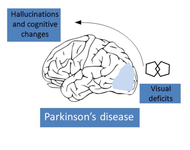
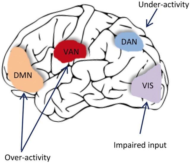
“The attention networks hypothesis for visual hallucinations in Parkinson’s disease (Shine et al., 2014).”
BackgroundNombela, C., Rowe, J. B., Winder‐Rhodes, S., Hampshire, A., Owen, A. M., Breen, D. P., Duncan, G. W., Khoo, T. K., Yarnall, A. J., Firbank, M., et al. (2014) · Brain · doi:10.1093/brain/awu201 · Cited by 162
Regan, D., & Neima, D. (1984) · British Journal of Ophthalmology · doi:10.1136/bjo.68.12.885 · Cited by 162 · cited 1×
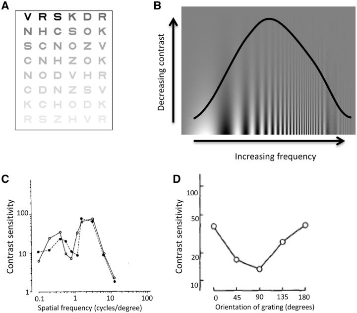
“In tests using vision charts with letters of the same size but reducing contrast, patients with Parkinson’s disease have normal acuity but impaired contrast detection (Regan and Neima, 1984) (Fig.”
BackgroundImpaired visual search in drivers with Parkinson's disease
Uc, E. Y., Rizzo, M., Anderson, S. W., Sparks, J., Rodnitzky, R. L., & Dawson, J. D. (2006) · Annals of Neurology · doi:10.1002/ana.20958 · Cited by 161 · cited 2×
“…sensitivity and driving performance in Parkinson’s disease and between visual perception and driving ability, particularly in tests measuring visual attention, motion perception and visuospatial construction (Uc et al., 2006, 2009; Devos et al., 2013; for a review see Crizzle et al., 2012).”
BackgroundCognitive slowing in Parkinson's disease fails to respond to levodopa treatment: The 15‐objects test
Pillon, B., Dubois, B., Bonnet, A.-M., Esteguy, M., Guimaraes, J., Vigouret, J.-M., Lhermitte, F., & Agid, Y. (1989) · Neurology · doi:10.1212/wnl.39.6.762 · Cited by 160 · cited 2×
“However, by mid-stage, they start to have difficulties in identifying overlapping objects (Pillon et al., 1989; Mosimann et al., 2004), with worse performance occurring in patients with left-sided motor symptoms (Levin et al., 1991).”
BackgroundPhosphorylated α-synuclein-immunoreactive retinal neuronal elements in Parkinson's disease subjects
Beach, T. G., Carew, J., Serrano, G. E., Adler, C. H., Shill, H. A., Sue, L. I., Sabbagh, M. N., Akiyama, H., & Cuenca, N. (2014) · Neuroscience Letters · doi:10.1016/j.neulet.2014.04.027 · Cited by 159 · cited 1×
“The recent finding in the inner retinal layer of misfolded -synuclein (Bodis-Wollner et al., 2014a) and of phosphorylated -synuclein (Beach et al., 2014), the pathological hallmark of Parkinson’s disease, is striking evidence of retinal involvement in Parkinson’s disease.”
BackgroundCerebral atrophy in Parkinson's disease patients with visual hallucinations
Ramírez‐Ruiz, B., Martí, M., TOLOSA, E. S., Giménez, M., Bargalló, N., Valldeoriola, F., & Junqué, C. (2007) · European Journal of Neurology · doi:10.1111/j.1468-1331.2007.01768.x · Cited by 159 · cited 1×
“Structural neuroimaging shows grey matter atrophy in the medial frontal lobe in Parkinson’s disease patients with visual hallucinations compared to those without (Ramirez-Ruiz et al., 2007; Goldman et al., 2014) as well as in visual processing regions.”
BackgroundSilva, M. F., Faria, P., Regateiro, F. S., Forjaz, V., Januário, C., Freire, A., & Castelo-Branco, M. S. (2005) · Brain · doi:10.1093/brain/awh581 · Cited by 157 · cited 5×
“However, abnormalities in colour vision can also be found using tests that are less susceptible to these confounds (Silva et al., 2005).”
BackgroundInfluentialPatients Homozygous and Heterozygous for SNCA Duplication in a Family With Parkinsonism and Dementia
Ikeuchi, T., Kakita, A., Shiga, A., Kasuga, K., Kaneko, H., Tan, C., Idezuka, J., Wakabayashi, K., Onodera, O., Iwatsubo, T., et al. (2008) · Archives of Neurology · doi:10.1001/archneur.65.4.514 · Cited by 154 · cited 2×
“Neuropathological studies of these patients show a greater -synuclein burden in the cerebral cortex than in idiopathic Parkinson’s disease (Ikeuchi et al., 2008; Obi et al., 2008) and fluorodeoxyglucose (FDG) signal is reduced in the occipital lobes (visual processing regions) of patients carrying…”
BackgroundNguyen-Legros, J. (1988) · Surgical and Radiologic Anatomy · doi:10.1007/bf02307822 · Cited by 153 · cited 2×
“, 1982) and patients with Parkinson’s disease have reduced dopamine innervation around the fovea (Nguyen-Legros, 1988).”
BackgroundProgressive Worsening of Spatial and Chromatic Processing Deficits in Parkinson Disease
Diederich, N. J., Raman, R., Leurgans, S. E., & Goetz, C. G. (2002) · Archives of Neurology · doi:10.1001/archneur.59.8.1249 · Cited by 150 · cited 5×
“Contrast sensitivity loss is partly reversible with L-DOPA treatment, in both early (Bulens et al., 1987) and mid-stage Parkinson’s disease (Bodis-Wollner et al., 1987) but it eventually deteriorates again as the disease advances (Diederich et al., 2002).”
BackgroundInfluentialNeuroanatomical substrate of visuospatial and visuoperceptual impairment in Parkinson's disease
Pereira, J. B., Junqué, C., Martı́, M. J., Ramírez‐Ruiz, B., Bargalló, N., & TOLOSA, E. S. (2009) · Movement Disorders · doi:10.1002/mds.22560 · Cited by 147 · cited 3×
“, 1991), with performance correlated with grey matter density in the fusiform face area, the region involved in face recognition in the heathy brain (Pereira et al., 2009).”
BackgroundExpanding the clinical phenotype of SNCA duplication carriers
Nishioka, K., Ross, O. A., Ishii, K., Kachergus, J. M., Ishiwata, K., Kitagawa, M., Kono, S., Obi, T., Mizoguchi, K., Inoue, Y., et al. (2009) · Movement Disorders · doi:10.1002/mds.22682 · Cited by 147 · cited 3×
“Patients with Parkinson’s disease carrying mutations in the gene for synuclein, SNCA, also show higher rates of cognitive deficits, REM sleep behaviour disorder (RBD) and hallucinations (Nishioka et al., 2009; Konno et al., 2016; Petrucci et al., 2016).”
BackgroundRoad safety in drivers with Parkinson disease
Uc, E. Y., Rizzo, M., Johnson, A. M., Dastrup, E., Anderson, S. W., & Dawson, J. D. (2009) · Neurology · doi:10.1212/wnl.0b013e3181c67b77 · Cited by 144
Insights into the neural control of locomotion from walking through doorways in Parkinson's disease
Cowie, D. A., Limousin, P. D., Peters, A., & Day, B. L. (2010) · Neuropsychologia · doi:10.1016/j.neuropsychologia.2010.05.022 · Cited by 143 · cited 2×
“In patients with freezing of gait, step length reduces on going through narrow doorways, suggesting a perceptual mechanism interfering with movement planning (Almeida and Lebold, 2010; Cowie et al., 2010).”
BackgroundTesting an aetiological model of visual hallucinations in Parkinson's disease
Gallagher, D. A., Parkkinen, L., O’Sullivan, S. S., Spratt, A., Shah, A., Davey, C. C., Bremner, F., Révész, T., Williams, D. R., Lees, A. J., et al. (2011) · Brain · doi:10.1093/brain/awr225 · Cited by 141 · cited 5×
“Visual dysfunction is more common in Parkinson’s disease patients experiencing hallucinations (Gallagher et al., 2011) and in neuroimaging studies, glucose consumption is reduced in occipito-temporal regions in Parkinson’s patients with visual hallucinations compared to those without (Boecker et al.”
BackgroundResultInfluentialSelf-citationDelayed psychosis after right temporoparietal stroke or trauma
Levine, D. N., & Finklestein, S. P. (1982) · Neurology · doi:10.1212/wnl.32.3.267 · Cited by 141 · cited 1×
“Previous models to explain these hallucinations include cortical irritation caused by electrical over-activity in brain regions containing images (Levine and Finklestein, 1982).”
BackgroundParkinson disease phenotype in Ashkenazi jews with and without LRRK2 G2019S mutations
Alcalay, R. N., Mirelman, A., Saunders-Pullman, R. J., Tang, M., Santana, H. M., Raymond, D., Roos, E., Orbe‐Reilly, M., Gurevich, T., Shira, A. B., et al. (2013) · Movement Disorders · doi:10.1002/mds.25647 · Cited by 139 · cited 2×
“, 2011) and fewer cognitive deficits than patients with idiopathic Parkinson’s disease (Alcalay et al., 2013) (Table 1).”
BackgroundAbe, Y. (2003) · Journal of Neurology Neurosurgery & Psychiatry · doi:10.1136/jnnp.74.4.419 · Cited by 139 · cited 2×
“…have been reported using PET in occipital cortex (Eberling et al., 1994; Bohnen et al., 1999) and occipital and parietal hypoperfusion in Parkinson’s disease can be detected by measuring regional cerebral blood flow (Abe et al., 2003), that worsens with disease progression (Nagamachi et al., 2008).”
BackgroundPredictors of mortality among nursing home residents with a diagnosis of Parkinson's disease.
Fernandez, H. H., & Lapane, K. L. (2002) · PubMed · Cited by 138
Motor correlates of occipital glucose hypometabolism in Parkinson’s disease without dementia
Bohnen, N. I., Minoshima, S., Giordani, B., Frey, K. A., & Kuhl, D. E. (1999) · Neurology · doi:10.1212/wnl.52.3.541 · Cited by 137 · cited 2×
“Metabolic deficits have been reported using PET in occipital cortex (Eberling et al., 1994; Bohnen et al., 1999) and occipital and parietal hypoperfusion in Parkinson’s disease can be detected by measuring regional cerebral blood flow (Abe et al., 2003), that worsens with disease progression…”
BackgroundPoor visual discrimination and visual hallucinations in Parkinson's disease.
Diederich, N. J., Goetz, C. G., Raman, R., Pappert, E. J., Leurgans, S. E., & Pieri, V. (1998) · PubMed · Cited by 137
Retinal nerve fiber layer thickness and visual hallucinations in Parkinson's Disease
Lee, J., Kim, J. M., Ahn, J., Kim, H., Jeon, B. S., & Kim, T. W. (2013) · Movement Disorders · doi:10.1002/mds.25543 · Cited by 134 · cited 2×
“Intriguingly, retinal thinning is associated with presence of minor hallucinations (Lee et al., 2014b), although this was not found for patients with complex visual hallucinations, consistent with the involvement of higher cognitive domains in these hallucinations.”
BackgroundResultGBA mutations are associated with Rapid Eye Movement Sleep Behavior Disorder
Gan‐Or, Z., Mirelman, A., Postuma, R. B., Arnulf, I., Bar‐Shira, A., Dauvilliers, Y. A., Désautels, A., Gagnon, J., Leblond, C. S., Frauscher, B., et al. (2015) · Annals of Clinical and Translational Neurology · doi:10.1002/acn3.228 · Cited by 133 · cited 2×
“…et al., 2011; Winder-Rhodes et al., 2013) and rapid eye movement (REM) sleep disorder than patients with idiopathic Parkinson’s disease (Beavan et al., 2015; Gan-Or et al., 2015) (see below for further discussion of clinical features associated with visual dysfunction in Parkinson’s disease).”
BackgroundAberrant cerebral network topology and mild cognitive impairment in early Parkinson's disease
Pereira, J. B., Aarsland, D., Ginestet, C. E., Lebedev, A. V., Wahlund, L., Simmons, A., Volpe, G., & Westman, E. (2015) · Human Brain Mapping · doi:10.1002/hbm.22822 · Cited by 129 · cited 2×
“In Parkinson’s disease patients with mild cognitive impairment, the efficiency of connections in parietal, as well as frontal regions is reduced (Pereira et al., 2015), consistent with involvement of visuo-perceptual processing brain regions in the early stages of dementia in Parkinson’s disease.”
BackgroundPhenotypic spectrum of alpha-synuclein mutations: New insights from patients and cellular models
Petrucci, S., Ginevrino, M., & Valente, E. M. (2015) · Parkinsonism & Related Disorders · doi:10.1016/j.parkreldis.2015.08.015 · Cited by 128 · cited 1×
“Patients with Parkinson’s disease carrying mutations in the gene for synuclein, SNCA, also show higher rates of cognitive deficits, REM sleep behaviour disorder (RBD) and hallucinations (Nishioka et al., 2009; Konno et al., 2016; Petrucci et al., 2016).”
BackgroundPharmacology of hallucinations induced by long-term drug therapy
Goetz, C. G., Tanner, C. M., & Klawans, H. L. (1982) · American Journal of Psychiatry · doi:10.1176/ajp.139.4.494 · Cited by 128 · cited 1×
“Anticholinergic drugs are a well-recognized cause of visual hallucinations and delirium in elderly patients with Parkinson’s disease (Goetz et al., 1982).”
BackgroundPsychosis in Parkinson's disease
Poewe, W. H. (2003) · Movement Disorders · doi:10.1002/mds.10567 · Cited by 127
Tranel, D., Vianna, E., Manzel, K. W., Damásio, H., & Grabowski, T. J. (2008) · Journal of Clinical and Experimental Neuropsychology · doi:10.1080/13803390802317542 · Cited by 125 · cited 1×
“Indeed, in lesion studies of non-parkinsonian patients, failure in orientation judgements is most strongly associated with parietal lobe lesions (Tranel et al., 2009).”
BackgroundMetabolic Alterations in Patients With Parkinson Disease and Visual Hallucinations
Boecker, H., Ceballos-Baumann, A., Volk, D., Conrad, B., Förstl, H., & Häussermann, P. (2007) · Archives of Neurology · doi:10.1001/archneur.64.7.984 · Cited by 123 · cited 2×
“, 2011) and in neuroimaging studies, glucose consumption is reduced in occipito-temporal regions in Parkinson’s patients with visual hallucinations compared to those without (Boecker et al., 2007).”
BackgroundVisual information channeling in normal and disordered vision.
Regan, D. M. (1982) · Psychological Review · doi:10.1037//0033-295x.89.4.407 · Cited by 119 · cited 1×
“This is more suggestive of a cortical rather than retinal or geniculate site of disruption as cortical neurons show stronger selectivity for different spatial frequencies than neurons in the retina or thalamus (Campbell and Robson, 1968; De Valois et al., 1982; Regan, 1982).”
BackgroundMental symptoms in Parkinson's disease during chronic treatment with levodopa
Sweet, R. D., MCDOWELL, F. H., FEIGENSON, J. S., Loranger, A. W., & Goodell, H. (1976) · Neurology · doi:10.1212/wnl.26.4.305 · Cited by 119 · cited 1×
“Early reports correlated the incidence of visual hallucinations with dose and duration of treatment with L-DOPA (Sweet et al., 1976; Weiner et al., 1980).”
BackgroundOphthalmological features of Parkinson disease
Safranow, K. (2014) · Medical Science Monitor · doi:10.12659/msm.890861 · Cited by 119 · cited 1×
“In addition, patients with Parkinson’s disease commonly show convergence insufficiency, that impacts on near activities and may lead to double vision on reading (Almer et al., 2012; Nowacka et al., 2014).”
BackgroundWilliams, D. R., Warren, J. D., & Lees, A. J. (2007) · Journal of Neurology Neurosurgery & Psychiatry · doi:10.1136/jnnp.2007.124677 · Cited by 118 · cited 2×
“Their presence is believed to be highly specific for Lewy body pathology (Williams et al., 2008), and can be helpful in distinguishing between Parkinson’s disease and other parkinsonian syndromes such as multiple system atrophy (MSA-P) and progressive supranuclear palsy (PSP-P) (Williams and Lees,…”
BackgroundSelf-citationFunctional connectome assessed using graph theory in drug-naive Parkinson’s disease
Luo, C. Y., Guo, X. Y., Song, W., Chen, Q., Cao, B., Yang, J., Gong, Q., & Shang, H. (2015) · Journal of Neurology · doi:10.1007/s00415-015-7750-3 · Cited by 118 · cited 2×
“nosis, with connectivity losses strongly correlated with reduced visuospatial performance (Luo et al., 2015).”
BackgroundGarcía‐Martín, E., Satué, M., Fuertes, I., Otín, S., Alarcia, R., Herrero, R., BAMBO, M., Fernández, J., & Pablo, L. E. (2012) · Ophthalmology · doi:10.1016/j.ophtha.2012.05.003 · Cited by 118 · cited 2×
“In Parkinson’s disease dementia, there is a correlation between retinal thickness and Mini-Mental State Examination scores (MorenoRamos et al., 2013), although this is not found in patients without dementia (Garcia-Martin et al., 2012).”
BackgroundClinical significance of REM sleep behavior disorder in Parkinson’s disease
Nomura, T., Inoue, Y., Kagimura, T., & Nakashima, K. (2012) · Sleep Medicine · doi:10.1016/j.sleep.2012.10.011 · Cited by 116 · cited 1×
“The presence of RBD in Parkinson’s disease is also predictive of progressive cognitive dysfunction (Sinforiani et al., 2008; Postuma et al., 2012; Nomura et al., 2013).”
BackgroundPathophysiology of slow vertical saccades in progressive supranuclear palsy
Bhidayasiri, R., Riley, D. E., Somers, J. T., Lerner, A. J., Büttner-Ennever, J. A., & Leigh, R. J. (2001) · Neurology · doi:10.1212/wnl.57.11.2070 · Cited by 113 · cited 1×
“Early decreased saccadic velocity, preservation of saccadic latency and paresis of vertical saccades favour a diagnosis of progressive supranuclear gaze palsy (Bhidayasiri et al., 2001).”
BackgroundVisual complaints and visual hallucinations in Parkinson's disease
Urwyler, P., Nef, T., Killen, A., Collerton, D., Thomas, A. J., Burn, D. J., McKeith, I. G., & Mosimann, U. P. (2014) · Parkinsonism & Related Disorders · doi:10.1016/j.parkreldis.2013.12.009 · Cited by 113 · cited 1×
“In questionnaire studies, 78% of patients with Parkinson’s disease report at least one visual symptom, including difficulty reading, sometimes with double vision, and misjudging objects and distances (Archibald et al., 2011; Urwyler et al., 2014).”
BackgroundNeuropsychological deficits in Parkinson's disease patients with visual hallucinations
Ramírez‐Ruiz, B., Junqué, C., Martı́, M. J., Valldeoriola, F., & TOLOSA, E. S. (2006) · Movement Disorders · doi:10.1002/mds.20965 · Cited by 112 · cited 2×
“…(Diederich et al., 1998) and patients with visual hallucinations show greater deficits in object and face identification compared to those without hallucinations (Barnes et al., 2003; Ramirez-Ruiz et al., 2006; Gallagher et al., 2011), reflecting involvement of higher cortical visual dysfunction.”
BackgroundThe pattern electroretinogram in Parkinson's disease reveals lack of retinal spatial tuning
Tagliati, M., Bodis-Wollner, I. G., & Yahr, M. D. (1996) · Electroencephalography and Clinical Neurophysiology/Evoked Potentials Section · doi:10.1016/0168-5597(95)00169-7 · Cited by 112 · cited 2×
“Retinal electrical activity is decreased compared to healthy controls, measured by electroretinogram (Tagliati et al., 1996; Moschos et al., 2011) and improves with LDOPA treatment (Peppe et al.”
BackgroundLoss of temporal retinal nerve fibers in Parkinson disease: a mitochondrial pattern?
La Morgia, C., Barboni, P., Rizzo, G., Carbonelli, M., Savini, G., Scaglione, C., Capellari, S., Bonazza, S., Giannoccaro, M. P., Calandra‐Buonaura, G., et al. (2013) · European Journal of Neurology · doi:10.1111/j.1468-1331.2012.03701.x · Cited by 110 · cited 3×
“Interocular differences are seen in retinal thickness in Parkinson’s disease (Shrier et al., 2012) with more thinning contralateral to the side of motor symptoms (La Morgia et al., 2013), consistent with an asymmetric process involving the eye on the same side as the affected substantia nigra.”
BackgroundResultImpairments of mental rotation in Parkinson's disease
Lee, A. C., Harris, J. P., & Calvert, J. E. (1998) · Neuropsychologia · doi:10.1016/s0028-3932(97)00017-1 · Cited by 109 · cited 1×
“In a previous small study, patients with Parkinson’s disease made more errors when matching objects in 3D space (Lee et al., 1998).”
BackgroundParkinson disease and driving: An evidence-based review
Crizzle, A. M., Classen, S., & Uc, E. Y. (2012) · Neurology · doi:10.1212/wnl.0b013e3182749e95 · Cited by 109
ORIENTATION-SELECTIVE VISUAL LOSS IN PATIENTS WITH PARKINSON'S DISEASE
Regan, D., & Maxner, C. E. (1987) · Brain · doi:10.1093/brain/110.2.415 · Cited by 108 · cited 3×
“…(Jones et al., 1992) and are not seen in early stage untreated patients (Biousse et al., 2004) or in some studies involving mid-stage patients (Regan and Maxner, 1987), although in many studies acuity is regarded as normal if it is 6/9 or better (Bodis-Wollner et al., 1987) or is considered…”
BackgroundCortical glucose metabolism in Parkinson's disease without dementia
Eberling, J. L., Richardson, B. C., Reed, B., Wolfe, N., & Jagust, W. (1994) · Neurobiology of Aging · doi:10.1016/0197-4580(94)90028-0 · Cited by 102 · cited 2×
“Metabolic deficits have been reported using PET in occipital cortex (Eberling et al., 1994; Bohnen et al., 1999) and occipital and parietal hypoperfusion in Parkinson’s disease can be detected by measuring regional cerebral blood flow (Abe et al., 2003), that worsens with disease progression…”
BackgroundBertrand, J., Bedetti, C., Postuma, R. B., Monchi, O., Marchand, D. G., Jubault, T., & Gagnon, J. (2012) · Movement Disorders · doi:10.1002/mds.25272 · Cited by 101 · cited 1×
“Colour discrimination, as measured in this way, correlates with cognitive performance and with white matter changes in posterior brain structures (Bertrand et al., 2012).”
BackgroundPredominant affection of the blue cone pathway in Parkinson's disease
Haug, B. A., Kolle, R., Trenkwalder, C., Oertel, W. H., & Paulus, W. (1995) · Brain · doi:10.1093/brain/118.3.771 · Cited by 101 · cited 2×
“, 2005), although other investigators have found greatest deficits in the tritan (blue–yellow) axis (Haug et al., 1995; Regan et al., 1998).”
BackgroundSieber, B.-A., Landis, S. C., Koroshetz, W. J., Bateman, R. J., Siderowf, A. D., Galpern, W. R., Dunlop, J., Finkbeiner, S. M., Sutherland, M. L., Wang, H., et al. (2014) · Annals of Neurology · doi:10.1002/ana.24261 · Cited by 98 · cited 2×
“There is an increasing acknowledgement of the clinical heterogeneity of idiopathic Parkinson’s disease (Selikhova et al., 2009; Halliday et al., 2011; Sieber et al., 2014) and differences in survival rates have been reported between clinical subtypes defined in longitudinal analysis (de Lau et al.”
BackgroundExecutive functions are impaired in patients with Parkinson's disease with visual hallucinations
Barnes, J., & Boubert, L. (2008) · Journal of Neurology Neurosurgery & Psychiatry · doi:10.1136/jnnp.2007.116202 · Cited by 98 · cited 2×
“…patients with hallucinations perform worse on tests of executive function compared to those without visual hallucinations (Grossi et al., 2005; Barnes and Boubert, 2008; Shin et al., 2012) and higher cortical Lewy body density is seen in the middle frontal gyrus in patients with visual…”
BackgroundCortical and amygdalar Lewy body burden in Parkinson's disease patients with visual hallucinations
Papapetropoulos, S., McCorquodale, D. S., Gonzalez, J., Jean-Gilles, L., & Mash, D. C. (2005) · Parkinsonism & Related Disorders · doi:10.1016/j.parkreldis.2005.10.005 · Cited by 98 · cited 2×
“…function compared to those without visual hallucinations (Grossi et al., 2005; Barnes and Boubert, 2008; Shin et al., 2012) and higher cortical Lewy body density is seen in the middle frontal gyrus in patients with visual hallucinations (Harding et al., 2002; Papapetropoulos et al., 2006).”
BackgroundDrug holiday and management of Parkinson disease
Weiner, W. J., Koller, W. C., Perlik, S., Nausieda, P. A., & Klawans, H. L. (1980) · Neurology · doi:10.1212/wnl.30.12.1257 · Cited by 98 · cited 11×
“Contrast sensitivity loss is partly reversible with L-DOPA treatment, in both early (Bulens et al., 1987) and mid-stage Parkinson’s disease (Bodis-Wollner et al., 1987) but it eventually deteriorates again as the disease advances (Diederich et al., 2002).”
BackgroundInfluentialShine, J. M., Keogh, R., O'Callaghan, C., Muller, A. J., Lewis, S. J. G., & Pearson, J. (2015) · Proceedings of the Royal Society B: Biological Sciences · doi:10.1098/rspb.2014.2047 · Cited by 97 · cited 1×
“Adapted from Shine et al. (2015) with permission. multiple comparisons was reported, making the data difficult to interpret.”
BackgroundAlegret, M., Vendrell, P., Junqué, C., Francesc, V., & Eduardo, T. (2001) · Journal of Clinical and Experimental Neuropsychology · doi:10.1076/jcen.23.5.592.1248 · Cited by 97 · cited 2×
“…comparing the ability to distinguish between lines of differing directions (Benton et al., 1978), several studies showed impaired performance in patients with Parkinson’s disease (Montse et al., 2001; Uc et al., 2005; Gullett et al., 2013) that correlated with duration and severity of disease.”
BackgroundResultCortical hypoperfusion in Parkinson's disease assessed using arterial spin labeled perfusion MRI
Fernández-Seara, M., Mengual, E., Vidorreta, M., Aznárez-Sanado, M., Loayza, F., Villagra, F., Irigoyen, J., & Pastor, M. (2012) · NeuroImage · doi:10.1016/j.neuroimage.2011.10.033 · Cited by 97
Visual Hallucinations and Delirium During Treatment with Amantadine (Symmetrel)
Postma, J. U., & van Tilburg, W. (1975) · Journal of the American Geriatrics Society · doi:10.1111/j.1532-5415.1975.tb00187.x · Cited by 96 · cited 2×
“Amantadine is also hallucinogenic and both drugs should be withdrawn when hallucinations occur (Postma and Van, 1975).”
BackgroundWang, C., Cai, Y., Gu, Z., Ma, J., Zheng, Z., Tang, B., Xu, Y., Zhou, Y., Feng, T., Wang, T., et al. (2013) · Neurobiology of Aging · doi:10.1016/j.neurobiolaging.2013.08.012 · Cited by 95 · cited 2×
“, 2014) and show a higher frequency of visual hallucinations (Neumann et al., 2009; Wang et al., 2014) than patients with idiopathic Parkinson’s disease.”
BackgroundOcular motor abnormalities in Parkinsonian syndromes
Pinkhardt, E. H., & Kassubek, J. (2010) · Parkinsonism & Related Disorders · doi:10.1016/j.parkreldis.2010.08.004 · Cited by 93 · cited 2×
“Cerebellar type eye movement abnormalities, such as gaze-evoked nystagmus, abnormal vestibulo-ocular-reflex suppression, down-beat nystagmus, and excessive square wave jerks are more suggestive of multiple system atrophy (for a review see Pinkhardt and Kassubek, 2011).”
BackgroundImpaired Visual Acuity as a Risk Factor for Visual Hallucinations in Parkinson’s Disease
Matsui, H., Udaka, F., Tamura, A., Oda, M., Kubori, T., Nishinaka, K., & Kameyama, M. (2006) · Journal of Geriatric Psychiatry and Neurology · doi:10.1177/0891988705284739 · Cited by 93 · cited 2×
“Visual hallucinations often occur in poor lighting and are associated with reduced visual acuity (Holroyd et al., 2001; Matsui et al., 2006; Archibald et al., 2011) but can also be seen in patients with normal visual acuity (Diederich et al., 1998; Biousse et al., 2004).”
BackgroundAkinetic Freezing and Trick Movements in Parkinson’s Disease
Stern, G. M., Lander, C. M., & Lees, A. J. (1980) · Springer eBooks · doi:10.1007/978-3-7091-8582-7_14 · Cited by 92 · cited 2×
“Patients with Parkinson’s disease carrying mutations in the gene for synuclein, SNCA, also show higher rates of cognitive deficits, REM sleep behaviour disorder (RBD) and hallucinations (Nishioka et al., 2009; Konno et al., 2016; Petrucci et al., 2016).”
BackgroundSelf-citationSarı, E. S., Koç, E. R., Yazıcı, A., Şahin, G., & Ermis, S. S. (2014) · Journal of Neuro-Ophthalmology · doi:10.1097/wno.0000000000000203 · Cited by 92 · cited 2×
“, 2015) and duration (Miri et al., 2015; Sari et al., 2015) has been reported in some but not all studies (La Morgia et al.”
ResultA neuropsychological longitudinal study in Parkinson's patients with and without hallucinations
Santangelo, G., Trojano, L., Vitale, C., Ianniciello, M., Amboni, M., Grossi, D., & Barone, P. (2007) · Movement Disorders · doi:10.1002/mds.21746 · Cited by 92 · cited 1×
“Visual hallucinations are strong predictors for nursing home placement (Goetz and Stebbins, 1993) and have been suggested as a harbinger for the onset of dementia in Parkinson’s disease (Santangelo et al., 2007).”
BackgroundOcular Motor and Sensory Function in Parkinson's Disease
Almer, Z. E., Klein, K. S., Marsh, L., Gerstenhaber, M., & Repka, M. X. (2011) · Ophthalmology · doi:10.1016/j.ophtha.2011.06.040 · Cited by 91 · cited 1×
“In addition, patients with Parkinson’s disease commonly show convergence insufficiency, that impacts on near activities and may lead to double vision on reading (Almer et al., 2012; Nowacka et al., 2014).”
BackgroundVisual short-term memory deficits associated with GBA mutation and Parkinson’s disease
Zokaei, N., McNeill, A., Proukakis, C., Beavan, M. S., Jarman, P., Korlipara, P., Hughes, D. A., Mehta, A. B., Hu, M. T. M., Schapira, A. H. V., et al. (2014) · Brain · doi:10.1093/brain/awu143 · Cited by 91 · cited 2×
“, 2012), have impaired visual memory (Zokaei et al., 2014) and show a higher frequency of visual hallucinations (Neumann et al.”
BackgroundDistorted color discrimination in ‘de nova’ parkinsonian patients
Büttner, T., Kuhn, W., Müller, T., Patzold, T., Heidbrink, K., & Przuntek, H. (1995) · Neurology · doi:10.1212/wnl.45.2.386 · Cited by 90 · cited 2×
“Published by Oxford University Press on behalf of the Guarantors of Brain.”
BackgroundEffect of levodopa treatment on contrast sensitivity in Parkinson's disease
Bulens, C., Meerwaldt, J. D., van der Wildt, G. J., & van Deursen, J. B. P. (1987) · Annals of Neurology · doi:10.1002/ana.410220313 · Cited by 90 · cited 5×
“Contrast sensitivity loss is partly reversible with L-DOPA treatment, in both early (Bulens et al., 1987) and mid-stage Parkinson’s disease (Bodis-Wollner et al., 1987) but it eventually deteriorates again as the disease advances (Diederich et al., 2002).”
BackgroundInfluentialSinforiani, E., Pacchetti, C., Zangaglia, R., Pasotti, C., Manni, R., & Nappi, G. (2008) · Movement Disorders · doi:10.1002/mds.22126 · Cited by 89 · cited 1×
“The presence of RBD in Parkinson’s disease is also predictive of progressive cognitive dysfunction (Sinforiani et al., 2008; Postuma et al., 2012; Nomura et al., 2013).”
BackgroundL-dopa improves colour vision in Parkinson's disease
Büttner, T., Kuhn, W., Patzold, T., & Przuntek, H. (1994) · Journal of Neural Transmission - Parkinsons Disease and Dementia Section · doi:10.1007/bf02252659 · Cited by 87 · cited 2×
“L-DOPA enhances colour vision and contrast sensitivity in Parkinson’s disease (Bulens et al., 1987; Buttner et al., 1994) and abnormal visual evoked potentials in patients with Parkinson’s disease improve with L-DOPA (BodisWollner and Yahr, 1978), supporting a dopaminergic basis for these deficits…”
BackgroundShin, S., Lee, J. E., Hong, J. Y., Sunwoo, M.-K., Sohn, Y. H., & Lee, P. H. (2012) · Journal of Neurology Neurosurgery & Psychiatry · doi:10.1136/jnnp-2012-303391 · Cited by 87 · cited 2×
“Executive dysfunction may also have a role in mediating visual hallucinations: Parkinson’s patients with hallucinations perform worse on tests of executive function compared to those without visual hallucinations (Grossi et al., 2005; Barnes and Boubert, 2008; Shin et al., 2012) and higher cortical Lewy body density i…”
BackgroundRole of dopamine receptor agonists in the treatment of early Parkinson's disease.
Bonuccelli, U., Dotto, P. D., & Rascol, O. (2009) · Parkinsonism & Related Disorders · doi:10.1016/s1353-8020(09)70835-1 · Cited by 87 · cited 11×
“Contrast sensitivity loss is partly reversible with L-DOPA treatment, in both early (Bulens et al., 1987) and mid-stage Parkinson’s disease (Bodis-Wollner et al., 1987) but it eventually deteriorates again as the disease advances (Diederich et al., 2002).”
BackgroundInfluentialGöker-Alpan, Ö., Masdeu, J. C., Kohn, P. D., Ianni, A. M., Lopez, G. J., Groden, C. A., Chapman, M. C., Cropp, B., Eisenberg, D. P., Maniwang, E., et al. (2012) · Brain · doi:10.1093/brain/aws174 · Cited by 86
Kulisevsky, J., & Pagonabarraga, J. (2010) · Drug Safety · doi:10.2165/11319860-000000000-00000 · Cited by 85 · cited 1×
“However, double-blind controlled trials with dopamine agonists consistently show a higher rate of hallucinations than on placebo or L-DOPA (Poewe, 2003; Baker et al., 2009; Bonuccelli et al., 2009; Kulisevsky and Pagonabarraga, 2010; Zhou et al., 2014).”
BackgroundCLINICOPATHOLOGIC STUDY OF A SNCA GENE DUPLICATION PATIENT WITH PARKINSON DISEASE AND DEMENTIA
Obi, T., Nishioka, K., Ross, O. A., Terada, T., Yamazaki, K., Sugiura, A., Takanashi, M., Mizoguchi, K., Mori, H., Mizuno, Y., et al. (2008) · Neurology · doi:10.1212/01.wnl.0000299387.59159.db · Cited by 85 · cited 2×
“…studies of these patients show a greater -synuclein burden in the cerebral cortex than in idiopathic Parkinson’s disease (Ikeuchi et al., 2008; Obi et al., 2008) and fluorodeoxyglucose (FDG) signal is reduced in the occipital lobes (visual processing regions) of patients carrying SNCA…”
BackgroundMelanopsin-expressing retinal ganglion cells: implications for human diseases
La Morgia, C., Ross-Cisneros, F. N., Hannibal, J., Montagna, P., Sadun, A. A., & Carelli, V. (2011) · Vision Research · doi:10.1016/j.visres.2010.07.023 · Cited by 84 · cited 2×
“…the projection of these cells to brain regions involved in circadian and sleep functions as well as to visual areas such as the lateral geniculate nucleus may explain some of the daytime-dependent (not just luminancedependent) visual symptoms of Parkinson’s disease (La Morgia et al., 2011).”
BackgroundResultImpairment of high‐contrast visual acuity in Parkinson's disease
Jones, R. D., Donaldson, I., & Timmings, P. L. (1992) · Movement Disorders · doi:10.1002/mds.870070308 · Cited by 83 · cited 3×
“Visual acuity may be impaired in Parkinson’s disease (Jones et al., 1992; Archibald et al., 2011), and this effect is not corrected by dopamine.”
BackgroundVisual Neglect in Parkinson's Disease
Villardita, C., Smirni, P., & Zappalà, G. (1983) · Archives of Neurology · doi:10.1001/archneur.1983.04050110055008 · Cited by 83 · cited 1×
“Similarly, in line cancellation tasks, patients with left-sided (but not rightsided) Parkinson’s disease show impaired line cancellation (Villardita et al., 1983).”
BackgroundAdaptive modification of saccade amplitude in Parkinson's disease
MacAskill, M. R. (2002) · Brain · doi:10.1093/brain/awf168 · Cited by 82 · cited 1×
“Latency and velocity are preserved but amplitude is reduced, producing hypometric movements (Ventre et al., 1992; Kimmig et al., 2002; MacAskill et al., 2002).”
BackgroundVisual alterations in de novo Parkinson's disease
Peppe, A., Stanzione, P., Pierelli, F., Angelis, D. D., Pierantozzi, M., & Bernardi, G. (1995) · Neurology · doi:10.1212/wnl.45.6.1144 · Cited by 80 · cited 2×
“Retinal electrical activity is decreased compared to healthy controls, measured by electroretinogram (Tagliati et al., 1996; Moschos et al., 2011) and improves with LDOPA treatment (Peppe et al., 1995).”
BackgroundEvidence from a line bisection task for visuospatial neglect in Left Hemiparkinson's disease
Lee, A. C., Harris, J. P., Atkinson, E. A., & Fowler, M. S. (2001) · Vision Research · doi:10.1016/s0042-6989(01)00129-8 · Cited by 80 · cited 3×
“In the healthy brain, object processing occurs in V2, V4 and the lateral occipital complex, especially in the right hemisphere (Grill-Spector et al., 1999; Kourtzi and Kanwisher, 2000; Large et al., 2007).”
BackgroundOptical Coherence Tomography in Parkinson's Disease: Is the Retina a Biomarker?
Lee, J., Ahn, J., Kim, T. W., & Jeon, B. S. (2014) · Journal of Parkinson s Disease · doi:10.3233/jpd-130306 · Cited by 78 · cited 2×
“Intriguingly, retinal thinning is associated with presence of minor hallucinations (Lee et al., 2014b), although this was not found for patients with complex visual hallucinations, consistent with the involvement of higher cognitive domains in these hallucinations.”
BackgroundResultWhat is pathological with gaze shift fragmentation in Parkinson's disease?
Kimmig, H., Haußmann, K., Mergner, T., & Lücking, C. H. (2002) · Journal of Neurology · doi:10.1007/s00415-002-0691-7 · Cited by 78 · cited 1×
“Latency and velocity are preserved but amplitude is reduced, producing hypometric movements (Ventre et al., 1992; Kimmig et al., 2002; MacAskill et al., 2002).”
BackgroundContrast sensitivity in Parkinson's disease
Bulens, C., Meerwaldt, J. D., van der Wildt, G. J., & Keemink, C. J. (1986) · Neurology · doi:10.1212/wnl.36.8.1121 · Cited by 77 · cited 4×
“Adapted from Bulens et al. (1986) with permission.”
BackgroundInfluentialAre patients with Parkinson’s disease blind to blindsight?
Diederich, N. J., Stebbins, G. T., Schiltz, C., & Goetz, C. G. (2014) · Brain · doi:10.1093/brain/awu094 · Cited by 77 · cited 3×
“The phenomenon of blindsight might also help to explain minor illusions in Parkinson’s disease (Diederich et al., 2014).”
BackgroundVolumetric correlates of cognitive functioning in nondemented patients with Parkinson's disease
Filoteo, J. V., Reed, J. D., Litvan, I., & Harrington, D. L. (2013) · Movement Disorders · doi:10.1002/mds.25633 · Cited by 76 · cited 1×
“However, no difference was seen by another group using a less detailed scoring system and fewer patients (Filoteo et al., 2014).”
BackgroundDay, B. L., & Guerraz, M. (2007) · The Journal of Physiology · doi:10.1113/jphysiol.2007.132092 · Cited by 74 · cited 2×
“The link between visual perceptual deficits and gait abnormalities may be partly explained by the critical role of vision in balance (Day and Guerraz, 2007).”
BackgroundInterocular Asymmetry of Foveal Thickness in Parkinson Disease
Shrier, E. M., Adam, C. R., Spund, B., Glazman, S., & Bodis-Wollner, I. G. (2012) · Journal of Ophthalmology · doi:10.1155/2012/728457 · Cited by 74 · cited 4×
“Interocular differences are seen in retinal thickness in Parkinson’s disease (Shrier et al., 2012) with more thinning contralateral to the side of motor symptoms (La Morgia et al., 2013), consistent with an asymmetric process involving the eye on the same side as the affected substantia nigra.”
BackgroundInfluentialGrossi, D., Trojano, L., Pellecchia, M. T., Amboni, M., Fragassi, N. A., & Barone, P. (2005) · International Journal of Geriatric Psychiatry · doi:10.1002/gps.1339 · Cited by 74 · cited 2×
“…Parkinson’s patients with hallucinations perform worse on tests of executive function compared to those without visual hallucinations (Grossi et al., 2005; Barnes and Boubert, 2008; Shin et al., 2012) and higher cortical Lewy body density is seen in the middle frontal gyrus in…”
BackgroundDopamine agonists in the treatment of early Parkinson's disease: a meta-analysis.
Baker, W., Silver, D., White, C., Kluger, J., Aberle, J., Patel, A. A., & Coleman, C. (2009) · Parkinsonism & Related Disorders · doi:10.1016/j.parkreldis.2008.07.004 · Cited by 74 · cited 11×
“Contrast sensitivity loss is partly reversible with L-DOPA treatment, in both early (Bulens et al., 1987) and mid-stage Parkinson’s disease (Bodis-Wollner et al., 1987) but it eventually deteriorates again as the disease advances (Diederich et al., 2002).”
BackgroundInfluentialEffect of stimulus orientation on contrast sensitivity in Parkinson's disease
Bulens, C., Meerwaldt, J. D., & van der Wildt, G. J. (1988) · Neurology · doi:10.1212/wnl.38.1.76 · Cited by 73 · cited 1×
“In addition to spatial frequency selectivity, contrast sensitivity in Parkinson’s disease is often orientation-specific, with greatest deficits for horizontal gratings (Regan and Maxner, 1987; Bulens et al., 1988; Trick et al., 1994) (Fig.”
BackgroundDirectional bias of initial visual exploration A symptom of neglect in Parkinson's disease
Ebersbach, G., Trottenberg, T., Hättig, H., Schelosky, L. D., Schrag, A. E., & Poewe, W. H. (1996) · Brain · doi:10.1093/brain/119.1.79 · Cited by 72 · cited 1×
“In eye movement analyses, when searching for a target, patients with left-sided Parkinson’s disease will explore the right hemifield first, in contrast with healthy controls and patients with right-sided Parkinson’s disease, who show a leftward bias (Ebersbach et al., 1996).”
BackgroundSelf-citationSpatiotemporal contrast sensitivity differs in normal aging and Parkinson's disease
Mestre, D., Blin, O. J., Serratrice, G., & Pailhous, J. (1990) · Neurology · doi:10.1212/wnl.40.11.1710 · Cited by 72 · cited 1×
“Patients have greater difficulty in detecting a moving grating than a static one, which is a reversal of the normal pattern where motion of a grating is detected before it can be identified (Mestre et al., 1990; Haug et al., 1994).”
BackgroundABNORMALITIES OF PREDICTIVE SACCADES IN HEMI-PARKINSON'S DISEASE
Ventre‐Dominey, J., Zee, D. S., Papageorgiou, H., & Reich, S. G. (1992) · Brain · doi:10.1093/brain/115.4.1147 · Cited by 68 · cited 2×
“Similarly, patients with left-sided Parkinson’s disease show longer latencies for saccades to left-sided targets compared to the right (Ventre et al., 1992).”
BackgroundMarquès, A., Dujardin, K., Boucart, M., Pins, D., Delliaux, M., Defebvre, L., Derambure, P., & Monaca, C. C. (2009) · Journal of Neurology · doi:10.1007/s00415-009-5328-7 · Cited by 67 · cited 4×
“The finding that performance improves in patients treated with L-DOPA is consistent with the involvement of dopaminergic neurons in this pathway (Sprengelmeyer et al., 2003).”
BackgroundResultInfluentialGoetz, C. G., Vaughan, C. L., Goldman, J. G., & Stebbins, G. T. (2013) · Movement Disorders · doi:10.1002/mds.25554 · Cited by 64 · cited 2×
“Hallucinations are difficult to capture using functional neuroimaging due to their transient nature, but a recent study has shown increased activation in frontal regions and decreased activation in visual cortex (Goetz et al., 2014), although no correction for Figure 3 Theories for visual hallucinations in Parkinson’s”
BackgroundDisruption of estimation of body-scaled aperture width in Hemiparkinson's disease
Lee, A. C., Harris, J. P., Atkinson, E. A., & Fowler, M. S. (2001) · Neuropsychologia · doi:10.1016/s0028-3932(01)00032-x · Cited by 63 · cited 3×
“In a related phenomenon, patients with left sided Parkinson’s disease perceive objects within their left visual field as smaller than on the right (Harris et al., 2003) and experience a reduced visual representation of doorways (Lee et al., 2001a).”
BackgroundVisual Impairment in Parkinson's Disease: Deficits in Orientation and Motion Discrimination
Trick, G. L., Kaskie, B. P., & Steinman, S. B. (1994) · Optometry and Vision Science · doi:10.1002/j.1538-9235.1994.tb03330.x · Cited by 63
Primary vision and facial emotion recognition in early Parkinson's disease
Hipp, G., Diederich, N. J., Pieria, V., & Vaillant, M. (2014) · Journal of the Neurological Sciences · doi:10.1016/j.jns.2013.12.047 · Cited by 62 · cited 3×
“Patients with early stage Parkinson’s disease are as good at recognizing common objects from silhouettes as healthy controls (Hipp et al., 2014).”
BackgroundVenturing into the no‐man's land of the retina in Parkinson's disease
Bodis-Wollner, I. G., Miri, S., & Glazman, S. (2013) · Movement Disorders · doi:10.1002/mds.25741 · Cited by 60 · cited 2×
“The recent finding in the inner retinal layer of misfolded -synuclein (Bodis-Wollner et al., 2014a) and of phosphorylated -synuclein (Beach et al., 2014), the pathological hallmark of Parkinson’s disease, is striking evidence of retinal involvement in Parkinson’s disease.”
BackgroundHaug, B. A., Trenkwalder, C., Arden, G. B., Oertel, W. H., & Paulus, W. (1994) · Movement Disorders · doi:10.1002/mds.870090510 · Cited by 60 · cited 1×
“Patients have greater difficulty in detecting a moving grating than a static one, which is a reversal of the normal pattern where motion of a grating is detected before it can be identified (Mestre et al., 1990; Haug et al., 1994).”
BackgroundRegan, B. C., Freudenthaler, N., Kolle, R., Mollon, J. D., & Paulus, W. (1998) · Vision Research · doi:10.1016/s0042-6989(97)00402-1 · Cited by 59 · cited 3×
“Colour discrimination may be confounded by cognitive or motor deficits in some studies (Regan et al., 1998), due to the length of time needed to administer the widely used Farnsworth-Munsell 100 Hue test.”
BackgroundMethodImpaired sense of smell and color discrimination in monogenic and idiopathic Parkinson's disease
Kertelge, L., Brüggemann, N., Schmidt, A., Tadić, V., Wisse, C., Dankert, S., Drude, L., van der Vegt, J., Siebner, H. R., Pawlack, H., et al. (2010) · Movement Disorders · doi:10.1002/mds.23272 · Cited by 57
Early impairment of cognitive functions in Parkinson's disease
Stella, F., Gobbi, L. T. B., Gobbi, S., Oliani, M. M., Tanaka, K., & Pieruccini‐Faria, F. (2007) · Arquivos de Neuro-Psiquiatria · doi:10.1590/s0004-282x2007000300008 · Cited by 55 · cited 2×
“In clock drawing tests, patients with Parkinson’s disease (and no dementia) make more errors than healthy controls (Dal et al., 1989; Stella et al., 2007; Saur et al., 2012) and errors in drawing houses are also reported in Parkinson’s disease (Kulkarni et al., 2013).”
BackgroundColor vision in Parkinson’s disease and essential tremor
Oh, Y., Kim, J.-S., Chung, S.-W., Song, I., Kim, Y.-D., Kim, Y.-I., & Lee, K.-S. (2010) · European Journal of Neurology · doi:10.1111/j.1468-1331.2010.03206.x · Cited by 54 · cited 2×
“Abnormalities in colour vision have also been correlated with axial motor symptoms (Oh et al., 2011), with motor speed (in untreated patients) (Muller et al.”
BackgroundCognitive and Motor Function in Long-DurationPARKIN-Associated Parkinson Disease
Alcalay, R. N., Caccappolo, E., Mejia‐Santana, H., Tang, M. X., Rosado, L., Reilly, M. O., Ruiz, D., Louis, E. D., Comella, C. L., Nance, M., et al. (2013) · JAMA Neurology · doi:10.1001/jamaneurol.2013.4498 · Cited by 53 · cited 1×
“Parkinson’s patients who carry PARK2 mutations, which are associated with dysfunction in the mitochondrial system and have a more restricted distribution of neuropathology, perform better in cognitive tests than patients with sporadic Parkinson’s disease (Alcalay et al., 2014).”
BackgroundPerceptual Abnormalities in Parkinson's Disease: Top-Down or Bottom-Up Processes?
Flowers, K. A., & Robertson, C. (1995) · Perception · doi:10.1068/p241201 · Cited by 53 · cited 3×
“In tasks requiring completion of patterns, patients with Parkinson’s disease perform worse than healthy controls (Flowers and Robertson, 1995).”
BackgroundDiscriminative power of different nonmotor signs in early Parkinson's disease. A case–control study
Diederich, N. J., Pieri, V., Hipp, G., Rufra, O., Blyth, S., & Vaillant, M. (2010) · Movement Disorders · doi:10.1002/mds.22963 · Cited by 52 · cited 1×
“In one study, deficits in colour and contrast sensitivity were shown to have better discriminatory power for early diagnosis of Parkinson’s disease than any other non-motor symptom, including hyposmia and sleep disturbance, in patients within 3 years of diagnosis (Diederich et al., 2010).”
BackgroundMiri, S., Glazman, S., Mylin, L. H., & Bodis-Wollner, I. G. (2015) · Parkinsonism & Related Disorders · doi:10.1016/j.parkreldis.2015.09.015 · Cited by 51 · cited 1×
“Recently, retinal morphology, measured using optical coherence tomography, combined with visual electrophysiology was shown to have high diagnostic yield in Parkinson’s disease (Miri et al., 2016).”
BackgroundSomme, J. H., Salazar, A. M., González, A., Tijero, B., Berganzo, K., Lezcano, E., Martínez, M. F., Zarranz, J. J., & Gómez‐Esteban, J. C. (2015) · Parkinsonism & Related Disorders · doi:10.1016/j.parkreldis.2015.02.019 · Cited by 51
Driving and off‐road impairments underlying failure on road testing in Parkinson's disease
Devos, H., Vandenberghe, W., Tant, M., Akinwuntan, A., Weerdt, W. D., Nieuwboer, A., & Uc, E. Y. (2013) · Movement Disorders · doi:10.1002/mds.25701 · Cited by 51 · cited 3×
“The postural instability and gait disorder phenotype is predictive of poorer onroad driving performance, beyond motor severity scores (Devos et al., 2013), consistent with the association of visuo-perceptual deficits with this phenotype.”
BackgroundThe role of retinal regulation of sleep in health and disease
Schmoll, C., Lascaratos, G., Dhillon, B., Skene, D. J., & Riha, R. L. (2010) · Sleep Medicine Reviews · doi:10.1016/j.smrv.2010.06.001 · Cited by 46 · cited 1×
“Dysfunction of these retinal ganglion cells, possibly by - synuclein deposition, or by a change in dopamine levels, causes unopposed melatonin production, with subsequent effects on sleep (for a review see Schmoll et al., 2011).”
BackgroundGullett, J. M., Price, C. C., Nguyen, P., Okun, M. S., Bauer, R. M., & Bowers, D. (2013) · The Clinical Neuropsychologist · doi:10.1080/13854046.2013.827744 · Cited by 46 · cited 4×
“…comparing the ability to distinguish between lines of differing directions (Benton et al., 1978), several studies showed impaired performance in patients with Parkinson’s disease (Montse et al., 2001; Uc et al., 2005; Gullett et al., 2013) that correlated with duration and severity of disease.”
BackgroundResultInfluentialHemispace differences in the visual perception of size in left hemiParkinson’s disease
Harris, J. P., Atkinson, E. A., Lee, A. C., Nithi, K. A., & Fowler, M. S. (2003) · Neuropsychologia · doi:10.1016/s0028-3932(02)00285-3 · Cited by 46 · cited 2×
“their left visual field as smaller than on the right (Harris et al., 2003) and experience a reduced visual representation of doorways (Lee et al.”
BackgroundStructural MRI correlates of the MMSE and pentagon copying test in Parkinson's disease
García-Díaz, A. I., Segura, B., Baggio, H. C., Martí, M. J., Valldeoriola, F., Compta, Y., Vendrell, P., Bargalló, N., TOLOSA, E. S., & Junqué, C. (2014) · Parkinsonism & Related Disorders · doi:10.1016/j.parkreldis.2014.10.014 · Cited by 43
The avascular zone and neuronal remodeling of the fovea in Parkinson disease
Miri, S., Shrier, E. M., Glazman, S., Ding, Y., Selesnick, I., Kozlowski, P. B., & Bodis-Wollner, I. G. (2015) · Annals of Clinical and Translational Neurology · doi:10.1002/acn3.146 · Cited by 42 · cited 3×
“, 2015) and duration (Miri et al., 2015; Sari et al., 2015) has been reported in some but not all studies (La Morgia et al.”
BackgroundResult
Research works printed but not linked: 3
- Ogden, 1990, Deficits on visuospatiaol tests involving forward planning in high-functioning Parkinsonians, Neuropsychiatry Neuropsychol Behav Neurol, 3, 125
- 10.1037/0033-295X.89.4.407 · doi:10.1037/0033-295x.89.4.407
- 10.1097/00006324-199404000-00002 · doi:10.1097/00006324-199404000-00002
Cited by
Abnormal intrinsic brain functional network dynamics in Parkinson’s disease
Kim, J., Criaud, M., Cho, S. S., Díez-Cirarda, M., Mihaescu, A., Coakeley, S., Ghadery, C., Valli, M., Jacobs, M. F., Houle, S., et al. (2017) · Brain · doi:10.1093/brain/awx233 · Cited by 369
“Visual function is also a major complex sensory domain affected by Parkinson’s disease (Mosimann et al., 2004; Weil et al., 2016).”
BackgroundDiederich, N. J., Goetz, C. G., & Stebbins, G. T. (2005) · Movement Disorders · doi:10.1002/mds.20308 · Cited by 290
Ko, F., Muthy, Z. A., Gallacher, J., Sudlow, C., Rees, G., Yang, Q., Keane, P. A., Petzold, A., Khaw, P. T., Reisman, C., et al. (2018) · JAMA Neurology · doi:10.1001/jamaneurol.2018.1578 · Cited by 241
Levodopa unresponsive symptoms in Parkinson disease: L-Dopa Unresponsive Symptoms in PD
Sethi, K. (2008) · Movement Disorders · doi:10.1002/mds.22049 · Cited by 175
Phosphorylated α‐synuclein in the retina is a biomarker of Parkinson's disease pathology severity
Ortuño‐Lizarán, I., Beach, T. G., Serrano, G. E., Walker, D. G., Adler, C. H., & Cuenca, N. (2018) · Movement Disorders · doi:10.1002/mds.27392 · Cited by 167
“Visual dysfunction and retinal changes in PD have been widely reported.(5,6) Patients suffering from PD have functional visual alterations such as reduced electroretinogram (ERG) responses and prolonged latency in visual evoked potentials.”
Background
Metadata
- Authors
- Weil, R. S., Schrag, A. E., Warren, J. D., Crutch, S. J., Lees, A. J., & Morris, H. R.
- Affiliations
- Royal Free London NHS Foundation Trust (GB)
- National Hospital for Neurology and Neurosurgery (GB)
- UCL Queen Square Institute of Neurology (GB)
- Corresponding author
- Weil, Rimona S.
- ORCID
- Venue
- Brain
- Publisher
- Oxford University Press (OUP)
- Published
- 2016-07-13
- Volume
- 139
- Issue
- 11
- Pages
- 2827-2843
- ISSN
- 0006-8950 (ISSN-L)
- 1460-2156
- PMC
- PMC5091042
- OpenAlex
- W2474900144
- Cited by
- 481
- References
- 232
- Citations by year
- 2026: 41 · 2025: 55 · 2024: 54 · 2023: 48 · 2022: 52 · 2021: 55 · 2020: 66 · 2019: 37 · 2018: 34 · 2017: 28 · 2016: 2
- FWCI
- 4.67
- Type
- journal article
- Language
- en
- Open access
- green
- www.ncbi.nlm.nih.gov
- Licence
- cc-by
- Locations
- Brain (publishedVersion)
- PubMed Central (publishedVersion, cc-by)
- PubMed (publishedVersion)
- UCL Discovery (University College London) (submittedVersion)
- HighWire Press Open Archive (submittedVersion)
- Funders
- National Institute for Health and Care Research
- Parkinson's UK (K-1501, J‐0804)
- Academy of Medical Sciences (AMS-SGCL13-Weil)
- Engineering and Physical Sciences Research Council (EP/M006093/1)
- Economic and Social Research Council (ES/L001810/1)
- Topics
- Health Sciences › Medicine › Clinical Neurology › Parkinson's Disease Mechanisms and Treatments
- Life Sciences › Neuroscience › Cognitive Neuroscience › Visual perception and processing mechanisms
- Life Sciences › Neuroscience › Cognitive Neuroscience › Hallucinations in medical conditions
- Keywords
- Parkinson's disease, visual dysfunction, contrast sensitivity, mental rotation, emotion recognition, visual neglect, motion processing, object processing, GBA mutations, LRRK2 mutations, REM sleep behavior disorder, postural instability, gait disturbance, visual hallucinations, biomarkers, dementia prediction
- MeSH terms
- Leucine-Rich Repeat Serine-Threonine Protein Kinase-2, beta-Glucosidase, Humans, Parkinson Disease, Vision Disorders, Visual Pathways, Visual Perception
About these checks
Registry, retraction, Cites and statistics results are computed, not judged, with one exception: a printed reference that no index lists is read once by a model before it counts as a citation error. Claims are extracted by a model and kept only when their quote appears verbatim in the text; each cited source is then checked, with a model’s help, for whether it says what the claim relies on. Those verdicts can be wrong — read the claim’s quote and the cited source before relying on one.
Statistics and arithmetic are recomputed, not judged.
Confidence bands are shown only for fields calibrated on our annotated test set. None is calibrated yet, so no band is shown.
What counts as a claim ›How we measure confidence ›How we check this ›
Are you an author, or do you see a problem with this page? Request a review
Data sources
Bibliographic data: Semantic Scholar, OpenAlex, Crossref.
Contains information from the Semantic Scholar Academic Graph Datasets, made available under the ODC Attribution License (ODC-BY 1.0).
Semantic Scholar’s data is described in: Kinney, R., et al. (2023). The Semantic Scholar Open Data Platform. arXiv:2301.10140.
