

Visual dysfunction in Parkinson’s disease
Open accessThe DOI matches its Crossref record; 233 of the 233 references it cites were found in a registry or an index.
Abstract
Patients with Parkinson's disease have a number of specific visual disturbances. These include changes in colour vision and contrast sensitivity and difficulties with complex visual tasks such as mental rotation and emotion recognition. We review changes in visual function at each stage of visual processing from retinal deficits, including contrast sensitivity and colour vision deficits to higher cortical processing impairments such as object and motion processing and neglect. We consider changes in visual function in patients with common Parkinson's disease-associated genetic mutations including GBA and LRRK2 . We discuss the association between visual deficits and clinical features of Parkinson's disease such as rapid eye movement sleep behavioural disorder and the postural instability and gait disorder phenotype. We review the link between abnormal visual function and visual hallucinations, considering current models for mechanisms of visual hallucinations. Finally, we discuss the role of visuo-perceptual testing as a biomarker of disease and predictor of dementia in Parkinson's disease.
Overview
The claims and statistics of this paper haven’t been checked yet.
Free: it reads the open-access text and checks the first 5 claims that cite a source we can check. Create a free account to check every claim.
Integrity signals
Registry
Cites
Statistics
Figures
Verification
Claims the paper makes
The claims and statistics of this paper haven’t been checked yet.
Free: it reads the open-access text and checks the first 5 claims that cite a source we can check. Create a free account to check every claim.
Self-check
Percentages, sums and reported differences re-computed from the paper’s own numbers.
The self-check needs the paper’s full text.
Citations
Cites
The precuneus: a review of its functional anatomy and behavioural correlates
Cavanna, A. E., & Trimble, M. R. (2006) · Brain · doi:10.1093/brain/awl004 · Cited by 5,128 · cited 2×
“Finally, in a structure from motion task, where volunteers detect an object that is formed from the contrast of moving dots, patients with Parkinson’s disease show higher thresholds for correctly identifying objects (Uc et al., 2005).”
BackgroundApplication of fourier analysis to the visibility of gratings
Campbell, F. W., & Robson, J. G. (1968) · The Journal of Physiology · doi:10.1113/jphysiol.1968.sp008574 · Cited by 3,019 · cited 3×
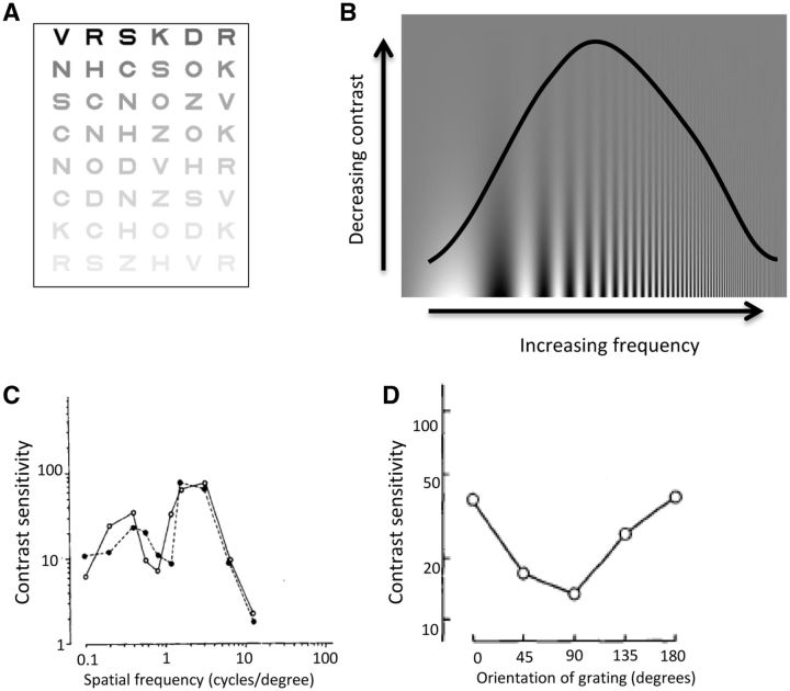
“This is more suggestive of a cortical rather than retinal or geniculate site of disruption as cortical neurons show stronger selectivity for different spatial frequencies than neurons in the retina or thalamus (Campbell and Robson, 1968; De Valois et al., 1982; Regan, 1982).”
BackgroundStages in the development of Parkinson’s disease-related pathology
Braak, H., Ghebremedhin, E., Rüb, U., Bratzke, H., & Del Tredici, K. (2004) · Cell and Tissue Research · doi:10.1007/s00441-004-0956-9 · Cited by 2,689 · cited 1×
“One theory of disease progression in Parkinson’s disease has suggested that the cerebral cortex is not damaged until the later stages of the disease (Braak et al., 2004).”
BackgroundThe Sydney multicenter study of Parkinson's disease: The inevitability of dementia at 20 years
Hely, M. A., Reid, W. G. J., Adena, M. A., Halliday, G. M., & Morris, J. G. L. (2008) · Movement Disorders · doi:10.1002/mds.21956 · Cited by 2,246 · cited 1×
“Visual hallucinations are also common in Parkinson’s disease, with a reported prevalence as high as 74% after 20 years of disease (Fenelon et al., 2000; Hely et al., 2008).”
BackgroundA direct demonstration of functional specialization in human visual cortex
Zeki, S., Watson, J., Lueck, C., Friston, K., Kennard, C., & Frackowiak, R. (1991) · The Journal of Neuroscience · doi:10.1523/jneurosci.11-03-00641.1991 · Cited by 1,716 · cited 2×
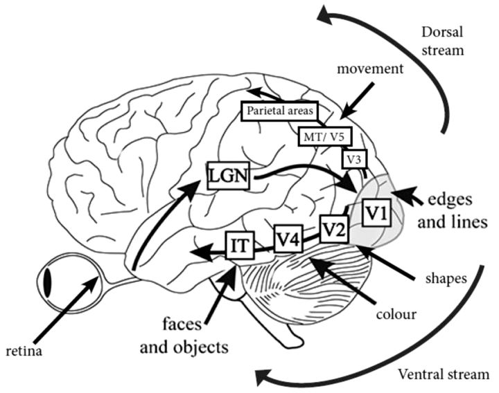
“IT = infero-temporal region; MT/V5 = motion processing region.”
BackgroundPharmacological Treatment of Parkinson Disease: A Review
Connolly, B. S., & Lang, A. E. (2014) · JAMA · doi:10.1001/jama.2014.3654 · Cited by 1,498 · cited 2×
“General advice is to consider visuo-perceptual ability and if there is any doubt, to refer the patient for a formal driving assessment via the national driving authority (Crizzle et al., 2012).”
BackgroundSpatial frequency selectivity of cells in macaque visual cortex
De Valois, R. L., Albrecht, D. G., & Thorell, L. G. (1982) · Vision Research · doi:10.1016/0042-6989(82)90113-4 · Cited by 1,489 · cited 2×
“This is more suggestive of a cortical rather than retinal or geniculate site of disruption as cortical neurons show stronger selectivity for different spatial frequencies than neurons in the retina or thalamus (Campbell and Robson, 1968; De Valois et al., 1982; Regan, 1982).”
Functional analysis of human MT and related visual cortical areas using magnetic resonance imaging
Tootell, R., Reppas, J., Kwong, K., Malach, R., Born, R., Brady, T., Rosen, B., & Belliveau, J. (1995) · The Journal of Neuroscience · doi:10.1523/jneurosci.15-04-03215.1995 · Cited by 1,415 · cited 2×
“IT = infero-temporal region; MT/V5 = motion processing region.”
BackgroundMazzulli, J. R., Xu, Y.-H., Sun, Y., Knight, A. L., McLean, P. J., Caldwell, G. A., Sidransky, E., Grabowski, G. A., & Krainc, D. (2011) · Cell · doi:10.1016/j.cell.2011.06.001 · Cited by 1,341 · cited 1×
“Both the GBA and SNCA genes are linked with the lysosomal pathway (Mazzulli et al., 2011), suggesting the visual deficits associated with these genes may share a pathophysiological mechanism, with lysosomal dysfunction associated with cortical visual dysfunction.”
BackgroundTHE DESIGN OF A NEW LETTER CHART FOR MEASURING CONTRAST SENSITIVITY
Pelli, D. G., Robson, J. G., & Wilkins, A. J. (1988) · Cited by 1,308
Research works printed but not linked: 3
- Ogden, 1990, Deficits on visuospatiaol tests involving forward planning in high-functioning Parkinsonians, Neuropsychiatry Neuropsychol Behav Neurol, 3, 125
- 10.1037/0033-295X.89.4.407 · doi:10.1037/0033-295x.89.4.407
- 10.1097/00006324-199404000-00002 · doi:10.1097/00006324-199404000-00002
Cited by
Abnormal intrinsic brain functional network dynamics in Parkinson’s disease
Kim, J., Criaud, M., Cho, S. S., Díez-Cirarda, M., Mihaescu, A., Coakeley, S., Ghadery, C., Valli, M., Jacobs, M. F., Houle, S., et al. (2017) · Brain · doi:10.1093/brain/awx233 · Cited by 369
“Visual function is also a major complex sensory domain affected by Parkinson’s disease (Mosimann et al., 2004; Weil et al., 2016).”
BackgroundDiederich, N. J., Goetz, C. G., & Stebbins, G. T. (2005) · Movement Disorders · doi:10.1002/mds.20308 · Cited by 290
Ko, F., Muthy, Z. A., Gallacher, J., Sudlow, C., Rees, G., Yang, Q., Keane, P. A., Petzold, A., Khaw, P. T., Reisman, C., et al. (2018) · JAMA Neurology · doi:10.1001/jamaneurol.2018.1578 · Cited by 241
Levodopa unresponsive symptoms in Parkinson disease: L-Dopa Unresponsive Symptoms in PD
Sethi, K. (2008) · Movement Disorders · doi:10.1002/mds.22049 · Cited by 175
Phosphorylated α‐synuclein in the retina is a biomarker of Parkinson's disease pathology severity
Ortuño‐Lizarán, I., Beach, T. G., Serrano, G. E., Walker, D. G., Adler, C. H., & Cuenca, N. (2018) · Movement Disorders · doi:10.1002/mds.27392 · Cited by 167
“Visual dysfunction and retinal changes in PD have been widely reported.(5,6) Patients suffering from PD have functional visual alterations such as reduced electroretinogram (ERG) responses and prolonged latency in visual evoked potentials.”
BackgroundPast, present and future role of retinal imaging in neurodegenerative disease
Kashani, A. H., Asanad, S., Chan, J. W., Singer, M. B., Zhang, J., Sharifi, M., Khansari, M. M., Abdolahi, F., Shi, Y., Biffi, A., et al. (2021) · Progress in Retinal and Eye Research · doi:10.1016/j.preteyeres.2020.100938 · Cited by 165
Ocular and visual disorders in Parkinson's disease: Common but frequently overlooked
Ekker, M. S., Janssen, S., Seppi, K., Poewe, W. H., de Vries, N. M., Theelen, T., Nonnekes, J. H., & Bloem, B. R. (2017) · Parkinsonism & Related Disorders · doi:10.1016/j.parkreldis.2017.02.014 · Cited by 161
Eye Movements in Parkinson’s Disease and Inherited Parkinsonian Syndromes
Pretegiani, E., & Optican, L. M. (2017) · Frontiers in Neurology · doi:10.3389/fneur.2017.00592 · Cited by 146
“, hallucinations and impairment of visual acuity, color and contrast sensitivity, motion perception, stereopsis), but whether and how they impact eye movement is difficult to establish (47, 48).”
BackgroundMorgia, C. L., Vito, L. D., Carelli, V., & Carbonelli, M. (2017) · Frontiers in Neurology · doi:10.3389/fneur.2017.00710 · Cited by 146
“Visual disturbances described in PD may range from reduced visual acuity and color vision to abnormal contrast sensitivity, dysfunction of eye movements and visual hallucinations (31, 32).”
BackgroundInternational Multicenter Analysis of Brain Structure Across Clinical Stages of Parkinson's Disease
Laansma, M. A., Bright, J. K., Al–Bachari, S., Anderson, T., Ard, T. D., Assogna, F., Baquero, K., Berendse, H. W., Blair, J. C., Cendes, F., et al. (2021) · Movement Disorders · doi:10.1002/mds.28706 · Cited by 133
“In addition, the implication of the occipital cortex may relate to compromised visual functions in early-stage PD patients.(23) The diffuse pattern of thinner cortices alongside smaller hippocampi and amygdalae in HY3 patients agrees with the more advanced symptomatic stages associated with PD dementia(24); we found t…”
BackgroundChrysou, A., Jansonius, N. M., & van Laar, T. (2019) · Parkinsonism & Related Disorders · doi:10.1016/j.parkreldis.2019.04.023 · Cited by 141
Alzheimer’s Retinopathy: Seeing Disease in the Eyes
Mirzaei, N., Shi, H., Oviatt, M., Doustar, J., Rentsendorj, A., Fuchs, D.-T. S., Sheyn, J., Black, K. L., Koronyo, Y., & Koronyo‐Hamaoui, M. (2020) · Frontiers in Neuroscience · doi:10.3389/fnins.2020.00921 · Cited by 145
Neuroimaging and fluid biomarkers in Parkinson’s disease in an era of targeted interventions
Zarkali, A., Thomas, G. E. C., Zetterberg, H., & Weil, R. S. (2024) · Nature Communications · doi:10.1038/s41467-024-49949-9 · Cited by 147
“The visual system is affected inPD, withworseperformanceofaffectedindividualsacrossarangeofvisual tasks: spatial navigation, line orientation, visual rotation, facial recognition, contrast sensitivity, colour vision 197 .”
BackgroundSelf-citationGlucocerebrosidase Mutations in Parkinson Disease
O’Regan, G. C., deSouza, R., Balestrino, R., & Schapira, A. H. V. (2017) · Journal of Parkinson s Disease · doi:10.3233/jpd-171092 · Cited by 136
McLean, G., Hindle, J. V., Guthrie, B., & Mercer, S. W. (2017) · BMC Neurology · doi:10.1186/s12883-017-0904-4 · Cited by 131
“The increased prevalence of visual dysfunction in the PD group may reflect the complex visual dysfunctions which are now known to be common in PD [27].”
BackgroundRetinal Ganglion Cells and Circadian Rhythms in Alzheimer’s Disease, Parkinson’s Disease, and Beyond
Morgia, C. L., Ross‐Cisneros, F. N., Sadun, A. A., & Carelli, V. (2017) · Frontiers in Neurology · doi:10.3389/fneur.2017.00162 · Cited by 125
Retinal α-synuclein deposits in Parkinson’s disease patients and animal models
Veys, L., Vandenabeele, M., Ortuño‐Lizarán, I., Baekelandt, V., Cuenca, N., Moons, L., & Groef, L. D. (2019) · Acta Neuropathologica · doi:10.1007/s00401-018-01956-z · Cited by 122
Sequence of clinical and neurodegeneration events in Parkinson’s disease progression
Oxtoby, N. P., Leyland, L., Aksman, L., Thomas, G. E. C., Bunting, E. L., Wijeratne, P. A., Young, A. L., Zarkali, A., Tan, M. M. X., Bremner, F., et al. (2021) · Brain · doi:10.1093/brain/awaa461 · Cited by 108
“Tests of verbal fluency and visuospatial function seem to be early indicators of an individual’s risk of developing dementia (Williams-Gray et al. , 2013), and visual processing regions are affected early in patients who develop cognitive impairment (Weil et al. , 2016).”
Self-citationThe Role of the GABAergic System in Diseases of the Central Nervous System
Zhang, W., Xiong, B., Zhang, L.-Q., Huang, X., Yuan, X., Tian, Y., & Tian, X. (2021) · Neuroscience · doi:10.1016/j.neuroscience.2021.06.037 · Cited by 101
Diagnostic biomarkers for Parkinson’s disease at a glance: where are we?
Cova, I., & Priori, A. (2018) · Journal of Neural Transmission · doi:10.1007/s00702-018-1910-4 · Cited by 102
Visual Dysfunction in Parkinson's Disease
Armstrong, R. A. (2017) · International review of neurobiology · doi:10.1016/bs.irn.2017.04.007 · Cited by 95
Oculo‐visual abnormalities in Parkinson's disease: Possible value as biomarkers
Guo, L., Normando, E. M., Shah, P. A., Groef, L. D., & Cordeiro, M. F. (2018) · Movement Disorders · doi:10.1002/mds.27454 · Cited by 105
Precision and False Perceptual Inference
Parr, T., Benrimoh, D. A., Vincent, P., & Friston, K. J. (2018) · Frontiers in Integrative Neuroscience · doi:10.3389/fnint.2018.00039 · Cited by 87
Ortuño‐Lizarán, I., Esquiva, G., Beach, T. G., Serrano, G. E., Adler, C. H., Lax, P., & Cuenca, N. (2018) · Acta Neuropathologica Communications · doi:10.1186/s40478-018-0596-z · Cited by 91
“Its main motor clinical features are rigidity, tremor and bradykinesia [19, 21, 46], but people with PD may also have several non-motor symptoms including cognitive decline and dementia [11], gastrointestinal and cardiovascular problems [43], mood disturbance [50], visual disruption [2, 55], impairment of the pupillar…”
BackgroundZarkali, A., McColgan, P., Leyland, L., Lees, A. J., Rees, G. E., & Weil, R. S. (2021) · Communications Biology · doi:10.1038/s42003-020-01622-9 · Cited by 91
“3 mm, (2) any FD > 5 mm, or (3) outliers >30% of the whole sample.”
BackgroundSelf-citationAdam, C. R., Shrier, E. M., Ding, Y., Glazman, S., & Bodis-Wollner, I. G. (2013) · Journal of Neuro-Ophthalmology · doi:10.1097/wno.0b013e31828c4e1a · Cited by 85
O’Callaghan, C., Hall, J. M., Tomassini, A., Muller, A. J., Walpola, I. C., Moustafa, A. A., Shine, J. M., & Lewis, S. J. G. (2017) · Biological Psychiatry Cognitive Neuroscience and Neuroimaging · doi:10.1016/j.bpsc.2017.04.007 · Cited by 92
Sha, Z., Xia, M., Lin, Q., Cao, M., Tang, Y., Xu, K., Song, H., Wang, Z., Wang, F., Fox, P. T., et al. (2017) · Cerebral Cortex · doi:10.1093/cercor/bhx273 · Cited by 91
Characterization of retinal architecture in Parkinson's disease
Chorostecki, J., Seraji‐Bozorgzad, N., Shah, A., Bao, F., Bao, G. W., George, E. B., Gorden, V., Caon, C., Frohman, E. M., Bhatti, M. T., et al. (2015) · Journal of the Neurological Sciences · doi:10.1016/j.jns.2015.05.007 · Cited by 86
“Improvements in retinal imaging have allowed for correlations between histopathology in post-mortem anatomy with in-vivo images [5].”
Retinal Thickness Predicts the Risk of Cognitive Decline in Parkinson Disease
Murueta‐Goyena, A., Pino, R. D., Galdós, M., Arana, B., Acera, M., Carmona‐Abellán, M., Fernández‐Valle, T., Tijero, B., Lucas‐Jiménez, O., Ojeda, N., et al. (2020) · Annals of Neurology · doi:10.1002/ana.25944 · Cited by 82
“Although abnormalities in perception and processing of visual information in PD might be in part related to the injury of cortical and subcortical areas of the brain, the involvement of the retina in visual cognition abnormalities of PD patients is firmly supported by several in vivo electrophysiological and OCT studi…”
ResultMultidisciplinary care for people with Parkinson’s disease: the new kids on the block!
Radder, D. L. M., de Vries, N. M., Riksen, N. P., Diamond, S. J., Gross, D. R., Gold, D. R., Heesakkers, J. P. F. A., Henderson, E. J., Hommel, A. L. A. J., Lennaerts, H. H., et al. (2018) · Expert Review of Neurotherapeutics · doi:10.1080/14737175.2019.1561285 · Cited by 77
“Also, levodopa may lead to some improvement in terms of visual acuity, although acuity, color and contrast deficits tend to be subtle to begin with [71].”
BackgroundArt therapy for Parkinson's disease
Cucca, A., Rocco, A. D., Acosta, I., Beheshti, M., Berberian, M., Bertisch, H. C., Droby, A., Ettinger, T., Hudson, T. E., Inglese, M., et al. (2021) · Parkinsonism & Related Disorders · doi:10.1016/j.parkreldis.2021.01.013 · Cited by 91
Cognitive associations with comprehensive gait and static balance measures in Parkinson's disease
Morris, R., Martini, D. N., Smulders, K., Kelly, V. E., Zabetian, C. P., Poston, K. L., Hiller, A., Chung, K. A., Yang, L., Hu, S., et al. (2019) · Parkinsonism & Related Disorders · doi:10.1016/j.parkreldis.2019.06.014 · Cited by 85
Association between ADHD and vision problems. A systematic review and meta-analysis
Bellato, A., Perna, J., Ganapathy, P. S., Solmi, M., Zampieri, A., Cortese, S., & Faraone, S. V. (2022) · Molecular Psychiatry · doi:10.1038/s41380-022-01699-0 · Cited by 86
“, schizophrenia [88, 93], and Parkinson’s [94, 95]).”
Murueta‐Goyena, A., Andikoetxea, A., Gómez‐Esteban, J. C., & Gabilondo, I. (2019) · Frontiers in Pharmacology · doi:10.3389/fphar.2019.01294 · Cited by 72
“Among primary visual functions, low-contrast visual acuity, contrast sensitivity and color vision are typically affected in PD (Weil et al., 2016).”
BackgroundVisual system manifestations of Alzheimer's disease
Kusne, Y. N., Wolf, A. B., Townley, K., Conway, M. D., & Peyman, G. A. (2016) · Acta Ophthalmologica · doi:10.1111/aos.13319 · Cited by 80
“However, it is again important to consider the specificity of these biomarkers as contrast sensitivity and colour vision, among other functional deficits, have been shown to be reduced in Parkinson’s disease patients as well (Weil et al. 2016).”
BackgroundStriatal dopamine denervation and sensory integration for balance in middle-aged and older adults
Cham, R., Perera, S., Studenski, S. A., & Bohnen, N. I. (2006) · Gait & Posture · doi:10.1016/j.gaitpost.2006.11.204 · Cited by 69
Orlosky, J., Itoh, Y., Ranchet, M., Kiyokawa, K., Morgan, J. C., & Devos, H. (2017) · IEEE Transactions on Visualization and Computer Graphics · doi:10.1109/tvcg.2017.2657018 · Cited by 85
“Whereas some other systems for evaluation utilize game performance to determine certain neurodegenerative characteristics like memory loss [4][27], we rely on the fact that the eye movements of patients with parkinsonism have consistently abnormal responses [37], which can even be used to aid in the differential diagn…”
BackgroundSeeing ophthalmologic problems in Parkinson disease
Borm, C. D. J. M., Visser, F., Werkmann, M., de Graaf, D., Putz, D., Seppi, K., Poewe, W. H., Vlaar, A. M. M., Hoyng, C. B., Bloem, B. R., et al. (2020) · Neurology · doi:10.1212/wnl.0000000000009214 · Cited by 67
“Alterations in the visual network from the retina to higher cortical brain regions have been found in PD, and may be the cause of ophthalmologic symptoms.(26) As many as 63% of patients reported a problem in the ocular surface domain.”
BackgroundLax, P., Ortuño‐Lizarán, I., Maneu, V., Vidal‐Sanz, M., & Cuenca, N. (2019) · International Journal of Molecular Sciences · doi:10.3390/ijms20133164 · Cited by 72
Increased weighting on prior knowledge in Lewy body-associated visual hallucinations
Zarkali, A., Adams, R. A., Psarras, S., Leyland, L., Rees, G. E., & Weil, R. S. (2019) · Brain Communications · doi:10.1093/braincomms/fcz007 · Cited by 72
“Hallucinations in LBD are usually progressive, starting as minor illusions or misperceptions before complex, detailed or animate hallucinations emerge (Mosimann et al., 2004; Weil et al., 2016).”
BackgroundInfluentialSelf-citationZarkali, A., McColgan, P., Ryten, M., Reynolds, R. H., Leyland, L., Lees, A. J., Rees, G. E., & Weil, R. S. (2020) · Brain · doi:10.1093/brain/awaa270 · Cited by 59
“Despite their impact, our understanding of how visual hallucinations are produced remains limited (Fénelon et al., 2000; Weil et al., 2016).”
BackgroundSelf-citationStructural and functional changes in the retina in Parkinson’s disease
Alves, J., Westner, B. U., Højlund, A., Weil, R. S., & Dalal, S. S. (2023) · Journal of Neurology Neurosurgery & Psychiatry · doi:10.1136/jnnp-2022-329342 · Cited by 71
Self-citationVision in laboratory rodents—Tools to measure it and implications for behavioral research
Leinonen, H., & Tanila, H. (2017) · Behavioural Brain Research · doi:10.1016/j.bbr.2017.07.040 · Cited by 75
“In particular, decreased spatial contrast sensitivity was a common clinical research finding already a few decades ago in PD patients [131-133].”
Li, Z., Yang, J., Wang, Y., Cai, M., Liu, X., & Lu, K. (2022) · Journal of Biomedical Informatics · doi:10.1016/j.jbi.2022.104085 · Cited by 78
Retinal involvement in dementia with Lewy bodies: A clue to hallucinations?
Maurage, C., Ruchoux, M., de Vos, R., Surguchov, A. P., & Destée, A. (2003) · Annals of Neurology · doi:10.1002/ana.10730 · Cited by 64
Parkinson disease: Not just a movement disorder
Pandya, M. A., Kubu, C. S., & Giroux, M. (2008) · Cleveland Clinic Journal of Medicine · doi:10.3949/ccjm.75a.07005 · Cited by 69
Basal ganglia atrophy–associated causal structural network degeneration in Parkinson's disease
Li, R., Zou, T., Wang, X., Wang, H., Hu, X., Xie, F., Li, M., & Chen, H. (2021) · Human Brain Mapping · doi:10.1002/hbm.25715 · Cited by 64
“color discrimination, complex visual hallucinations (Archibald, Clarke, Mosimann, & Burn, 2009; Weil et al., 2016), and cognitive impairments including executive dysfunction and problems with working memory (Emre, Ford, Bilgic, & Ey, 2014; Halliday, Leverenz,”
BackgroundGalasko, D. R. (2017) · Neurologic Clinics · doi:10.1016/j.ncl.2017.01.004 · Cited by 72
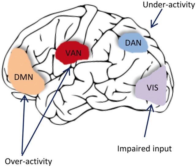
“The anatomic basis of hallucinations is not clear, although weakening of interactions between neural networks for attention and conscious perception may be important.(22) The burden of LB in the inferior temporal lobe plus cholinergic deficits in the temporal lobe and other cortical areas may play a role.”
BackgroundThe use of optical coherence tomography in neurology: a review
Xie, J. S., Donaldson, L., & Margolin, E. (2022) · Brain · doi:10.1093/brain/awac317 · Cited by 60
Visual Dysfunction in Parkinson’s Disease
Nieto-Escámez, F. A., Obrero‐Gaitán, E., & Cortés‐Pérez, I. (2023) · Brain Sciences · doi:10.3390/brainsci13081173 · Cited by 74
“tasks requiring high-order processing capabilities, such as object mental rotation [15], perception of space [16], spatial maps representation, visuospatial working memory, effective navigation, and target localization [17–19], which can be considered as preclinical markers and predictors of disease development [20].”
BackgroundInfluentialLiao, J., He, W., Li, L., Wang, J., Gong, L., Zhang, Q., & Lin, Z. (2025) · Cell Biomaterials · doi:10.1016/j.celbio.2025.100016 · Cited by 77
Functional and structural brain network correlates of visual hallucinations in Lewy body dementia
Mehraram, R., Peraza, L. R., Murphy, N., Cromarty, R. A., Graziadio, S., O'Brien, J. T., Killen, A., Colloby, S. J., Firbank, M., Su, L., et al. (2022) · Brain · doi:10.1093/brain/awac094 · Cited by 62
Does retina play a role in Parkinson's Disease?
Devi, S. M., Iyer, M., Aswathy, N. P., Dhivya, V., & Vellingiri, B. (2020) · Acta Neurologica Belgica · doi:10.1007/s13760-020-01274-w · Cited by 56
Dopamine, Alpha-Synuclein, and Mitochondrial Dysfunctions in Parkinsonian Eyes
Indrieri, A., Pizzarelli, R., Franco, B., & Leonibus, E. D. (2020) · Frontiers in Neuroscience · doi:10.3389/fnins.2020.567129 · Cited by 63
“The data reporting visual and retinal abnormalities in PD patients have been extensively reviewed recently (Weil et al., 2016; Guo et al., 2018).”
BackgroundThe Pharmacology of Visual Hallucinations in Synucleinopathies
Russo, M., Carrarini, C., Dono, F., Rispoli, M. G., Pietro, M. D., Stefano, V. D., Ferri, L., Bonanni, L., Sensi, S. L., & ONOFRJ, M. C. (2019) · Frontiers in Pharmacology · doi:10.3389/fphar.2019.01379 · Cited by 60
Bernasconi, F., Blondiaux, E., Potheegadoo, J., Stripeikyte, G., Pagonabarraga, J., Bejr‐Kasem, H., Bassolino, M., Akselrod, M., Martínez‐Horta, S., Sampedro, F., et al. (2021) · Science Translational Medicine · doi:10.1126/scitranslmed.abc8362 · Cited by 51
“We note, however, that these studies mostly focused on patients with structured visual hallucinations (20) or did 1Laboratory of Cognitive Neuroscience, Center for Neuroprosthetics & Brain Mind Institute, Ecole Polytechnique Fédérale de Lausanne (EPFL), 1202 Geneva, Switzerland.”
Neuropathology of Nonmotor Symptoms of Parkinson's Disease
Jellinger, K. A. (2017) · International review of neurobiology · doi:10.1016/bs.irn.2017.05.005 · Cited by 55
Prognostic potentials of AI in ophthalmology: systemic disease forecasting via retinal imaging
Tan, Y. Y., Kang, H. G., Lee, C. J., Kim, S. S., Park, S., Thakur, S., Soh, Z. D., Cho, Y., Peng, Q., Lee, K., et al. (2024) · Eye and Vision · doi:10.1186/s40662-024-00384-3 · Cited by 66
Kawabata, K., Watanabe, H., Hara, K., Bagarinao, E., Yoneyama, N., Ogura, A., Imai, K., Masuda, M., Yokoi, T., Ohdake, R., et al. (2018) · Journal of Neurology · doi:10.1007/s00415-018-8755-5 · Cited by 46
“Patients with PD present several visual perceptual disturbances, including color discrimination, contrast sensitivity, and difficulties with complex visual tasks, such as mental rotation and emotion recognition [67].”
BackgroundZhu, H., Huang, J., Deng, L., He, N., Cheng, L., Shu, P., Yan, F., Tong, S., Sun, J., & Ling, H. (2019) · Frontiers in Neuroscience · doi:10.3389/fnins.2019.00080 · Cited by 47
“Indeed, visual network is another major complex sensory domain affected by PD, and PD may lead to visual hallucinations, perceptual, executive and sleep dysfunction (Archibald et al., 2011; Wu et al., 2011; Weil et al., 2016).”
BackgroundPieperhoff, P., Südmeyer, M., Dinkelbach, L., Hartmann, C. J., Ferrea, S., Moldovan, A. S., Minnerop, M., Diaz-Pier, S., Schnitzler, A., & Amunts, K. (2022) · Cortex · doi:10.1016/j.cortex.2022.03.009 · Cited by 57
Cognitive effort and active inference
Parr, T., Holmes, E., Friston, K. J., & Pezzulo, G. (2023) · Neuropsychologia · doi:10.1016/j.neuropsychologia.2023.108562 · Cited by 66
“However, the same principles are likely to apply to modified versions of this task, such as the auditory Stroop task which has been deployed in Parkinson ’ s disease (Janssen et al., 2019), a condition associated with visual dysfunction (Weil et al., 2016).”
BackgroundPsychosis in Parkinson's Disease
Ffytche, D. H., & Aarsland, D. (2017) · International review of neurobiology · doi:10.1016/bs.irn.2017.04.005 · Cited by 46
“PD is associated with low-level visual dysfunction (for review, see Weil et al., 2016) helping account for the predominantly visual nature of PD psychosis symptoms.”
BackgroundDefining visual illusions in Parkinson's disease: Kinetopsia and object misidentification illusions
Nishio, Y., Yokoi, K., Hirayama, K., Ishioka, T., Hosokai, Y., Gang, M., Uchiyama, M., Baba, T., Suzuki, K., Takeda, A., et al. (2018) · Parkinsonism & Related Disorders · doi:10.1016/j.parkreldis.2018.05.023 · Cited by 45
Yakubovich, S., Israeli‐Korn, S., Halperin, O., Yahalom, G., Hassin‐Baer, S., & Zaidel, A. (2020) · Brain Communications · doi:10.1093/braincomms/fcaa035 · Cited by 48
“…disease, including delays in visual evoked responses, abnormalities in contrast, spatiotemporal and colour sensitivity and altered perception of visual orientation (Bodis and Yahr, 1978; Bodis-Wollner et al. , 1987; Montse et al. , 2001; Gullett et al. , 2013; Weil et al. , 2016, 2017, 2018).”
Stuart, S., Galna, B., Delicato, L. S., Lord, S., & Rochester, L. (2017) · European Journal of Neuroscience · doi:10.1111/ejn.13589 · Cited by 47
“Visual deficits are also common in PD and range from impaired visual functions such as visual acuity (VA) and contrast sensitivity (CS) to more complex processes such as depth or motion perception (Weil et al., 2016).”
Circadian Rhythm Abnormalities in Parkinson’s Disease from Humans to Flies and Back
Lazzari, F. D., Bisaglia, M., Zordan, M. A., & Sandrelli, F. (2018) · International Journal of Molecular Sciences · doi:10.3390/ijms19123911 · Cited by 46
“Among these is the optomotor response [138,139], the study of which might constitute a useful phenotypic output for the symptoms related to visual acuity and contrast sensitivity detected in some PD patients and fly PD models [16,140,141].”
BackgroundWalpola, I. C., Muller, A. J., Hall, J. M., Andrews‐Hanna, J. R., Irish, M., Lewis, S. J. G., Shine, J. M., & O’Callaghan, C. (2020) · Cortex · doi:10.1016/j.cortex.2019.12.023 · Cited by 46
“This may be further exacerbated in Parkinson’s disease due to the numerous sources of visual and attentional dysfunction that render incoming sensory information less reliable (Weil et al., 2016).”
BackgroundYue, Y., Jiang, Y., Shen, T., Pu, J., Lai, H., & Zhang, B. (2020) · Frontiers in Neuroscience · doi:10.3389/fnins.2020.00141 · Cited by 45
Vignando, M., ffytche, D., Lewis, S. J. G., Lee, P. H., Chung, S. J., Weil, R. S., Hu, M. T., Mackay, C. E., Griffanti, L., Pins, D., et al. (2022) · Nature Communications · doi:10.1038/s41467-022-28087-0 · Cited by 42
Self-citationDonato, L., Scimone, C., Alibrandi, S., Abdalla, E. M., Nabil, K. M., D’Angelo, R., & Sidoti, A. (2020) · International Journal of Molecular Sciences · doi:10.3390/ijms22010070 · Cited by 42
“Mutations in LRRK2 were associated with better color discrimination [40] but, probably due to the contemporary presence of variants in DRD4 and CHRNA7, as well as the homozygous one in CNGB3, cone neurotransmission was prevalent.”
BackgroundHou, Y., Ou, R., Yang, J., Song, W., Gong, Q., & Shang, H. (2018) · Neuroradiology · doi:10.1007/s00234-018-2101-6 · Cited by 40
Birba, A., Fittipaldi, S., Escobar, J. C., Campo, C. G., Legaz, A., Galiani, A., Rivera, M. D., Martorell, M., Alifano, F., Piña‐Escudero, S. D., et al. (2021) · Cerebral Cortex · doi:10.1093/cercor/bhab421 · Cited by 48
Structural Brain Correlations of Visuospatial and Visuoperceptual Tests in Parkinson’s Disease
Garcia-Diaz, A. I., Segura, B., Baggio, H. C., Marti, M. J., Valldeoriola, F., Compta, Y., Bargallo, N., Uribe, C., Campabadal, A., Abos, A., et al. (2018) · Journal of the International Neuropsychological Society · doi:10.1017/s1355617717000583 · Cited by 39
Thomas, G. E. C., Zeidman, P., Sultana, T., Zarkali, A., Razi, A., & Weil, R. S. (2022) · Brain Communications · doi:10.1093/braincomms/fcac329 · Cited by 38
“Until recently, models for how VH are generated within the brain remained somewhat limited and disparate, despite the high prevalence of VH and the clear burden they have on patients and their carers.(6)”
BackgroundSelf-citationSelf‐motion perception in Parkinson's disease
Halperin, O., Israeli‐Korn, S., Yakubovich, S., Hassin‐Baer, S., & Zaidel, A. (2020) · European Journal of Neuroscience · doi:10.1111/ejn.14716 · Cited by 40
Guan, X., Zeng, Q., Guo, T., Wang, J., Xuan, M., Gu, Q., Wang, T., Huang, P., Xu, X., & Zhang, M. (2017) · Frontiers in Aging Neuroscience · doi:10.3389/fnagi.2017.00360 · Cited by 43
“Weil et al. (2016) concluded in a review that patients with increased tremor perform better on vision-related tasks and the presence of tremor is associated with fewer color vision abnormalities.”
BackgroundCucinotta, F. A., & Cacao, E. (2020) · Life Sciences in Space Research · doi:10.1016/j.lssr.2019.10.004 · Cited by 34
Treatment of Nonmotor Symptoms Associated with Parkinson Disease
Goldman, J. G., & Guerra, C. M. (2020) · Neurologic Clinics · doi:10.1016/j.ncl.2019.12.003 · Cited by 40
Harnessing Visual Imagery and Oculomotor Behaviour to Understand Prospection.
Conti, F., & Irish, M. (2021) · Trends in Cognitive Sciences · doi:10.1016/j.tics.2021.01.009 · Cited by 32
Ho, M. W.-R., Chien, S. H., Lu, M., Chen, J., Aoh, Y., Chen, C., Lane, H., & Tsai, C. (2020) · Scientific Reports · doi:10.1038/s41598-020-61310-w · Cited by 36
Huang, L., Wang, C., Wang, W. “., Wang, Y., & Zhang, R. (2020) · Journal of Neurology · doi:10.1007/s00415-020-10094-0 · Cited by 38
Deep learning based diagnosis of Parkinson’s Disease using diffusion magnetic resonance imaging
Zhao, H., Tsai, C., Zhou, M., Liu, Y., Chen, Y.-L., Huang, F., Lin, Y., & Wang, J. (2022) · Brain Imaging and Behavior · doi:10.1007/s11682-022-00631-y · Cited by 40
“Although PD is characterized by specific motor disability, non-motor abnormalities may aggravate motor symptoms (Weil et al., 2016).”
BackgroundHamilton, C. A., Matthews, F. E., Donaghy, P. C., Taylor, J., O'Brien, J. T., Barnett, N. A., Olsen, K., Lloyd, J. J., Petrides, G. S., McKeith, I. G., et al. (2020) · American Journal of Geriatric Psychiatry · doi:10.1016/j.jagp.2020.07.018 · Cited by 38
Retinal imaging biomarkers of neurodegenerative diseases
Christinaki, E., Kulenovic, H., Hadoux, X., Baldassini, N., Eijgen, J. V., Groef, L. D., Stalmans, I., & van Wijngaarden, P. (2021) · Clinical and Experimental Optometry · doi:10.1080/08164622.2021.1984179 · Cited by 35
Non-motor Symptoms and Treatments in Parkinson’s Disease
Goldman, J. G. (2025) · Neurologic Clinics · doi:10.1016/j.ncl.2024.12.008 · Cited by 45
Fundarò, C., Maestri, R., Ferriero, G., Chimento, P., Taveggia, G., & Casale, R. (2019) · European Journal of Physical and Rehabilitation Medicine · doi:10.23736/s1973-9087.18.05368-6 · Cited by 33
Retinal Degeneration: A Window to Understand the Origin and Progression of Parkinson’s Disease?
Zhang, Y., Zhang, X., Yue, Y., & Tian, T. (2022) · Frontiers in Neuroscience · doi:10.3389/fnins.2021.799526 · Cited by 36
“, 2016), misfolded a-synuclein (Weil et al., 2016), retinal ganglion cells loss (Malmfors, 1963), thinning of retinal nerve fiber layer (Frederick et al.”
BackgroundInfluentialCompensatory Neural Responses to Cognitive Fatigue in Young and Older Adults
Samuel, I. B. H., Wang, C., Burke, S., Kluger, B. M., & Ding, M. (2019) · Frontiers in Neural Circuits · doi:10.3389/fncir.2019.00012 · Cited by 36
Visual dysfunction is a better predictor than retinal thickness for dementia in Parkinson’s disease
Hannaway, N., Zarkali, A., Leyland, L., Bremner, F., Nicholas, J. M., Wagner, S. K., Roig, M., Keane, P. A., Toosy, A., Chataway, J., et al. (2023) · Journal of Neurology Neurosurgery & Psychiatry · doi:10.1136/jnnp-2023-331083 · Cited by 32
Self-citationVeetil, I. K., Chowdary, D. E., Chowdary, P. N., Sowmya, V., & Gopalakrishnan, E. A. (2024) · Digital Signal Processing · doi:10.1016/j.dsp.2024.104407 · Cited by 36
Neurovascular coupling dysfunction of visual network organization in Parkinson's disease
Li, T., Liu, T., Zhang, J., Ma, Y., Wang, G., Suo, D., Yang, B., Wang, X., Funahashi, S., Zhang, K., et al. (2023) · Neurobiology of Disease · doi:10.1016/j.nbd.2023.106323 · Cited by 33
“…Barzgari et al., 2019; Fernandez-Seara et al., 2012; Shang et al., 2021a), especially in the motor cortices and parieto-occipital cortices, which are correlated with the visual network (Cardoso et al., 2010; Weil et al., 2016; Melzer et al., 2011; Madhyastha et al., 2015; De Micco et al., 2021).”
BackgroundInfluentialRetinal Alterations Predict Early Prodromal Signs of Neurodegenerative Disease
Casciano, F., Zauli, E., Celeghini, C., Caruso, L., Gonelli, A., Zauli, G., & Pignatelli, A. (2024) · International Journal of Molecular Sciences · doi:10.3390/ijms25031689 · Cited by 38
“While retinal DA deficiency may be the primary cause of these visual disorders, the orientation-specific impairment due to the cerebral cortex should not be excluded as the loss of dopaminergic neuronal projection [50,247–249].”
BackgroundWhat can we learn from fMRI capture of visual hallucinations in Parkinson’s disease?
Dujardin, K., Roman, D., Baille, G., Pins, D., Lefebvre, S., Delmaire, C., Defebvre, L., & Jardri, R. (2019) · Brain Imaging and Behavior · doi:10.1007/s11682-019-00185-6 · Cited by 30
Boertien, J., van der Zee, S., Chrysou, A., Gerritsen, M., Jansonius, N., Spikman, J., van Laar, T., Verwey, N., van Harten, B., Portman, A., et al. (2020) · BMC Neurology · doi:10.1186/s12883-020-01811-3 · Cited by 28
Benvenutti, R., Marcon, M., Reis, C. G., Nery, L. R., Miguel, C., Herrmann, A. P., Vianna, M. R. M. R., & Piato, Â. (2018) · PeerJ · doi:10.7717/peerj.4957 · Cited by 28
“These patients have difficulty performing complex visual tasks such as mental rotation and emotion recognition (Weil et al., 2016).”
BackgroundImaging Markers of Progression in Parkinson's Disease
Strafella, A. P., Bohnen, N. I., Pavese, N., Vaillancourt, D. E., van Eimeren, T., Politis, M., Tessitore, A., Ghadery, C., Lewis, S. J. G., & on behalf of IPMDS‐Neuroimaging Study Group (2018) · Movement Disorders Clinical Practice · doi:10.1002/mdc3.12673 · Cited by 35
Tang, X., Zhang, Y., Liu, D., Hu, Y., Jiang, L., & Zhang, J. (2021) · Neurology · doi:10.1212/wnl.0000000000011894 · Cited by 30
“Patients with the AR subtype also showed a decreased LGI in the cuneus and nearby visual cortices, which may relate to visual disturbances in these patients.(30) Taken together, our findings of widespread LGI decreases might indicate the anatomical substrates underlying the motor and nonmotor impairments in patients w…”
BackgroundNeural mechanisms of psychosis vulnerability and perceptual abnormalities in the ALS‐FTD spectrum
Devenney, E. M., Tu, S., Caga, J., Ahmed, R. M., Ramsey, E., Zoing, M. C., Kwok, J. B., Halliday, G. M., Piguet, O., Hodges, J. R., et al. (2021) · Annals of Clinical and Translational Neurology · doi:10.1002/acn3.51363 · Cited by 26
“In the neurodegenerative literature, abnormal perceptual experiences related to vision and the formation of visual hallucinations have been frequently explored in Parkinson’s disease.(64) A number of theories exist for the generation of hallucinations in Parkinson’s disease that could be relevant here.”
Background
Metadata
- Authors
- Weil, R. S., Schrag, A. E., Warren, J. D., Crutch, S. J., Lees, A. J., & Morris, H. R.
- Affiliations
- Royal Free London NHS Foundation Trust (GB)
- National Hospital for Neurology and Neurosurgery (GB)
- UCL Queen Square Institute of Neurology (GB)
- Corresponding author
- Weil, Rimona S.
- ORCID
- Venue
- Brain
- Publisher
- Oxford University Press (OUP)
- Published
- 2016-07-13
- Volume
- 139
- Issue
- 11
- Pages
- 2827-2843
- ISSN
- 0006-8950 (ISSN-L)
- 1460-2156
- PMC
- PMC5091042
- OpenAlex
- W2474900144
- Cited by
- 481
- References
- 232
- Citations by year
- 2026: 41 · 2025: 55 · 2024: 54 · 2023: 48 · 2022: 52 · 2021: 55 · 2020: 66 · 2019: 37 · 2018: 34 · 2017: 28 · 2016: 2
- FWCI
- 4.67
- Type
- journal article
- Language
- en
- Open access
- green
- www.ncbi.nlm.nih.gov
- Licence
- cc-by
- Locations
- Brain (publishedVersion)
- PubMed Central (publishedVersion, cc-by)
- PubMed (publishedVersion)
- UCL Discovery (University College London) (submittedVersion)
- HighWire Press Open Archive (submittedVersion)
- Funders
- National Institute for Health and Care Research
- Parkinson's UK (K-1501, J‐0804)
- Academy of Medical Sciences (AMS-SGCL13-Weil)
- Engineering and Physical Sciences Research Council (EP/M006093/1)
- Economic and Social Research Council (ES/L001810/1)
- Topics
- Health Sciences › Medicine › Clinical Neurology › Parkinson's Disease Mechanisms and Treatments
- Life Sciences › Neuroscience › Cognitive Neuroscience › Visual perception and processing mechanisms
- Life Sciences › Neuroscience › Cognitive Neuroscience › Hallucinations in medical conditions
- Keywords
- Parkinson's disease, visual dysfunction, contrast sensitivity, mental rotation, emotion recognition, visual neglect, motion processing, object processing, GBA mutations, LRRK2 mutations, REM sleep behavior disorder, postural instability, gait disturbance, visual hallucinations, biomarkers, dementia prediction
- MeSH terms
- Leucine-Rich Repeat Serine-Threonine Protein Kinase-2, beta-Glucosidase, Humans, Parkinson Disease, Vision Disorders, Visual Pathways, Visual Perception
About these checks
Registry, retraction, Cites and statistics results are computed, not judged, with one exception: a printed reference that no index lists is read once by a model before it counts as a citation error. Claims are extracted by a model and kept only when their quote appears verbatim in the text; each cited source is then checked, with a model’s help, for whether it says what the claim relies on. Those verdicts can be wrong — read the claim’s quote and the cited source before relying on one.
Statistics and arithmetic are recomputed, not judged.
Confidence bands are shown only for fields calibrated on our annotated test set. None is calibrated yet, so no band is shown.
What counts as a claim ›How we measure confidence ›How we check this ›
Are you an author, or do you see a problem with this page? Request a review
Data sources
Bibliographic data: Semantic Scholar, OpenAlex, Crossref.
Contains information from the Semantic Scholar Academic Graph Datasets, made available under the ODC Attribution License (ODC-BY 1.0).
Semantic Scholar’s data is described in: Kinney, R., et al. (2023). The Semantic Scholar Open Data Platform. arXiv:2301.10140.
