

The DOI matches its Crossref record; 54 of the 54 references it cites were found in a registry or an index.
Human Amygdala Sensitivity to the Pupil Size of Others
Open accessAbstract
Stimulation of the amygdala produces pupil dilation in animal and human subjects. The present study examined whether the amygdala is sensitive to variations in the pupil size of others. Male subjects underwent event-related functional magnetic resonance imaging while passively viewing unfamiliar female faces whose pupils were either unaltered (natural variations in large and small pupils) or altered to be larger or smaller than their original size. Results revealed that the right amygdala and left amygdala/substantia innominata were sensitive to the pupil size of others, exhibiting increased activity for faces with relatively large pupils. Upon debrief, no subject reported being aware that the pupils had been manipulated. These results suggest a function for the amygdala in the detection of changes in pupil size, an index of arousal and/or interest on the part of a conspecific, even in the absence of explicit knowledge.
Overview
The claims and statistics of this paper haven’t been checked yet.
Run the check — free, no account needed. It reads the open-access text and checks the sources it cites.
Integrity signals
Registry
Cites
Statistics
Figures
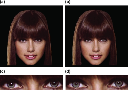
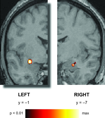
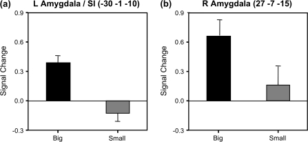
Verification
Claims the paper makes
The claims and statistics of this paper haven’t been checked yet.
Run the check — free, no account needed. It reads the open-access text and checks the sources it cites.
Claims other papers make about this one
Each is checked against this paper’s own text: does it say what the citing paper says it does?
The source backs part of the claim, not all of it — or backs it for a different population, condition or comparison.
In the citing paper
“For example, people with large pupils are commonly rated as more attractive and activate neural responses in the amygdala [35,36], and a recent study demonstrates that human participants evaluate those with dilating pupils as more friendly, attractive and trustworthy and behave more cooperatively with them in the trust game [37].”
From Are dark-eyed dogs favoured by humans? Domestication as a potential driver of iris colour difference between dogs and wolves (2023) · cites this paper as [36]
Experimental — accuracy not yet measuredThe source backs part of the claim, not all of it — or backs it for a different population, condition or comparison.
In the citing paper
“humans have a cognitive tendency to perceive emotional arousal based on iris–pupil contrast and make inferences about a person's personality [33–37].”
From Are dark-eyed dogs favoured by humans? Domestication as a potential driver of iris colour difference between dogs and wolves (2023) · cites this paper as [36]
Experimental — accuracy not yet measuredIn the citing paper
“Seeing others' dilated pupils also causes significantly higher amygdala activation than viewing undilated pupils, an effect that nonetheless appears to be unrelated to subjective reports of attractiveness (Demos et al., 2008;Amemiya and Ohtomo, 2012).”
From Oxytocin enhances pupil dilation and sensitivity to 'hidden' emotional expressions (2013) · cites this paper as Demos et al., 2008
Self-check
Percentages, sums and reported differences re-computed from the paper’s own numbers.
The self-check needs the paper’s full text.
Citations
Cites
Co-Planar Stereotaxic Atlas of the Human Brain
Talairach, J., & Tournoux, P. (1988) · Medical Entomology and Zoology · Cited by 11,798
Co-planar stereotaxic atlas of the human brain
Laitinen, L. (1989) · Clinical Neurology and Neurosurgery · doi:10.1016/0303-8467(89)90128-5 · Cited by 7,565
The amygdala: vigilance and emotion
Davis, M., & Whalen, P. J. (2001) · Molecular Psychiatry · doi:10.1038/sj.mp.4000812 · Cited by 2,882
Self-citationCohen, J., MacWhinney, B., Flatt, M., & Provost, J. (1993) · Behavior Research Methods, Instruments, & Computers · doi:10.3758/bf03204507 · Cited by 2,802
Whalen, P. J., Rauch, S. L., Etcoff, N. L., McInerney, S. C., Lee, M. B., & Jenike, M. A. (1998) · The Journal of Neuroscience · doi:10.1523/jneurosci.18-01-00411.1998 · Cited by 2,205
Self-citationA differential neural response in the human amygdala to fearful and happy facial expressions
Morris, J. S., Frith, C. D., Perrett, D. I., Rowland, D., Young, A. W., Calder, A. J., & Dolan, R. J. (1996) · Nature · doi:10.1038/383812a0 · Cited by 2,013
Emotion and Cognition: Insights from Studies of the Human Amygdala
Phelps, E. A. (2006) · Annual Review of Psychology · doi:10.1146/annurev.psych.56.091103.070234 · Cited by 1,798 · cited 2×
“In this way, amygdala activity potentiates neuronal responses in sensory cortical regions that may, in turn, facilitate subsequent information processing (Amaral et al. 1992; Kapp et al. 1992; Whalen 1998; Morris et al. 2001; Pessoa et al. 2006; Phelps 2006 ).”
BackgroundResponse and Habituation of the Human Amygdala during Visual Processing of Facial Expression
Breiter, H. C., Etcoff, N. L., Whalen, P. J., Kennedy, W. A., Rauch, S. L., Buckner, R. L., Strauss, M. M., Hyman, S. E., & Rosen, B. R. (1996) · Neuron · doi:10.1016/s0896-6273(00)80219-6 · Cited by 1,711
Self-citationThe amygdala: Neurobiological aspects of emotion, memory, and mental dysfunction.
Aggleton, J. P. (1992) · Wiley-Liss eBooks · Cited by 1,536
LaBar, K. S., Gatenby, J. C., Gore, J. C., LeDoux, J. E., & Phelps, E. A. (1998) · Neuron · doi:10.1016/s0896-6273(00)80475-4 · Cited by 1,402
Research works printed but not linked: 1
- Amaral, 1992, Anatomical organization of the primate amygdaloid complex, 1
Cited by
Sorted by citations
Items 101–111 of 111
Azzouni, J. (2025) · doi:10.1093/9780197790151.003.0011
Azzouni, J. (2025) · doi:10.1093/9780197790151.003.0014
The Language of Visual Phenomenology
Azzouni, J. (2025) · doi:10.1093/9780197790151.003.0004
Azzouni, J. (2025) · doi:10.1093/9780197790151.003.0009
Azzouni, J. (2025) · doi:10.1093/9780197790151.003.0008
The Phenomenology of Inference
Azzouni, J. (2025) · doi:10.1093/9780197790151.003.0010
Teikari, P. (2012) · theses.fr (ABES) · doi:10.70675/96362824z0120z4ed7z94c7zb60f3d7e5f50 · Cited by 2
(2026) · Wiley eBooks · doi:10.1002/9781394461714.ch18
Metadata
- Authors
- Demos, K. E., Kelley, W. M., Ryan, S. L., Davis, F. C., & Whalen, P. J.
- Affiliations
- Dartmouth College (US)
- Corresponding author
- Demos, K.E.
- ORCID
- Demos, K.E.
- Venue
- Cerebral Cortex
- Publisher
- Oxford University Press (OUP)
- Published
- 2008-03-27
- Volume
- 18
- Issue
- 12
- Pages
- 2729-2734
- ISSN
- 1047-3211 (ISSN-L)
- 1460-2199
- PMC
- PMC2583162
- OpenAlex
- W2119582005
- Cited by
- 112
- References
- 53
- Citations by year
- 2026: 1 · 2025: 14 · 2024: 4 · 2023: 4 · 2022: 6 · 2021: 5 · 2020: 6 · 2019: 7 · 2018: 4 · 2017: 5 · 2016: 5 · 2015: 5 · 2014: 4 · 2013: 9 · 2012: 4
- FWCI
- 5.01
- Type
- journal article
- Language
- en
- Open access
- hybrid
- doi.org
- Licence
- cc-by-nc
- Locations
- Cerebral Cortex (publishedVersion, cc-by-nc)
- PubMed Central (publishedVersion, cc-by-nc)
- PubMed (publishedVersion)
- HighWire Press Open Archive (submittedVersion)
- Funders
- Dartmouth College
- U.S. Public Health Service (080716, 069315)
- National Institute of Mental Health (R01MH080716, 080716, 069315)
- Topics
- Life Sciences › Neuroscience › Cognitive Neuroscience › Face Recognition and Perception
- Social Sciences › Psychology › Experimental and Cognitive Psychology › Evolutionary Psychology and Human Behavior
- Life Sciences › Neuroscience › Cognitive Neuroscience › Memory and Neural Mechanisms
- Keywords
- amygdala, pupil size, social perception, facial expressions, arousal detection, substantia innominata, face perception, unconscious processes, pupillometry
- MeSH terms
- Adolescent, Amygdala, Arousal, Cues, Facial Expression, Female, Humans, Functional Laterality, Male, Pupil, Visual Acuity, Visual Perception, Knowledge, Young Adult
About these checks
Registry, retraction, Cites and statistics results are computed, not judged, with one exception: a printed reference that no index lists is read once by a model before it counts as a citation error. Claims are extracted by a model and kept only when their quote appears verbatim in the text; each cited source is then checked, with a model’s help, for whether it says what the claim relies on. Those verdicts can be wrong — read the claim’s quote and the cited source before relying on one.
Statistics and arithmetic are recomputed, not judged.
Confidence bands are shown only for fields calibrated on our annotated test set. None is calibrated yet, so no band is shown.
What counts as a claim ›How we measure confidence ›How we check this ›
Are you an author, or do you see a problem with this page? Request a review
