

The DOI matches its Crossref record; 26 of the 26 references it cites were found in a registry or an index.
3′ Branch ligation: a novel method to ligate non-complementary DNA to recessed or internal 3′OH ends in DNA or RNA
Published version of a preprint
Abstract
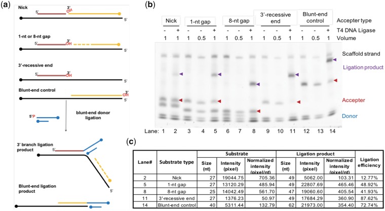
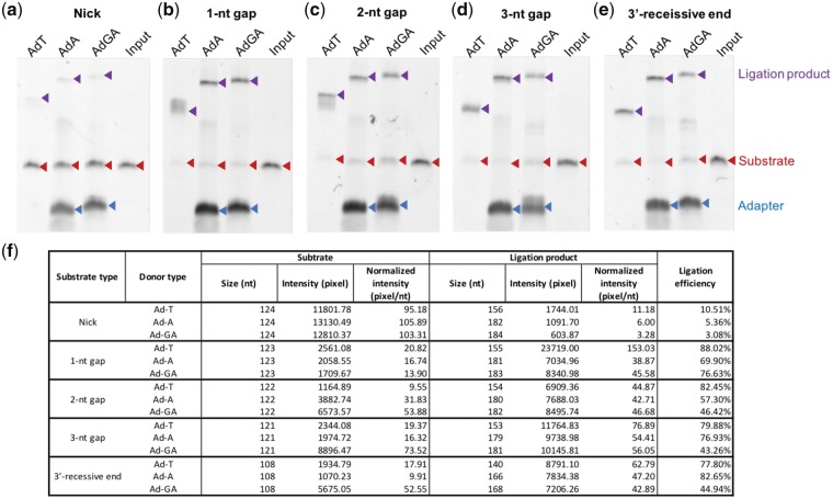
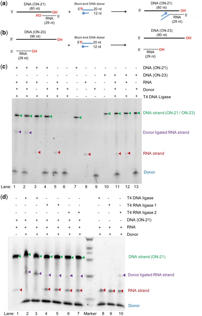
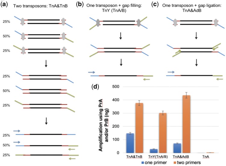
Abstract Nucleic acid ligases are crucial enzymes that repair breaks in DNA or RNA during synthesis, repair and recombination. Various molecular tools have been developed using the diverse activities of DNA/RNA ligases. Herein, we demonstrate a non-conventional ability of T4 DNA ligase to join 5’ phosphorylated blunt-end double-stranded DNA to DNA breaks at 3’ recessive ends, gaps, or nicks to form a 3’ branch structure. Therefore, this base pairing-independent ligation is termed 3’ branch ligation (3’BL). In an extensive study of optimal ligation conditions, similar to blunt-end ligation, the presence of 10% PEG-8000 in the ligation buffer significantly increased ligation efficiency. A low level of nucleotide preference was observed at the junction sites using different synthetic DNAs. Furthermore, we discovered that T4 DNA ligase efficiently ligated DNA to the 3’ recessed end of RNA, not to that of DNA, in a DNA/RNA hybrid, whereas RNA ligases are less efficient in this reaction. These novel properties of T4 DNA ligase can be utilized as a broad molecular technique in many important applications. We performed a proof-of-concept study of a new directional tagmentation protocol for next generation sequencing (NGS) library construction that eliminates inverted adapters and allows sample barcode insertion adjacent to genomic DNA. 3’BL after single transposon tagmentation can theoretically achieve 100% usable template, and our empirical data demonstrate that the new approach produced higher yield compared with traditional double transposon or Y transposon tagmentation. We further explore the potential use of 3’BL for preparing targeted RNA NGS libraries with mitigated structure-based bias and adapter dimer problems.
Overview
The claims and statistics of this paper haven’t been checked yet.
Run the check — free, no account needed. It reads the open-access text and checks the sources it cites.
Integrity signals
Registry
Cites
Statistics
Figures
Verification
Claims the paper makes
The claims and statistics of this paper haven’t been checked yet.
Run the check — free, no account needed. It reads the open-access text and checks the sources it cites.
Claims other papers make about this one
Each is checked against this paper’s own text: does it say what the citing paper says it does?
The cited source says what the claim attributes to it.
In the citing paper
“Even though this recommendation is not tested in the current design of the study, it is well-known that T4 RNA ligase 1 prefers single stranded RNA as a substrate (36,37) and therefore, 5′ overhang of acceptor RNA in ‘blocked’ RNA:DNA hybrid might result in adapter ligation and in this way may reduce blocking efficiency of the oligonucleotides.”
From Depletion of erythropoietic miR-486-5p and miR-451a improves detectability of rare microRNAs in peripheral blood-derived small RNA sequencing libraries (2020) · cites this paper as [36]
Self-check
Percentages, sums and reported differences re-computed from the paper’s own numbers.
The self-check needs the paper’s full text.
Citations
Cites
Bushati, N., & Cohen, S. M. (2007) · Annual Review of Cell and Developmental Biology · doi:10.1146/annurev.cellbio.23.090506.123406 · Cited by 2,750
Functions of microRNAs and related small RNAs in plants
Mallory, A. C., & Vaucheret, H. (2006) · Nature Genetics · doi:10.1038/ng1791 · Cited by 830
Lehman, I. R. (1974) · Science · doi:10.1126/science.186.4166.790 · Cited by 655
DNA Ligases: Structure, Reaction Mechanism, and Function
Tomkinson, A. E., Vijayakumar, S., Pascal, J. M., & Ellenberger, T. (2006) · Chemical Reviews · doi:10.1021/cr040498d · Cited by 297
Structure and function of mammalian DNA ligases
Tomkinson, A. E., & Mackey, Z. B. (1998) · Mutation Research/DNA Repair · doi:10.1016/s0921-8777(97)00050-5 · Cited by 200
DNA Ligases: Progress and Prospects
Shuman, S. (2009) · Journal of Biological Chemistry · doi:10.1074/jbc.r900017200 · Cited by 189
DNA ligases in the repair and replication of DNA
Timson, D. J., Singleton, M. R., & Wigley, D. B. (2000) · Mutation Research/DNA Repair · doi:10.1016/s0921-8777(00)00033-1 · Cited by 175
Ho, C. K., & Shuman, S. (2002) · Proceedings of the National Academy of Sciences · doi:10.1073/pnas.192184699 · Cited by 173
Structure and Mechanism of RNA Ligase
Ho, C. K., Wang, L., Lima, C. D., & Shuman, S. (2004) · Structure · doi:10.1016/j.str.2004.01.011 · Cited by 148
Self-citationUltra-low-input, tagmentation-based whole-genome bisulfite sequencingPreprint
Adey, A., & Shendure, J. (2012) · Genome Research · doi:10.1101/gr.136242.111 · Cited by 128
Sgaramella, V., & Khorana, H. G. (1972) · Journal of Molecular Biology · doi:10.1016/0022-2836(72)90155-6 · Cited by 123
Direct comparison of nick-joining activity of the nucleic acid ligases from bacteriophage T4
Bullard, D. R., & Bowater, R. P. (2006) · Biochemical Journal · doi:10.1042/bj20060313 · Cited by 114
DNA and RNA ligases: structural variations and shared mechanisms
Pascal, J. M. (2008) · Current Opinion in Structural Biology · doi:10.1016/j.sbi.2007.12.008 · Cited by 101
Enhanced DNA sequencing by hybridization.
Broude, N. E., Sano, T., Smith, C. L., & Cantor, C. R. (1994) · Proceedings of the National Academy of Sciences · doi:10.1073/pnas.91.8.3072 · Cited by 98
Mutation Detection by Ligation to Complete n-mer DNA ArraysPreprint
Gunderson, K. L., Huang, X. C., Morris, M. S., Lipshutz, R. J., Lockhart, D. J., & Chee, M. S. (1998) · Genome Research · doi:10.1101/gr.8.11.1142 · Cited by 91
Template‐independent ligation of single‐stranded DNA by T4 DNA ligase
Kuhn, H., & Frank-Kamenet︠s︡kiĭ, M. D. (2005) · FEBS Journal · doi:10.1111/j.1742-4658.2005.04954.x · Cited by 89
Determination of the Free-Energy Change for Repair of a DNA Phosphodiester Bond
Dickson, K. S., Burns, C. M., & Richardson, J. P. (2000) · Journal of Biological Chemistry · doi:10.1074/jbc.m910044199 · Cited by 78
Ligation Bias in Illumina Next-Generation DNA Libraries: Implications for Sequencing Ancient Genomes
Seguin-Orlando, A., Schubert, M., Clary, J., Stagegaard, J., Alberdi, M. T., Prado, J. L., Prieto, A., Willerslev, E., & Orlando, L. (2013) · PLoS ONE · doi:10.1371/journal.pone.0078575 · Cited by 76
Sealing of gaps in duplex DNA by T4 DNA ligase
Nilsson, S. V., & Magnusson, G. (1982) · Nucleic Acids Research · doi:10.1093/nar/10.5.1425 · Cited by 74
Goffin, C., Bailly, V., & Verly, W. G. (1987) · Nucleic Acids Research · doi:10.1093/nar/15.21.8755 · Cited by 59
Haplotype phasing of whole human genomes using bead-based barcode partitioning in a single tube
Zhang, F., Christiansen, L., Thomas, J., Pokholok, D., Jackson, R., Morrell, N., Zhao, Y., Wiley, M., Welch, E., Jaeger, E. B., et al. (2017) · Nature Biotechnology · doi:10.1038/nbt.3897 · Cited by 48
Sgaramella, V., & Ehrlich, S. D. (1978) · European Journal of Biochemistry · doi:10.1111/j.1432-1033.1978.tb12336.x · Cited by 46
Alexander, R. C. (2003) · Nucleic Acids Research · doi:10.1093/nar/gkg415 · Cited by 36
Mendel-Hartvig, M. (2004) · Nucleic Acids Research · doi:10.1093/nar/gnh011 · Cited by 18
Characterization of Bacteriophage T3 DNA Ligase
Cai, L. (2004) · The Journal of Biochemistry · doi:10.1093/jb/mvh047 · Cited by 17
Improved Transposon-Based Library Preparation for the Ion Torrent Platform
Gorbacheva, T., Quispe‐Tintaya, W., Popov, V. N., Vijg, J., & Maslov, A. Y. (2015) · BioTechniques · doi:10.2144/000114277 · Cited by 12
Research works printed but not linked: 1
- Goryshin, 2016, Oligonucleotide replacement for di-tagged and directional libraries
Cited by
Juzenas, S., Lindqvist, C. M., Ito, G., Dolshanskaya, Y., Halfvarson, J., Franke, A., & Hemmrich-Stanisak, G. (2020) · NAR Genomics and Bioinformatics · doi:10.1093/nargab/lqaa008 · Cited by 17
Metadata
- Authors
- Wang, L., Xi, Y., Zhang, W., Wang, W., Shen, H., Wang, X., Zhao, X., Alexeev, A., Peters, B. A., Albert, A., Xu, X., Ren, H., Wang, O., Kirkconnell, K., Perazich, H., Clark, S., Hurowitz, E., Chen, A., Xu, X., Drmanac, R., & Jiang, Y.
- Affiliations
- Advanced Genomics Technology Lab, Complete Genomics Inc., 2904 Orchard Pkwy, San Jose, California, USA
- Institute of Biochemistry, BGI-Shenzhen, Shenzhen, China
- ORCID
- Jiang, Yuan
- Venue
- DNA Research
- Publisher
- Oxford University Press (OUP)
- Published
- 2018-11-13
- Volume
- 26
- Issue
- 1
- Pages
- 45-53
- ISSN
- 1340-2838
- 1756-1663
- PMC
- PMC6379041
- Cited by
- 13
- References
- 26
- Language
- en
- Funders
- Shenzhen Peacock Plan (KQTD20150330171505310)
About these checks
Registry, retraction, Cites and statistics results are computed, not judged, with one exception: a printed reference that no index lists is read once by a model before it counts as a citation error. Claims are extracted by a model and kept only when their quote appears verbatim in the text; each cited source is then checked, with a model’s help, for whether it says what the claim relies on. Those verdicts can be wrong — read the claim’s quote and the cited source before relying on one.
Statistics and arithmetic are recomputed, not judged.
Confidence bands are shown only for fields calibrated on our annotated test set. None is calibrated yet, so no band is shown.
What counts as a claim ›How we measure confidence ›How we check this ›
Are you an author, or do you see a problem with this page? Request a review
Data sources
Bibliographic data: Semantic Scholar, OpenAlex, Crossref.
Contains information from the Semantic Scholar Academic Graph Datasets, made available under the ODC Attribution License (ODC-BY 1.0).
Semantic Scholar’s data is described in: Kinney, R., et al. (2023). The Semantic Scholar Open Data Platform. arXiv:2301.10140.
