

Genomic Insights into Plastid Evolution
Open accessThe DOI matches its Crossref record; 114 of the 116 references it cites were found in a registry or an index; 46 of the 106 sources its claims cite were checked for support.
Abstract
The origin of plastids (chloroplasts) by endosymbiosis stands as one of the most important events in the history of eukaryotic life. The genetic, biochemical, and cell biological integration of a cyanobacterial endosymbiont into a heterotrophic host eukaryote approximately a billion years ago paved the way for the evolution of diverse algal groups in a wide range of aquatic and, eventually, terrestrial environments. In this review, we provide an overview of recent advances in our understanding of the origin and spread of plastids from the perspective of comparative genomics. Comparative genomics has also allowed us to better understand how endosymbionts become organelles.
Overview
CoChat Summary
## TL;DR
This is a review article summarizing genomic evidence on how plastids originated, spread, were replaced, and were lost across eukaryotes. It argues that comparative genomics has clarified many major transitions but that key questions about the number and partners of endosymbioses remain unresolved.
## Key findings
- Primary plastids are described as originating once from a cyanobacterial endosymbiont, likely at least ~900 Ma, in the ancestor of green algae/land plants, red algae, and glaucophytes.
- Comparative genomics indicates that endosymbiotic gene transfer was extensive: less than 5% of genes thought present in the cyanobacterial progenitor typically remain in plastid genomes, and 7–15% of plastid-targeted proteins in diverse lineages are noncyanobacterial in origin.
- The paper states that only three unambiguous cases of complete plastid loss have been documented so far, all in parasitic species: Cryptosporidium, certain gregarines, and Hematodinium.
- It reports several specific plastid replacement / complex-plastid examples, including at least 11 different diatom species found across closely related dinoflagellates, 90 haptophyte-to-dinoflagellate EGTs in Karenia brevis and Karlodinium veneficum, and only 17 of 433 nucleus-encoded chromatophore-targeted proteins in Paulinella chromatophora appearing to come from α-cyanobacteria.
## Methods
- Narrative review of recent genomics-based studies of plastid evolution, with emphasis on comparative genomics, phylogenomics, transcriptomics, genome sequencing, and proteomics.
- Synthesizes evidence from multiple taxa, including algae, plants, dinoflagellates, apicomplexans, Paulinella, and other protists, using published genome and transcriptome datasets.
- Main analysis is interpretive: the authors compare phylogenetic, genomic, and cell-biological evidence to reconstruct plastid gains, losses, replacements, and endosymbiotic gene transfer.
## Limitations
- The paper explicitly says that important details are still missing and that “we are still in the dark” about how many eukaryote–eukaryote endosymbioses occurred and, in many cases, who the partner cells were.
- It notes that phylogenetic reconstruction is limited by conflicting signals, methodological artifacts, and incomplete taxonomic sampling. (stated in the paper)
- It also states that increased plastid-genome sampling alone may not resolve some questions because of limited phylogenetic signal; nuclear genomes and better taxon sampling are needed. (stated in the paper)
Model-written summary. Check it against the paper before relying on it.
Integrity signals
Registry
Cites
Statistics
Figures
Verification
Claims the paper makes
31 Valid · 2 Not valid · 42 Not checked · 30 Own results and interpretations
Some cited sources support this claim and some don’t — read each source’s verdict below.
In the paper
“Less than 5% of the genes thought to have been present in the cyanobacterial progenitor of the plastid typically remain in the organellar genome, and hundreds of cyanobacterial genes can be found in the nuclear genomes of extant algae and plants (Dagan et al. 2013, Qiu, Yoon, et al. 2013).”
§ Genomic Insights into Plastid Evolution > The Endosymbiont-to-Organelle Transition
What the sources say
- Disputedcites Dagan et al. 2013 sourceExperimental — accuracy not yet measured
The Introduction gives a different figure: modern plastid genomes contain 5–10% as many genes as free-living cyanobacteria, not less than 5%. It also says many ancestral genes were lost or transferred to the nucleus, but that does not support the claimed percentage. (LLM, conf=0.88)
Checked by a reasoning model — may be wrong.
- Partly supportedcites Qiu, Yoon, et al. 2013 sourceExperimental — accuracy not yet measured
The section on endosymbiotic/horizontal gene transfer of prokaryotic genes in Plantae says modern plastid genomes are highly reduced and encode fewer than 250 genes. It does not give the estimated gene count of the cyanobacterial progenitor or state that less than 5% remain, so the claimed proportion is unsupported by this paper. (LLM, conf=0.85)
plain- Asserted: passes — The authors put it forward as true.
- Checkable: passes — It can be checked against evidence.
- Load-bearing: unclear — It is unclear whether the paper’s conclusions depend on it.
- Based: passes — The paper gives a reason why it holds.
- Disputed
At least one cited source doesn’t support this claim or points to a different paper, and none confirmed it — see the verdicts below.
In the paper
“Approximately half of the described dinoflagellate species maintain the peridinin-pigmented plastid thought to have been present in their common ancestor (Yoon et al. 2002).”
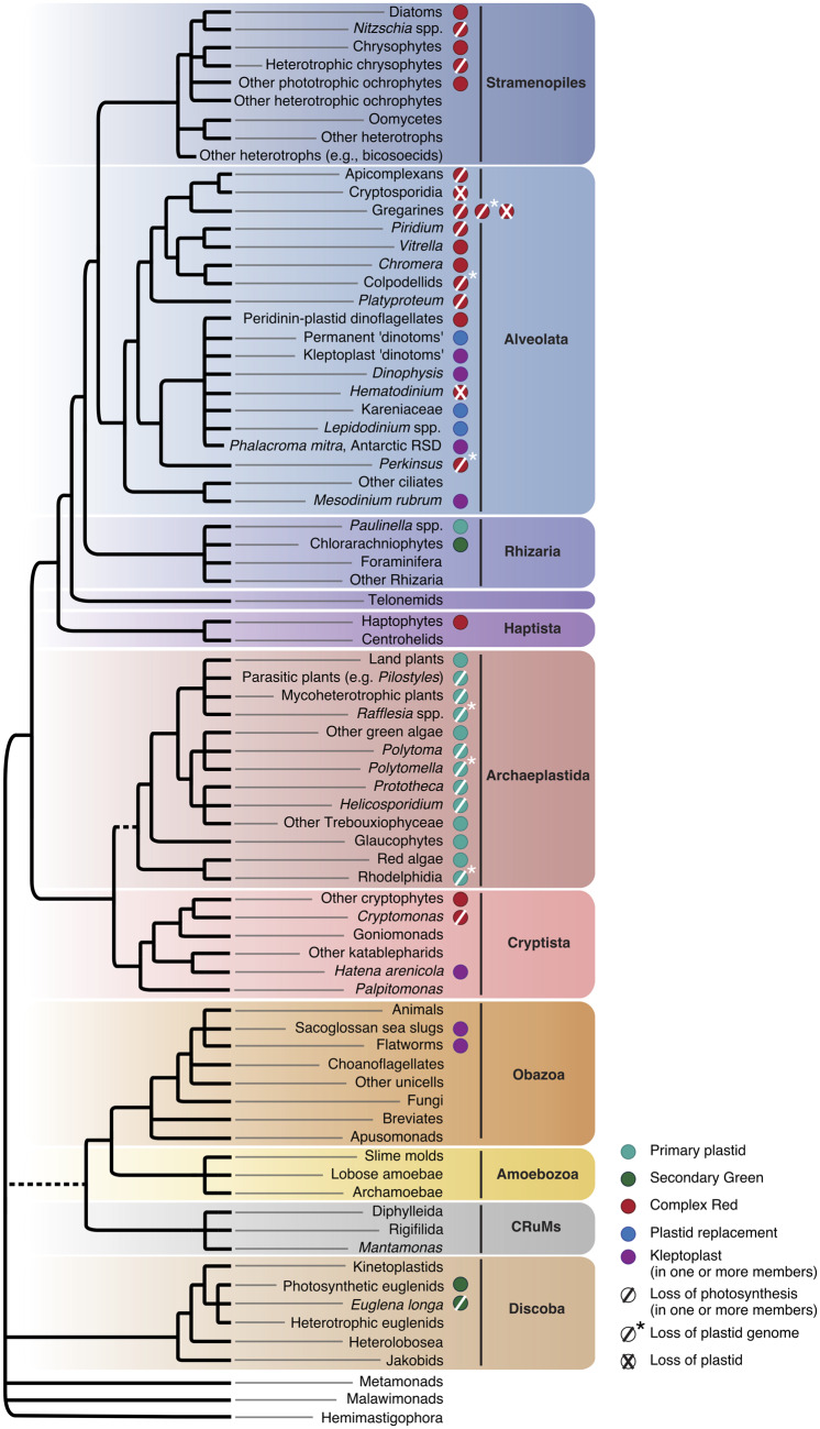
§ Genomic Insights into Plastid Evolution > Kleptomania Thus far, we have focused on the origins and fates of permanently integrated organelles. But there is also much to learn from the study of more transient associations between organisms in nature, the phenomenon of kleptoplasty—“plastid stealing”—being one such an example. Kleptoplasty is surprisingly common, having been observed in a variety of protist lineages (particularly foraminiferans, dinoflagellates, and ciliates) and even some animals (sea slugs and some flat worms). These stolen plastids come from varying sources and exhibit a wide range of retention times. In sea slugs, algal plastids are harvested, sequestered, and retained for periods of time ranging from days to weeks and months before being lost (Händeler et al. 2009). Even though only the plastid remains from its algal prey, the kleptoplasts remain photosynthetically active. Transcriptome and genome sequencing have failed to provide evidence of gene transfer from algae to the sacoglossan sea slug nuclear genome, despite suggestions that this had occurred (see Bhattacharya et al. 2013; Rauch et al. 2015 and references therein). In a recently described case of kleptoplasty in two flatworm species, sequestered plastids are stolen from different diatom species and retained for a couple of weeks (Steenkiste et al. 2019). In this instance, transcriptome sequencing suggests that kleptoplast genes are actively expressed, whereas algal nuclear genes are not, supporting the observation that only the plastid is retained and is functional (Steenkiste et al. 2019). Kleptoplastic tendencies are frequently observed in single-celled eukaryotes and are especially common among dinoflagellates. Approximately half of the described dinoflagellate species maintain the peridinin-pigmented plastid thought to have been present in their common ancestor (Yoon et al. 2002). Many dinoflagellates harbor kleptoplastids of various origins, whereas others have permanently replacing their ancestral plastid (see below). In addition as discussed above, still others completely lack photosynthetic abilities and appear to no longer have a plastid (e.g., Hematodinium; Gornik et al. 2015). Haptophyte-derived kleptoplastids are found in Phalacroma mitra (Koike et al. 2005) and the Antarctic Ross Sea (ARS) dinoflagellate (Sellers et al. 2014; Hehenberger et al. 2019). The ARS dinoflagellate is known not only to retain a relic version of its ancestral peridinin plastid but also to be obligately kleptoplastidic and reliant on its haptophyte prey for photosynthesis, which it can maintain for at least 30 months (Gast et al. 2007). Transcriptomics of the ARS dinoflagellate by Hehenberger et al. (2019) showed that plastid functions are split between the relic peridinin plastid and the kleptoplast, with photosynthesis related functions occurring there. Bioinformatic analyses suggest that many nucleus-encoded proteins are targeted to the kleptoplast, most of which do not appear to be derived from the plastid donor itself. Notably, the ARS dinoflagellate is closely related to other dinoflagellate lineages, specifically Karenia and Karlodinium, who harbor fully integrated, permanent haptophyte-derived plastids. Comparative genomics between the species with permanent haptophyte plastids and the haptophyte-derived kleptoplasts of the ARS dinoflagellate showed a number of shared gene transfers whose protein products are targeted to the plastid/kleptoplast, suggesting that, in some cases at least, a certain level of genetic integration can precede permanent organelle integration (Hehenberger et al. 2019). Other dinoflagellates have cryptophyte- or diatom-derived kleptoplasts. In one of the most intricate cases, Dinophysis obtains its kleptoplast by preying on a ciliate (Mesodinium rubrum) which itself harbors a kleptoplast of cryptophyte origin (either Geminigera cryophila [Johnson et al. 2006] or Teleaulax amphioxeia [Nishitani et al. 2010]). In Mesodinium rubrum, the kleptoplast is transcriptionally active and has been observed to be retained for up to a month with continual replacement (Johnson et al. 2007). Transcriptomics and phylogenetic analysis of plastid-related genes in Dinophysis fortii revealed that most such genes are of apparent peridinin plastid origin and thus represent EGTs from the original dinoflagellate plastid (Hongo et al. 2019). Intriguingly, the rest of the plastid-associated genes were not only of cryptophyte kleptoplast origin but also of haptophyte origin—including genes related to those of dinoflagellates with permanent tertiary haptophyte plastids (i.e., fucoxanthin dinoflagellates). This suggests that the ancestors of extant Dinophysis engaged in haptophyte kleptoplasty at some point during their evolutionary history (Hongo et al. 2019). Finally, dinoflagellates are known to harbor plastids derived from diatoms. These so-called “dinotoms” are retained permanently in the cytosol and the evidence suggests that the host cell exhibits some level of control over their division (e.g., Hehenberger et al. 2016; Yamada et al. 2017). To date, there is only one known example of a kleptoplasty-derived dinotom. In this case, the dinoflagellate Durinskia capensis appears to retain its diatom only temporarily, for ∼2 months, and does not appear to control its cell division (Yamada et al. 2019). Further genomic and transcriptomic investigations of the myriad ways in which dinoflagellates acquire and recycle their plastids will no doubt continue to provide insight into the molecular, biochemical, and cell biological factors underlying the endosymbiont-to-organelle transition.
What the sources say
- Disputedcites Yoon et al. 2002 sourceExperimental — accuracy not yet measured
The Discussion (page not provided) calls fucoxanthin the primitive plastid condition and peridinin a derived state, contrary to the claim that the peridinin-pigmented plastid was present in the common ancestor. The introductory passage says about half of dinoflagellates are photosynthetic; it does not say that half of described species retain a peridinin plastid. (LLM, conf=0.88)
Checked by a reasoning model — may be wrong.
plain- Asserted: passes — The authors put it forward as true.
- Checkable: passes — It can be checked against evidence.
- Load-bearing: unclear — It is unclear whether the paper’s conclusions depend on it.
- Based: passes — The paper gives a reason why it holds.
- Disputed
The cited sources support only part of this claim — read each source’s verdict below.
In the paper
“Recent molecular clock analyses estimate that the Paulinella chromatophore evolved a mere 90–140 Ma (Delaye et al. 2016).”
§ Genomic Insights into Plastid Evolution > The Chromatophores of Paulinella
What the sources say
- Partly supportedcites Delaye et al. 2016 sourceExperimental — accuracy not yet measured
The Abstract reports molecular-clock estimates of roughly 90–140 million years for the divergence of P. chromatophora from heterotrophic relatives. It calls that divergence a maximum date for the chromatophore’s origin, not an estimate that the chromatophore itself evolved within that interval. (LLM, conf=0.88)
hedged- Asserted: passes — The authors put it forward as true.
- Checkable: passes — It can be checked against evidence.
- Load-bearing: unclear — It is unclear whether the paper’s conclusions depend on it.
- Based: passes — The paper gives a reason why it holds.
- Partly supported
The sources with a verdict support this claim; the rest weren’t checked yet, have no full text we could read, or their verdicts aren’t rated yet.
In the paper
“a trend that has nevertheless emerged is plastid trees that unite haptophytes and cryptophytes to the exclusion of all other complex algae; see, e.g., Janouškovec et al. 2010; Ševčíková et al. 2015; Kim et al. 2017 and references therein for discussion”
§ Genomic Insights into Plastid Evolution > The Complexity of Complex Plastids As important as it was, the primary endosymbiotic origin of plastids accounts for only a fraction of the diversity of plant and algal life on Earth. Multiple higher-order endosymbiotic events—mergers between two eukaryotic cells—have taken place. In some photosynthetic lineages, the details are reasonably well understood, whereas in others they are completely uncertain. So-called “secondary” plastids of green algal origin arose on two separate occasions, one in the chlorarachniophytes and the other in the euglenids. We can infer that these correspond to two separate secondary endosymbioses because the organisms belong to different eukaryotic supergroups, Rhizaria in the case of chlorarachniophytes and Discoba in the case of euglenids (fig. 1). Efforts to pinpoint the precise green algal source for each of these plastids have brought us closer to an answer as more genomic data have become available. For example, recent targeted sequencing of algal lineages closely related to potential plastid donors (based on previous sequencing and phylogenetic analysis) and taxon-rich phylogenetic analysis of their plastid genomes pinned down their sources to be a precursor of siphonous green algae (Bryopsidales) in the case of chlorarachniophytes, and a prasinophyte from the order pyramimonadales in euglenids (Jackson et al. 2018). Although progress has been made, Jackson et al. (2018) suggest that increased sampling of plastid genomes of secondary green plastid relatives may not resolve the question much further due to limitations in phylogenetic signal. Improved resolution may be obtained by increased nuclear genome sequencing of these taxa and investigation of endosymbiont-derived nuclear genes. However, we must also acknowledge that in the case of chlorarachniophytes and/or euglenids, the plastid donor could have been an unknown lineage or a distant relative of extant taxa that has gone extinct. Complex plastids of red algal origin (including those acquired from secondary, tertiary, and possibly even higher-order endosymbiotic events) are found across an even wider diversity of eukaryotes, including cryptophytes and haptophytes, as well as some alveolates and stramenopiles (including diatoms and brown/golden algae) (fig. 1). Despite decades of study, many uncertainties still surround the origin(s) and evolution of red algal complex plastids—how many times they were established and how often they were horizontally spread (and between whom) is unclear. Why is the evolutionary history of such plastids so difficult to discern? Part of the problem lies in incongruences between phylogenies of plastid and nuclear genes. Plastid multigene phylogenies typically place complex red plastid-containing algae in a monophyletic clade that branches from within the red algae, albeit with internal tree topologies that are sensitive to phylogenetic method and taxon sampling (a trend that has nevertheless emerged is plastid trees that unite haptophytes and cryptophytes to the exclusion of all other complex algae; see, e.g., Janouškovec et al. 2010; Ševčíková et al. 2015; Kim et al. 2017 and references therein for discussion). Such phylogenies are consistent with the idea of a single secondary endosymbiotic origin of complex red alga-derived plastids, although it is important to note that, in isolation, they are also consistent with multiple independent endosymbioses involving closely related red algal endosymbionts. The precise nature of the algal donor is similarly unclear. At present, the data suggest that red alga-type complex plastids share more recent common ancestry with those of mesophilic red algae such as Porphyra and Chondrus than they do with the plastids of extremophiles such as Cyanidioschyzon and Galdieria (e.g., Ševčíková et al. 2015; Kim et al. 2017; Muñoz-Gómez et al. 2017). Host nuclear gene phylogenies, however, generally do not resolve the various algae containing complex red plastids as a monophyletic group (e.g., Strassert et al. 2019); the organisms are found in several different eukaryotic supergroups, with intervening heterotrophic lineages in which there is no evidence of there ever having been a plastid (fig. 1). This discordance makes it unclear whether complex plastids of red algal origin were established once, vertically inherited, and differentially lost, or whether they were horizontally transferred from one lineage to another on multiple occasions. One of the most prominent and controversial hypotheses of the past two decades of research in this area is the chromalveolate hypothesis (Cavalier-Smith 1999), which suggests that all red algal complex plastids are derived from a single secondary endosymbiosis with a red alga in the common ancestor of all taxa who possess them (i.e., some stramenopiles, some alveolates, haptophytes, and cryptophytes). Enthusiasm for the chromalveolate hypothesis has diminished in recent years, in part due to the results of large-scale phylogenomic analyses. The hypothesis was initially founded on the premise that the number of inferred plastid establishments should be minimized due to the perceived difficulties associated with evolving an organelle, including the nucleus-to-nucleus transfer of hundreds to thousands of genes and the establishment of a functional protein import apparatus with each secondary endosymbiosis (Cavalier-Smith 1999). However, given our current understanding of the structure of the eukaryotic tree of life (e.g., Burki et al. 2016; Strassert et al. 2019), the chromalveolate hypothesis is unparsimonious in the sense that it requires extensive plastid loss in numerous, phylogenetically intervening, heterotrophic lineages (fig. 1; see below). Recent large-scale phylogenomic studies of nuclear genes place haptophytes and the heterotrophic centrohelids together (Haptista) as sister to the SAR assemblage (stramenopiles, alveolates, and rhizarians), whereas Cryptista (to which cryptophytes belong) branches completely separate in a highly supported relationship with Archaeplastida (e.g., Burki et al. 2016; Strassert et al. 2019). As Burki et al. (2016) pointed out, this branching pattern altogether rules out the chromalveolate hypothesis as red alga-derived secondary plastids would have had to originate before red algal plastids themselves even existed. Although the genomes of red alga-derived complex plastids appear monophyletic (see above), the conflicting evolutionary histories of the plastid and nucleus have prompted alternative (and generally quite similar) hypotheses centered around scenarios involving a single secondary endosymbiosis followed by additional higher-order endosymbiotic events (e.g., Sanchez-Puerta and Delwiche 2008; Bodył et al. 2009; Petersen et al. 2014; Stiller et al. 2014; Ševčíková et al. 2015; Burki 2017; Bodył 2018). Stiller et al. (2014) proposed one such scenario, where the initial secondary endosymbiosis occurred in a heterotrophic ancestor of cryptophytes. The secondary plastid in cryptophytes is then suggested to have spread to an ancestor of photosynthetic stramenopiles via a tertiary endosymbiosis, and then into haptophytes by a quaternary endosymbiotic event with a stramenopile. This “cryptophyte first” model places the initial secondary event in the ancestor of cryptophytes on the basis of plastid gene phylogenies, statistical analyses of EGTs in ochrophytes, haptophytes, and cryptophytes, and the preservation of a relic of the primary red algal nucleus in cryptophytes (i.e., the nucleomorph; Stiller et al. 2014). Ševčíková et al. (2015) further expanded phylogenetic analysis to include the plastid genome of a chromerid (a representative of alveolates, which were not included in the analyses of Stiller et al. [2014]). In doing so, these authors proposed that the chromerid plastid, and perhaps other plastid-bearing alveolates, emerged from a tertiary or quaternary endosymbiotic event with an early ochrophyte, with the exact type of endosymbiosis depending on the evolutionary origin of the ochrophyte plastid itself (see below for discussion). As its name suggests, the “cryptophyte first” scenario places cryptophytes at the origin of red algal complex plastids, making these algae and the rest of Cryptista of particular interest for the study of how these secondary plastids evolved. Cryptista is generally thought to be an ancestrally nonphotosynthetic clade due to a lack of molecular evidence for a cryptic plastid or plastid-derived genes in the katablepharids (Burki et al. 2012), one of the early diverging plastid-lacking clades within this phylum. Until recently, it was not known if the closest heterotrophic lineage to the cryptophytes—the goniomonads—was ancestrally heterotrophic, or if it once had a red alga-derived plastid. In the absence of cytological evidence for the existence of a vestigial plastid in the goniomonad species Goniomonas avonlea (Kim and Archibald 2013), Cenci et al. (2018) searched for a genomic footprint of red algal endosymbiosis (i.e., EGTs) and past plastid ancestry. Consistent with a previous transcriptome-based survey of Goniomonas pacifica (Yabuki et al. 2014), the genomic survey of G. avonlea found no convincing evidence for the previous existence of a red alga-derived plastid in goniomonads, suggesting that the red algal plastid in cryptophytes was established after their divergence from goniomonads (Cenci et al. 2018). Intriguingly, Cenci et al. (2018) identified genes in G. avonlea suggesting that it grazes not just on bacteria but eukaryotes as well, including algae and specifically red algae (via the identification of an agarase gene encoding a protein with a signal peptide). It is thus not a stretch to imagine a scenario in which a red algal cell was phagocytosed by a Goniomonas-like ancestor but not digested, giving rise to the first photosynthetic cryptophytes.
What the sources say
- Not checked yetcites Janouškovec et al. 2010 source
Checked before — its earlier verdict is held back until a new check runs.
- Could not verifycites Ševčíková et al. 2015 sourceExperimental — accuracy not yet measured
The section “Phylogenomic analyses of plastid genomes support the Limnista clade” reports that haptophyte and cryptophyte plastids are each monophyletic, but does not say they form a clade together. Searches for their sister relationship did not find a passage in our copy establishing that plastid trees unite them to the exclusion of other complex algae. The Abstract’s discussion of their uncertain affiliations concerns host lineages, not this plastid-tree claim; no page numbers were available. (LLM, conf=0.70)
- Supportedcites Kim et al. 2017 source
In Results and Discussion (Phylogenetic Relationships among Major Red-Algal-Derived Plastids; page not provided), the plastid-genome tree strongly groups cryptophytes with haptophytes, with that pair sister to stramenopiles and alveolates. The paper also says these two lineages consistently cluster together in its plastid-genome trees. (LLM, conf=0.93)
hedgedstates an association- Asserted: passes — The authors put it forward as true.
- Checkable: passes — It can be checked against evidence.
- Load-bearing: unclear — It is unclear whether the paper’s conclusions depend on it.
- Based: passes — The paper gives a reason why it holds.
- Not checked yet
Every source the paper cites for this claim was checked and supports it.
In the paper
“Less than 5% of the genes thought to have been present in the cyanobacterial progenitor of the plastid typically remain in the organellar genome, and hundreds of cyanobacterial genes can be found in the nuclear genomes of extant algae and plants (Dagan et al. 2013, Qiu, Yoon, et al. 2013).”
§ Genomic Insights into Plastid Evolution > The Endosymbiont-to-Organelle Transition
What the sources say
- Supportedcites Dagan et al. 2013 source
The Results section, “The Nature of the Plastid Ancestor” (page not provided), identifies 611 plant nuclear gene families as plastid acquisitions and reports cyanobacterial homologs among nuclear genes in the sampled photosynthetic eukaryotes. This supports the claim that hundreds of cyanobacterial genes are found in the nuclear genomes of extant algae and plants. (LLM, conf=0.94)
- Supportedcites Qiu, Yoon, et al. 2013 source
The section “Endosymbiotic/Horizontal Gene Transfer of Prokaryotic Genes in Plantae” states that hundreds of cyanobacterium-derived genes moved to host nuclear genomes and identifies living algae and land plants among the lineages with these genes. This supports the attributed claim. (LLM, conf=0.94)
plain- Asserted: passes — The authors put it forward as true.
- Checkable: passes — It can be checked against evidence.
- Load-bearing: passes — The paper’s conclusions depend on it.
- Based: passes — The paper gives a reason why it holds.
- Supported
39 more claims · 9 with their cited sources checked
Create a free account to see all 105 claims and to verify the 24 references still open.
- It may state two separate claims as one. 54
- Probably not a substantial claim. 11
- The quote only partly states this claim. 1
Claims other papers make about this one
Each is checked against this paper’s own text: does it say what the citing paper says it does?
The cited source says what the claim attributes to it.
In the citing paper
“CASH lineages have plastids related to red algae, but the mechanisms by which they were acquired and the ancestors involved remain unclear [1–3].”
From Decontamination, pooling and dereplication of the 678 samples of the Marine Microbial Eukaryote Transcriptome Sequencing Project (2021) · cites this paper as [3]
Citations
Cites
Shinozaki, K., Ohme, M., Tanaka, M., Wakasugi, T., Hayashida, N., Matsubayashi, T., Zaita, N., Chunwongse, J., Obokata, J., Yamaguchi-Shinozaki, K., et al. (1986) · The EMBO Journal · doi:10.1002/j.1460-2075.1986.tb04464.x · Cited by 2,403
Endosymbiotic gene transfer: organelle genomes forge eukaryotic chromosomes
Timmis, J. N., Ayliffe, M. A., Huang, C. Y., & Martin, W. (2004) · Nature Reviews Genetics · doi:10.1038/nrg1271 · Cited by 1,568
Ohyama, K., Fukuzawa, H., Kohchi, T., Shirai, H., Sano, T., Sano, S., Umesono, K., Shiki, Y., Takeuchi, M., Chang, Z., et al. (1986) · Nature · doi:10.1038/322572a0 · Cited by 1,446
Burki, F., Roger, A. J., Brown, M. W., & Simpson, A. G. B. (2020) · Trends in Ecology & Evolution · doi:10.1016/j.tree.2019.08.008 · Cited by 913
Estimating the timing of early eukaryotic diversification with multigene molecular clocks
Parfrey, L. W., Lahr, D. J. G., Knoll, A. H., & Katz, L. A. (2011) · Proceedings of the National Academy of Sciences · doi:10.1073/pnas.1110633108 · Cited by 899
Improving the coverage of the cyanobacterial phylum using diversity driven genome sequencing
Shih, P. M., Wu, D., Latifi, A., Axen, S. D., Fewer, D. P., Talla, E., Calteau, A., Cai, F., Tandeau de Marsac, N., Rippka, R., et al. (2013) · Proceedings of the National Academy of Sciences · doi:10.1073/pnas.1217107110 · Cited by 800
Gould, S. B., Waller, R. F., & McFadden, G. I. (2008) · Annual Review of Plant Biology · doi:10.1146/annurev.arplant.59.032607.092915 · Cited by 634
CAVALIER‐SMITH, T. (1999) · Journal of Eukaryotic Microbiology · doi:10.1111/j.1550-7408.1999.tb04614.x · Cited by 621 · cited 2×
“One of the most prominent and controversial hypotheses of the past two decades of research in this area is the chromalveolate hypothesis (Cavalier-Smith 1999), which suggests that all red algal complex plastids are derived from a single secondary endosymbiosis with a red alga in the common ancestor…”
BackgroundPan genome of the phytoplankton Emiliania underpins its global distribution
Emiliania huxleyi Annotation Consortium, Read, B. A., Kegel, J., Klute, M. J., Kuo, A., Lefebvre, S. C., Maumus, F., Mayer, C., Miller, J., Monier, A., et al. (2013) · Nature · doi:10.1038/nature12221 · Cited by 516
A common red algal origin of the apicomplexan, dinoflagellate, and heterokont plastids
Janouškovec, J., Horák, A., Oborník, M., Lukeš, J., & Keeling, P. J. (2010) · Proceedings of the National Academy of Sciences · doi:10.1073/pnas.1003335107 · Cited by 489
Research works printed but not linked: 1
- Lauterborn, 1895, Protozoenstudien II. Paulinella chromatophora nov gen., nov. spec., ein beschalter Rhizopode des Süßwassers mit blaugrünen chromatophorenartigen Einschlüssen., Z Wiss Zool, 59, 537
Cited by
Tiara: deep learning-based classification system for eukaryotic sequences
Karlicki, M., Antonowicz, S., & Karnkowska, A. (2021) · Bioinformatics · doi:10.1093/bioinformatics/btab672 · Cited by 159
Single cell genomics reveals plastid-lacking Picozoa are close relatives of red algae
Schön, M. E., Zlatogursky, V. V., Singh, R. P., Poirier, C. L., Wilken, S., Mathur, V., Strassert, J. F. H., Pinhassi, J., Worden, A. Z., Keeling, P. J., et al. (2021) · Nature Communications · doi:10.1038/s41467-021-26918-0 · Cited by 118
Green diatom mutants reveal an intricate biosynthetic pathway of fucoxanthin
Bai, Y. J., Cao, T., Dautermann, O., Buschbeck, P., Cantrell, M., Chen, Y., Lein, C. D., Shi, X., Ware, M. A., Yang, F., et al. (2022) · Proceedings of the National Academy of Sciences · doi:10.1073/pnas.2203708119 · Cited by 121
“Peridinin and the various fucoxanthin species are present only in algae with so-called complex plastids that were acquired by secondary or higher-order endosymbiosis involving an early red alga (8, 9).”
BackgroundUwizeye, C., Decelle, J., Jouneau, P., Flori, S., Gallet, B., Keck, J.-B., Bo, D. D., Moriscot, C., Seydoux, C., Chevalier, F., et al. (2021) · Nature Communications · doi:10.1038/s41467-021-21314-0 · Cited by 114
Biomolecular Basis of Cellular Consciousness via Subcellular Nanobrains
Baluška, F., Miller, W. B., & Reber, A. S. (2021) · International Journal of Molecular Sciences · doi:10.3390/ijms22052545 · Cited by 99
Plant genome sequence assembly in the era of long reads: Progress, challenges and future directions
Pucker, B., Irisarri, I., de Vries, J., & Xu, B. (2022) · Quantitative Plant Biology · doi:10.1017/qpb.2021.18 · Cited by 113
“Additionally, many other eukaryotic groups secondarily acquired plastids by eukaryote–eukaryote endosymbioses, including brown and golden algae or diatoms, among many others (Keeling, 2013; Sibbald & Archibald, 2020; Strassert et al., 2021).”
BackgroundCyanoseq : A database of cyanobacterial 16S rRNA gene sequences with curated taxonomy
Lefler, F. W., Berthold, D. E., & Laughinghouse, H. D. (2023) · Journal of Phycology · doi:10.1111/jpy.13335 · Cited by 74
“These organisms were also subject to several endosymbiotic events by the ancestor to the Archaeplastida ~ 1 Ga ago, ultimately becoming chloroplasts (Sagan, 1967; Sibbald & Archibald, 2020).”
BackgroundKleptoplasty: Getting away with stolen chloroplasts
Cruz, S., & Cartaxana, P. (2022) · PLoS Biology · doi:10.1371/journal.pbio.3001857 · Cited by 67
“Secondary and tertiary plastid acquisition events, among several eukaryote lineages, gave rise to the diversity of algae found in aquatic ecosystems [7,8].”
BackgroundPlastids: diving into their diversity, their functions, and their role in plant development
Sierra, J., Escobar‐Tovar, L., & León, P. (2023) · Journal of Experimental Botany · doi:10.1093/jxb/erad044 · Cited by 65
The Microalgae Chlamydomonas for Bioremediation and Bioproduct Production
Bellido-Pedraza, C. M., Torres, M. J., & Llamas, Á. (2024) · Cells · doi:10.3390/cells13131137 · Cited by 68
“This process has resulted in the emergence of a wide range of colorful and metabolically diverse algal groups, such as diatoms and dinoflagellates [4].”
BackgroundA glossary of plant cell structures: Current insights and future questions
Kang, B., Anderson, C. T., Arimura, S., Bayer, E., Bezanilla, M., Botella, M. A., Brandizzí, F., Burch‐Smith, T. M., Chapman, K. D., Dünser, K., et al. (2021) · The Plant Cell · doi:10.1093/plcell/koab247 · Cited by 58
The World of Algae Reveals a Broad Variety of Cryptochrome Properties and Functions
Petersen, J., Rredhi, A., Szyttenholm, J., Oldemeyer, S., Kottke, T., & Mittag, M. (2021) · Frontiers in Plant Science · doi:10.3389/fpls.2021.766509 · Cited by 56
“Recently, it was shown that tertiary and possibly even higher-order endosymbiotic events occurred (Sibbald and Archibald, 2020), but these will not be part of this review.”
BackgroundOrganellar transcripts dominate the cellular mRNA pool across plants of varying ploidy levels
Forsythe, E. S., Grover, C. E., Miller, E. R., Conover, J. L., Arick, M. A., Chavarro, C. M., Leal‐Bertioli, S. C. M., Peterson, D. G., Sharbrough, J., Wendel, J. F., et al. (2022) · Proceedings of the National Academy of Sciences · doi:10.1073/pnas.2204187119 · Cited by 43
“The endosymbiotic acquisition of mitochondria and plastids was instrumental in the early evolution and diversification of eukaryotes (1, 2).”
BackgroundEuglenozoan kleptoplasty illuminates the early evolution of photoendosymbiosis
Karnkowska, A., Yubuki, N., Maruyama, M., Yamaguchi, A., Kashiyama, Y., Suzaki, T., Keeling, P. J., Hampl, V., & Leander, B. S. (2023) · Proceedings of the National Academy of Sciences · doi:10.1073/pnas.2220100120 · Cited by 40
Rappemonads are haptophyte phytoplankton
Kawachi, M., Nakayama, T., Kayama, M., Nomura, M., Miyashita, H., Bojo, O., Rhodes, L., Sym, S. D., Pienaar, R. N., Probert, I., et al. (2021) · Current Biology · doi:10.1016/j.cub.2021.03.012 · Cited by 37
Eco-Evolutionary Perspectives on Mixoplankton
Mansour, J. S., & Anestis, K. (2021) · Frontiers in Marine Science · doi:10.3389/fmars.2021.666160 · Cited by 39
Apomixis and the paradox of sex in plants
Hörandl, E. (2024) · Annals of Botany · doi:10.1093/aob/mcae044 · Cited by 41
Decelle, J., Kayal, E., Bigeard, E., Gallet, B., Bougoure, J., Clode, P. L., Schieber, N. L., Templin, R., Hehenberger, E., Prensier, G., et al. (2022) · The ISME Journal · doi:10.1038/s41396-022-01274-z · Cited by 36
“in all species described so far including Amoebophrya and Hematodinium), and there is no evidence of a vestigial organelle in their cytoplasm [10–12], like the apicoplast found in apicomplexans [13, 14].”
BackgroundCalatrava, V., Tejada‐Jiménez, M., Sanz‐Luque, E., Fernández, E. M., Galván, A., & Llamas, Á. (2023) · Plants · doi:10.3390/plants12040788 · Cited by 33
Integrated overview of stramenopile ecology, taxonomy, and heterotrophic origin
Jírsová, D., & Wideman, J. G. (2024) · The ISME Journal · doi:10.1093/ismejo/wrae150 · Cited by 34
“They represent a clade of extremely diverse eukaryotic autotrophs [65, 66], which includes photosynthetic nano-and pico-algae, giant kelps, and even derived heterotrophs that secondarily lost their photosynthetic capabilities [45, 48, 67–71].”
BackgroundVan Vlierberghe, M., Di Franco, A., Philippe, H., & Baurain, D. (2021) · BMC Research Notes · doi:10.1186/s13104-021-05717-2 · Cited by 27
Evolution of plant metabolism: the state-of-the-art
Fernie, A. R., de Vries, S., & de Vries, J. (2024) · Philosophical Transactions of the Royal Society B Biological Sciences · doi:10.1098/rstb.2023.0347 · Cited by 34
“Here, a free-living cyanobacterium was engulfed by a heterotrophic protist, giving rise to the lineage of Archaeplastida, whose last common ancestor likely had most features of a typical extant alga [27–29].”
BackgroundSchreiber, M., Rensing, S. A., & Gould, S. B. (2022) · Trends in Plant Science · doi:10.1016/j.tplants.2022.05.005 · Cited by 27
IRplus: An Augmented Tool to Detect Inverted Repeats in Plastid Genomes
Menéndez, C. D., Poczai, P., Williams, B., Myllys, L., & Amiryousefi, A. (2023) · Genome Biology and Evolution · doi:10.1093/gbe/evad177 · Cited by 26
“The endosymbiosis occurred repeatedly, and plastid-bearing lineages spread across the eukaryotic tree of life ( Sibbald and Archibald 2020).”
BackgroundSystems Biology of Streptophyte Cell Evolution
Goldbecker, E. S., & de Vries, J. (2025) · Annual Review of Plant Biology · doi:10.1146/annurev-arplant-083123-060254 · Cited by 32
Sørensen, M., Zlatogursky, V. V., Brännström, I. O., Walraven, A., Foster, R. A., & Burki, F. (2023) · Current Biology · doi:10.1016/j.cub.2023.07.017 · Cited by 27
“The evolution of plastids involved gains, losses, and replacements, spreading photosynthetic capabilities to most eukaryotic supergroups and creating a complicated pattern across the tree of life.(1) Eukaryotes first acquired photosynthesis in an ancestor of Archaeplastida through endosymbiosis with cyanobacteria, est…”
An Enigmatic Stramenopile Sheds Light on Early Evolution in Ochrophyta Plastid Organellogenesis
Azuma, T., Pánek, T., Tice, A. K., Kayama, M., Kobayashi, M., Miyashita, H., Suzaki, T., Yabuki, A., Brown, M. W., & Kamikawa, R. (2022) · Molecular Biology and Evolution · doi:10.1093/molbev/msac065 · Cited by 26
“Ochrophyta is one of the most diverse groups of photo-synthetic eukaryotes (Adl et al. 2019; Sibbald and Archibald 2020), including nonphotosynthetic species that have lost photosynthesis secondarily (Kamikawa et al. 2017; Dorrell et al. 2019; Kayama et al. 2020).”
BackgroundEndosymbioses Have Shaped the Evolution of Biological Diversity and Complexity Time and Time Again
Bennett, G. M., Kwak, Y., & Maynard, R. H. (2024) · Genome Biology and Evolution · doi:10.1093/gbe/evae112 · Cited by 31
“A similar process permitted the capture and establishment of chloroplasts (Sibbald and Archibald 2020), as well as the continued acquisition of plastids through secondary endosymbioses (Ponce-Toledo et al. 2018).”
BackgroundKan, J., Nie, L., Wang, M., Tiwari, R. P., Tembrock, L. R., & Wang, J. (2024) · Genomics Communications · doi:10.48130/gcomm-0024-0004 · Cited by 27
Protist genomics: key to understanding eukaryotic evolution
Schoenle, A., Francis, O., Archibald, J. M., Burki, F., de Vries, J., Dumack, K., Eme, L., Florent, I., Hehenberger, E., Hoffmeyer, T. T., et al. (2025) · Trends in Genetics · doi:10.1016/j.tig.2025.05.004 · Cited by 40
“These include cryptophytes, haptophytes, alveolates, and stramenopiles [16,24], and lineages that still remain completely uncultivated, such as the ‘ deep-branching plastid lineages ’ [72], some of which are of high ecological importance or biotechnological relevance.”
Self-citationDe Novo Transcriptome Meta-Assembly of the Mixotrophic Freshwater Microalga Euglena gracilis
Córdoba, J., Perez, E., Vlierberghe, M. V., Bertrand, A. R., Lupo, V., Cardol, P., & Baurain, D. (2021) · Genes · doi:10.3390/genes12060842 · Cited by 24
“gracilis bears a complex plastid [16], derived from a green alga belonging to Pyramimonadales, and acquired by a free living phagotrophic eukaryovorous euglenid ancestor [17–19].”
BackgroundA new lineage of non-photosynthetic green algae with extreme organellar genomes
Pánek, T., Barcytė, D., Treitli, S. C., Záhonová, K., Sokol, M., Ševčíková, T., Zadrobílková, E., Jaške, K., Naoji Yubuki, A., Čepička, I., et al. (2022) · BMC Biology · doi:10.1186/s12915-022-01263-w · Cited by 17
“Background Secondary loss of photosynthesis has occurred numerous times across the diversity of plastid-bearing eukaryotes, including land plants [1, 2].”
BackgroundOlisthodiscus represents a new class of Ochrophyta
Barcytė, D., Eikrem, W., Engesmo, A., Seoane, S., Wohlmann, J., Horák, A., Yurchenko, T., & Eliáš, M. (2021) · Journal of Phycology · doi:10.1111/jpy.13155 · Cited by 21
“They most likely emerged by a higher-order (secondary or tertiary) endosymbiotic event featuring a heterotrophic stramenopile host and an endosymbiont that was a red alga or an organism with a rhodophyte-derived secondary plastid (Dorrell et al. 2017, Sibbald and Archibald 2020).”
BackgroundMiyagishima, S. (2023) · Communications Biology · doi:10.1038/s42003-023-05544-0 · Cited by 37
Genome Architecture and Speciation in Plants and Animals
Wang, S., Mank, J. E., Ortíz-Barrientos, D., & Rieseberg, L. H. (2025) · Molecular Ecology · doi:10.1111/mec.70004 · Cited by 27
“…a barrier, as the Weeping Willow is an allopolyploid resulting from two lineages with XY sex chromosomes, but on different linkage groups (He et al. 2024). eukaryotic host engulfed a cyanobacterium, the progenitor of all chloroplasts, founding the plant kingdom (Sibbald and Archibald 2020).”
Evolving Perspective on the Origin and Diversification of Cellular Life and the Virosphere
Spang, A., Mahendrarajah, T., Offre, P., & Stairs, C. W. (2022) · Genome Biology and Evolution · doi:10.1093/gbe/evac034 · Cited by 21
Harada, R., Hirakawa, Y., Yabuki, A., Kim, E., Yazaki, E., Kamikawa, R., Nakano, K., Eliáš, M., & Inagaki, Y. (2024) · Molecular Biology and Evolution · doi:10.1093/molbev/msae014 · Cited by 17
“In primary plastid endosymbiosis, the host and the endosymbiont were the common ancestor of Archaeplastida and a cyanobacterium, respectively (Sibbald and Archibald 2020).”
BackgroundMolecular evolution and diversification of phytoene synthase (PSY) gene family
Lisbôa, M. P., Canal, D., Filgueiras, J. P. C., & Turchetto‐Zolet, A. C. (2022) · Genetics and Molecular Biology · doi:10.1590/1678-4685-gmb-2021-0411 · Cited by 17
“Our results corroborate with other studies, such those that have demonstrated that the ochrophytahas a red alga-derived plastid through eukaryote– eukaryote endosymbiosis (Ševčíková et al ., 2015; Sibbald and Archibald, 2020; Azuma et al ., 2022).”
Uthanumallian, K., Iha, C., Repetti, S. I., Chan, C. X., Bhattacharya, D., Duchêne, S., & Verbruggen, H. (2021) · Molecular Biology and Evolution · doi:10.1093/molbev/msab295 · Cited by 18
Matsuo, E., Morita, K., Nakayama, T., Yazaki, E., Sarai, C., Takahashi, K., Iwataki, M., & Inagaki, Y. (2022) · Frontiers in Plant Science · doi:10.3389/fpls.2022.918543 · Cited by 17
“Peridinin plastids were one of the “secondary plastids” derived from red algal endosymbionts and are believed to be established prior to the divergence of the extant dinoflagellates (Archibald, 2009; Keeling, 2010; Sibbald and Archibald, 2020).”
BackgroundAdaptive evolution of chloroplast division mechanisms during plant terrestrialization
Liu, M., Yu, J. Q., Yang, M. Y., Cao, L., & Chen, C. (2024) · Cell Reports · doi:10.1016/j.celrep.2024.113950 · Cited by 19
Lower Statistical Support with Larger Data Sets: Insights from the Ochrophyta Radiation
Franco, A. D., Baurain, D., Glöckner, G., Melkonian, M., & Philippe, H. (2021) · Molecular Biology and Evolution · doi:10.1093/molbev/msab300 · Cited by 21
Kayama, M., Chen, J., Nakada, T., Nishimura, Y., Shikanai, T., Azuma, T., Miyashita, H., Takaichi, S., Kashiyama, Y., & Kamikawa, R. (2020) · BMC Biology · doi:10.1186/s12915-020-00853-w · Cited by 15
“non-photosynthetic algal/plant lineages [11–19] and have been reviewed in recent papers [20, 21].”
BackgroundComparative Plastid Genomics of Non-Photosynthetic Chrysophytes: Genome Reduction and Compaction
Kim, J. I., Jeong, M., Archibald, J. M., & Shin, W. (2020) · Frontiers in Plant Science · doi:10.3389/fpls.2020.572703 · Cited by 19
“The loss of photosynthesis in autotrophic organisms containing primarily and secondarily derived plastids has occurred multiple times during the course of eukaryotic evolution (Kim and Archibald, 2009; Hadariová et al., 2018; Maciszewski and Karnkowska, 2019; Sibbald and Archibald, 2020).”
BackgroundSelf-citationCastagna, A., Dierssen, H. M., Organelli, E., Bogorad, M., Mortelmans, J., Vyverman, W., & Sabbe, K. (2021) · Frontiers in Marine Science · doi:10.3389/fmars.2021.770340 · Cited by 15
Bontemps, Z., Paranjape, K., & Guy, L. (2024) · FEMS Microbiology Reviews · doi:10.1093/femsre/fuae021 · Cited by 16
“It is widel y accepted that mitoc hondria and plastids, in early eukary otes, w er e deriv ed fr om an alpha pr o-teobacterial endosymbiont and a c y anobacterial endosymbiont, r espectiv el y (Sa gan 1967 , Huang and Gogarten 2007 , Gray 2012 , Martijn et al. 2018 , Sibbald and Archibald 2020 ).”
BackgroundRegulation of Microalgal Photosynthetic Electron Transfer
Milrad, Y., Mosebach, L., & Buchert, F. E. (2024) · Plants · doi:10.3390/plants13152103 · Cited by 18
“…1, based on recent studies [9,17,18]), presenting the most relevant model organisms of eukaryotic microbial phototrophs. we included a simpli fi ed phylogenetic tree (Figure 1, based on recent studies [9,17,18]), presenting the most relevant model organisms of eukaryotic microbial phototrophs.”
BackgroundFates of Sec, Tat, and YidC Translocases in Mitochondria and Other Eukaryotic Compartments
Petrů, M., Dohnálek, V., Füssy, Z., & Doležal, P. (2021) · Molecular Biology and Evolution · doi:10.1093/molbev/msab253 · Cited by 15
Light-driven processes: key players of the functional biodiversity in microalgae
Falciatore, A., Bailleul, B., Boulouis, A., Bouly, J., Bujaldon, S., Cheminant-Navarro, S., Choquet, Y., de Vitry, C., Eberhard, S., Jaubert, M., et al. (2022) · Comptes Rendus Biologies · doi:10.5802/crbiol.80 · Cited by 14
Mistletoe-Extract Drugs Stimulate Anti-Cancer Vγ9Vδ2 T Cells
Ma, L., Phalke, S., Stévigny, C., Souard, F., & Vermijlen, D. (2020) · Cells · doi:10.3390/cells9061560 · Cited by 13
A mysterious cloak: the peptidoglycan layer of algal and plant plastids
MacLeod, A. I., Knopp, M. R., & Gould, S. B. (2023) · PROTOPLASMA · doi:10.1007/s00709-023-01886-y · Cited by 15
“The increase in the availability of chloroplastidal genome and transcriptome assemblies, coinciding with advances in phylogenomic and bioinformatics methods, have provided new opportunities to study the evolution and diversity of the PG layer in the Chloroplastida.”
BackgroundInfluentialMarine Protists: A Hitchhiker’s Guide to their Role in the Marine Microbiome
Bachy, C., Hehenberger, E., Ling, Y.-C., Needham, D. M., Strauss, J., Wilken, S., & Worden, A. Z. (2022) · The microbiomes of humans, animals, plants, and the environment · doi:10.1007/978-3-030-90383-1_4 · Cited by 20
Plastid Nucleoids: Insights into Their Shape and Dynamics
Nishimura, Y. (2023) · Plant and Cell Physiology · doi:10.1093/pcp/pcad090 · Cited by 11
Pyrih, J., Žárský, V., Fellows, J. D., Grosche, C., Włoga, D., Striepen, B., Maier, U. G., & Tachezy, J. (2021) · BMC Ecology and Evolution · doi:10.1186/s12862-021-01777-x · Cited by 10
Goodenough, U. W., Roth, R. A., & Lee, J.-H. (2025) · Proceedings of the National Academy of Sciences · doi:10.1073/pnas.2522352122 · Cited by 10
“Crystal - containing vacuoles are also present in several complex microalgae whose plastids derive from endosymbioses with red algae ( 13 ).”
A cargo sorting receptor mediates chloroplast protein trafficking through the secretory pathway
Liu, J., Chen, H., Liu, L. X., Meng, X., Liu, Q., Ye, Q., Wen, J., Wang, T., & Dong, J. (2024) · The Plant Cell · doi:10.1093/plcell/koae197 · Cited by 10
A genomic view of Earth’s biomes
Szabó, G., Eloe‐Fadrosh, E. A., Pett‐Ridge, J., & Woyke, T. (2025) · Nature Reviews Genetics · doi:10.1038/s41576-025-00888-1 · Cited by 15
“Additionally, such analyses provided insights into the symbiotic origins and gene contents of mitochondria, plastids and other organelles 120,122 .”
Kim, J. I., Tanifuji, G., Jeong, M., Shin, W., & Archibald, J. M. (2022) · BMC Biology · doi:10.1186/s12915-022-01429-6 · Cited by 10
“In addition to cryptophytes, this includes the haptophytes, ochrophytes (plastid-bearing stramenopiles), chlorarachniophytes, and some dinoflagellates [3, 4].”
BackgroundSelf-citationEditorial: Algal symbiotic relationships in freshwater and marine environments
Tirichine, L., & Piganeau, G. (2023) · Frontiers in Plant Science · doi:10.3389/fpls.2023.1155759 · Cited by 8
“Although actively debated with numerous theories on the origin of eukaryotes, it is widely accepted that a metabolic symbiosis and successive endosymbioses were important in the evolution of eukaryotes and their diversification (Hartman and Fedorov, 2002; Sapp, 2004; Embley and Martin, 2006; Cenci et al., 2017; Sibbal…”
BackgroundRampant intraspecific variation of plastid genomes in Gentiana section Chondrophyllae
Sun, S., Pan, Z., Fu, Y., et al. (2024) · Ecology and Evolution · doi:10.1002/ece3.70239 · Cited by 8
Modes and mechanisms for the inheritance of mitochondria and plastids in pathogenic protists
Collier, S. L., Farrell, S. N., Goodman, C. D., & McFadden, G. I. (2025) · PLoS Pathogens · doi:10.1371/journal.ppat.1012835 · Cited by 9
Chen, M., Koszti, S. G., Bonavoglia, A., Maco, B., von Rohr, O., Peng, H., Soldati‐Favre, D., & Kloehn, J. (2025) · Nature Communications · doi:10.1038/s41467-025-57302-x · Cited by 11
Acquired Phototrophy as an Evolutionary Path to Mixotrophy
Brown, A. L., Casarez, G. A., & Moeller, H. V. (2023) · The American Naturalist · doi:10.1086/725918 · Cited by 7
“Second, because plastid genomes are reduced and incomplete (Archibald and Keeling 2002), a number of genes must be obtained by the new host in order to fully incorporate the plastid (Sibbald and Archibald 2020).”
Cooperative motility, force generation and mechanosensing in a foraging non-photosynthetic diatom
Zheng, P., Kumadaki, K., Quek, C., Lim, Z. H., Ashenafi, Y., Yip, Z. T., Newby, J., Alverson, A. J., Yan, J., & Jedd, G. (2023) · Open Biology · doi:10.1098/rsob.230148 · Cited by 6
Vlierberghe, M. V., Philippe, H., & Baurain, D. (2021) · BMC Research Notes · doi:10.1186/s13104-021-05553-4 · Cited by 9
Hao, H.-M., Bi, Y., Wei, N.-N., Lin, P.-C., Mei, S.-H., & Zhou, Z. (2023) · Journal of Applied Phycology · doi:10.1007/s10811-023-03088-8 · Cited by 9
“…was engulfed by a non-photosynthetic protist via primary endosymbiosis to give rise to three photosynthetic lineages, i.e. red algae, glaucophyte algae, and green algae and their land plant descendants (Moreira et al. 2000; Rodríguez-Ezpeleta et al. 2005; Keeling 2010; Sibbald and Archibald 2020).”
BackgroundGenomes of nitrogen-fixing eukaryotes reveal an alternate path for organellogenesis
Frail, S., Steele-Ogus, M. C., Doenier, J., Moulin, S., Braukmann, T., Xu, S., & Yeh, E. (2025) · Proceedings of the National Academy of Sciences · doi:10.1073/pnas.2507237122 · Cited by 15
“Despite their genetic integration, these kleptoplasts cannot replicate in the host cell and are not required for host cell survival, indicating that genetic integration is not sufficient to achieve stable integration of the endosymbiont ( 21 – 23 ).”
Photoperiodic control of growth and reproduction in non-flowering plants
Biswal, D. P., & Panigrahi, K. C. S. (2024) · Journal of Experimental Botany · doi:10.1093/jxb/erae471 · Cited by 8
Transition dynamics in plastid interconversion in land plants
Altamura, M. M., Piacentini, D., Rovere, F. D., Fattorini, L., Valletta, A., & Falasca, G. (2024) · Plant Biosystems - An International Journal Dealing with all Aspects of Plant Biology · doi:10.1080/11263504.2024.2375333 · Cited by 6
“The two organisms became inextricably linked due to several processes occurring during their co-evolution (Ševčíková et al. 2015; Sibbald and Archibald 2020).”
Global metagenomics reveals plastid diversity and unexplored algal lineages
Shrestha, B., Romero, M. F., Villada, J. C., Mcmahon, K., Stepanauskas, R., Buchan, A., Möck, T., Fisher, K. M., Slonczewski, J. L., Ward, L. M., et al. (2026) · Nature Communications · doi:10.1038/s41467-026-68871-w · Cited by 8
“Despite these competing hypotheses, empirical evidence supporting them remains limited 27 .”
Stettler, J. M., Stevens, M. R., Meservey, L. M., et al. (2021) · PLoS ONE · doi:10.1371/journal.pone.0261143 · Cited by 6
“The overall chloroplast genome size is greatly reduced from the ancestral cyanobacteria species, which have up to 12,000 CDS genes, compared to a chloroplast’s 80–230, primarily through horizontal gene transfer [54, 55].”
BackgroundIs an archaeon the ancestor of eukaryotes?
Spang, A. (2022) · Environmental Microbiology · doi:10.1111/1462-2920.16323 · Cited by 8
Lower statistical support with larger datasets: insights from the Ochrophyta radiationPreprint
Franco, A. D., Baurain, D., Glöckner, G., et al. (2021) · bioRxiv (Cold Spring Harbor Laboratory) · doi:10.1101/2021.01.14.426536 · Cited by 6
Deciphering plastid retrograde signals and their roles in plant development
León, P., Sierra, J., & McQuinn, R. P. (2025) · Current Opinion in Plant Biology · doi:10.1016/j.pbi.2025.102813 · Cited by 5
Okada, K., Fujiwara, T., Hirooka, S., Kobayashi, Y., Onuma, R., & Miyagishima, S. (2025) · Science Advances · doi:10.1126/sciadv.adz0004 · Cited by 8
The ecology of horizontal gene transfer
Etten, J. V., & Johnson, M. D. (2026) · Trends in Genetics · doi:10.1016/j.tig.2026.02.002 · Cited by 5
Lim, Z. H., Zheng, P., Quek, C., Nowrousian, M., Aachmann, F. L., & Jedd, G. (2025) · PLoS Biology · doi:10.1371/journal.pbio.3003038 · Cited by 5
Schagerl, M. (2022) · Practice, progress, and proficiency in sustainability · doi:10.4018/978-1-6684-2438-4.ch001 · Cited by 4
Katz, L. A., Cote‐L’Heureux, A., Leleu, M., Ani, G., & Gawron, R. (2025) · mBio · doi:10.1128/mbio.01770-25 · Cited by 8
“Given the many papers demonstrating a substantial effect of EGT from plastid genomes to nuclear genomes (reviewed in reference 60), we conducted a final analysis by removing gene families that may have been affected by primary and/or secondary EGT.”
Hornberger, L. O., Maggard, I. J., Matthews, R. A., et al. (2023) · Phycologia · doi:10.1080/00318884.2023.2202069 · Cited by 4
“2These ASVs did not clearly associate within phylogenetic clades of Cryptomonas species with archived ITS2 sequences but appear to occur within the genus. with relatively conserved gene content (Yu et al. 2018; Crowell et al. 2019; Kim et al. 2019; Sibbald & Archibald 2020; Gastineau et al. 2021).”
BackgroundTian, X., Guo, J., Song, Y., et al. (2024) · Ecology and Evolution · doi:10.1002/ece3.11119 · Cited by 4
Maciszewski, K., Wilga, G., Jagielski, T., Bakuła, Z., Gawor, J., Gromadka, R., & Karnkowska, A. (2024) · BMC Biology · doi:10.1186/s12915-024-02089-4 · Cited by 4
“Despite their small size and modest genetic repertoire, the genomes of colorless plastids can be important sources of insight into the evolutionary history and lifestyle of their host, as well as, in case of plastid-bearing parasites, even their vulnerabilities [3, 11, 15, 16].”
Shang, S., Liu, R., Luo, L., Li, X., Zhang, S., Zhang, Y., Zheng, P., Chen, Z., & Wang, B. (2022) · Frontiers in Marine Science · doi:10.3389/fmars.2022.874448 · Cited by 4
The Game of Evolution Is Won by Competitive Cheating
Hurst, C. J. (2021) · Advances in environmental microbiology · doi:10.1007/978-3-030-63512-1_26 · Cited by 4
Magdy, M., Alwutayd, K. M., Mansour, H., Alamri, A. M., ALshamrani, S. M., Aboud, N. M. A., & El-Moneim, D. A. (2025) · Genetic Resources and Crop Evolution · doi:10.1007/s10722-025-02456-2 · Cited by 3
“The LSC and SSC in chloroplast genomes typically house essential genes, and their conservation is of paramount importance (Ruhlman and Jansen 2014; Rogalski et al. 2015; Henriquez et al. 2020; Sibbald and Archibald 2020).”
Complex Plastids Across the Eukaryotes: An Overview of Inherited and Convergently Evolved Characters
Vanclová, A. M. G. N., & Dorrell, R. G. (2024) · Springer eBooks · doi:10.1007/978-3-031-57446-7_2 · Cited by 7
Yamashita, S., Hirooka, S., Fujiwara, T., Zhou, B., Yagisawa, F., Tamashiro, K., Murakami, H., Awai, K., & Miyagishima, S. (2025) · Communications Biology · doi:10.1038/s42003-025-08284-5 · Cited by 2
Highlight: The Colorful History of Plastids
McGrath, C. (2020) · Genome Biology and Evolution · doi:10.1093/gbe/evaa116 · Cited by 3
Nucleomorph: A Fascinating Remnant of Endosymbiosis
Mori, S., Sumiya, N., & Matsunaga, S. (2022) · CYTOLOGIA · doi:10.1508/cytologia.87.203 · Cited by 3
Evolutionary Dynamics and Lateral Gene Transfer in Raphidophyceae Plastid Genomes
Kim, J. I., Jo, B. Y., Park, M. G., Yoo, Y. D., Shin, W., & Archibald, J. M. (2022) · Frontiers in Plant Science · doi:10.3389/fpls.2022.896138 · Cited by 3
“6 with manual refinement (Smith et al., 1994).”
Self-citationKleiner, F. H., Vesteg, M., & Steiner, J. M. (2021) · Journal of Cell Science · doi:10.1242/jcs.255901 · Cited by 3
Euglenozoan kleptoplasty illuminates the early evolution of photoendosymbiosisPreprint
Karnkowska, A., Yubuki, N., Maruyama, M., et al. (2022) · bioRxiv (Cold Spring Harbor Laboratory) · doi:10.1101/2022.11.29.517283 · Cited by 3
Algae from Secondary Endosymbiosis
Friedl, T. (2024) · Springer eBooks · doi:10.1007/978-3-662-65712-6_5 · Cited by 3
Kleptoplasty: Letting the cart lead the horse
Sprecher, B. N., & Johnson, M. D. (2025) · Current Biology · doi:10.1016/j.cub.2025.04.068 · Cited by 2
Diatom Biosilica: Synthesis, Characteristics, Modification, and Application
Golubeva, A., & Kulikovskiy, M. S. (2025) · Microbiology · doi:10.1134/s0026261724610108 · Cited by 4
“The chimeric genome of these microalgae contains the genes obtained in a series of endosymbiotic events from bacteria and red and green algae (Petersen et al., 2006; Stiller et al., 2014; Sibbald and Archibald, 2020).”
Carretero‐Paulet, L., Marczuk‐Rojas, J. P., & Gálvez-Salido, A. (2026) · The Plant Journal · doi:10.1111/tpj.70685 · Cited by 5
“As a result of EGT, plastid genomes in plants are substantially reduced compared to their free-living cyanobacterial ancestors, retaining fewer than 5 – 10% of the ancestral genes (Sibbald & Archibald, 2020; Timmis et al., 2004).”
Evolution of a chloroplast subcompartment housing plastid DNA inside a cup-like starch granule
Zheng, P., Lim, Z. H., Ettinger, C. L., Quek, C., Nguyen, T. A., Stajich, J., & Jedd, G. (2026) · Current Biology · doi:10.1016/j.cub.2026.03.033 · Cited by 2
Translocation of Proteins into Four Membrane-Bound Complex Plastids of Red Algal Origin
Gruber, A., & Kroth, P. G. (2024) · Springer eBooks · doi:10.1007/978-3-031-57446-7_15 · Cited by 2
Ultrastructure of the Endoplasmic Reticulum in Eukaryotic Microalgae
Goodenough, U. W., & Roth, R. A. (2025) · Journal of Eukaryotic Microbiology · doi:10.1111/jeu.70030 · Cited by 2
“The “primary” chloroplast of green and red microalgae derives from an endosymbiotic cyanobacteria that was ingested and “domesticated” by a eukaryotic host (Archibald 2015; Sibbald and Archibald 2020).”
The photoaerogens: algae and plants reunited conceptually
Sanders, W. B. (2022) · American Journal of Botany · doi:10.1002/ajb2.1828 · Cited by 2
Metadata
- Authors
- Sibbald, S. J., & Archibald, J. M.
- Affiliations
- Dalhousie University (CA)
- Corresponding author
- Archibald, John M
- Venue
- Genome Biology and Evolution
- Publisher
- Oxford University Press (OUP)
- Published
- 2020-05-13
- Volume
- 12
- Issue
- 7
- Pages
- 978-990
- ISSN
- 1759-6653 (ISSN-L)
- PMC
- PMC7348690
- OpenAlex
- W3024872414
- Cited by
- 161
- References
- 116
- Citations by year
- 2026: 26 · 2025: 31 · 2024: 34 · 2023: 17 · 2022: 22 · 2021: 27 · 2020: 4
- FWCI
- 1.68
- Type
- journal article
- Language
- en
- Open access
- gold
- doi.org
- Licence
- Locations
- Genome Biology and Evolution (publishedVersion, cc-by-nc)
- PubMed Central (publishedVersion, cc-by-nc)
- PubMed (publishedVersion)
- Funders
- Natural Sciences and Engineering Research Council of Canada (05871-2014)
- Gordon and Betty Moore Foundation (GBMF5782)
- Natural Sciences and Engineering Research Council of Canada and Killam Trusts
- Topics
- Life Sciences › Agricultural and Biological Sciences › Ecology, Evolution, Behavior and Systematics › Protist diversity and phylogeny
- Life Sciences › Biochemistry, Genetics and Molecular Biology › Molecular Biology › Photosynthetic Processes and Mechanisms
- Life Sciences › Biochemistry, Genetics and Molecular Biology › Genetics › Chromosomal and Genetic Variations
- Keywords
- plastid evolution, chloroplast origin, endosymbiosis, secondary endosymbiosis, comparative genomics, photosynthetic eukaryotes, organelle evolution, cyanobacterial endosymbiont, algal phylogeny
- MeSH terms
- Amoeba, Chromatophores, Biological Evolution, Photosynthesis, Symbiosis, Diatoms, Plastids, Genomics
About these checks
Registry, retraction, Cites and statistics results are computed, not judged, with one exception: a printed reference that no index lists is read once by a model before it counts as a citation error. Claims are extracted by a model and kept only when their quote appears verbatim in the text; each cited source is then checked, with a model’s help, for whether it says what the claim relies on. Those verdicts can be wrong — read the claim’s quote and the cited source before relying on one.
The claim verdicts this report counts are experimental: their accuracy is not yet measured.
A language model drew the claims from the paper on 2026-10-10, under version 3 of the claim definition (model deepseek-v4.1-flash, prompt extract-v3).
A second model labelled each claim on 2026-10-10 (model jev-1.13-20260917, definition revision gold-guideline-v0.2).
Cited sources are checked with a language model’s help (gpt-6-sol).
Statistics and arithmetic are recomputed, not judged.
Confidence bands are shown only for fields calibrated on our annotated test set. None is calibrated yet, so no band is shown.
What counts as a claim ›How we measure confidence ›How we check this ›
Are you an author, or do you see a problem with this page? Request a review
Data sources
Bibliographic data: Semantic Scholar, OpenAlex, Crossref.
Contains information from the Semantic Scholar Academic Graph Datasets, made available under the ODC Attribution License (ODC-BY 1.0).
Semantic Scholar’s data is described in: Kinney, R., et al. (2023). The Semantic Scholar Open Data Platform. arXiv:2301.10140.
