Cited by 780Uterine Myomas and TreatmentsReproductive Health and ContraceptionGynecological conditions and treatments

The DOI matches its Crossref record; 223 of the 223 references it cites were found in a registry or an index.

Uterine fibroid management: from the present to the future

Open access
Donnez, Jacques, Dolmans, Marie-Madeleine·2016·Human Reproduction Update

Abstract

Uterine fibroids (also known as leiomyomas or myomas) are the most common form of benign uterine tumors. Clinical presentations include abnormal bleeding, pelvic masses, pelvic pain, infertility, bulk symptoms and obstetric complications.Almost a third of women with leiomyomas will request treatment due to symptoms. Current management strategies mainly involve surgical interventions, but the choice of treatment is guided by patient's age and desire to preserve fertility or avoid 'radical' surgery such as hysterectomy. The management of uterine fibroids also depends on the number, size and location of the fibroids. Other surgical and non-surgical approaches include myomectomy by hysteroscopy, myomectomy by laparotomy or laparoscopy, uterine artery embolization and interventions performed under radiologic or ultrasound guidance to induce thermal ablation of the uterine fibroids.There are only a few randomized trials comparing various therapies for fibroids. Further investigations are required as there is a lack of concrete evidence of effectiveness and areas of uncertainty surrounding correct management according to symptoms. The economic impact of uterine fibroid management is significant and it is imperative that new treatments be developed to provide alternatives to surgical intervention.There is growing evidence of the crucial role of progesterone pathways in the pathophysiology of uterine fibroids due to the use of selective progesterone receptor modulators (SPRMs) such as ulipristal acetate (UPA). The efficacy of long-term intermittent use of UPA was recently demonstrated by randomized controlled studies.The need for alternatives to surgical intervention is very real, especially for women seeking to preserve their fertility. These options now exist, with SPRMs which are proven to treat fibroid symptoms effectively. Gynecologists now have new tools in their armamentarium, opening up novel strategies for the management of uterine fibroids.

Overview

The claims and statistics of this paper haven’t been checked yet.

Run the check — free, no account needed. It reads the open-access text and checks the sources it cites.

A few free checks a day · papers already verified open instantly · results are public

Integrity signals

Registry

Crossref · 10.1093/humupd/dmw023

Cites

223 of 223 references verified0 citation errors · 0 not checkable

Statistics

Figures

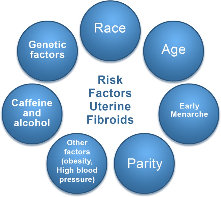
Figure 1 Risk factors for uterine fibroid. These include race, age, delayed pregnancy, early menarche, parity (protective effect), caffeine, genetic alterations, and others, such as obesity and a diet rich in red meat.
Figure 1 Risk factors for uterine fibroid. These include race, age, delayed pregnancy, early menarche, parity (protective effect), caffeine, genetic alterations, and others, such as obesity and a diet rich in red meat.CC BY · PubMed Central
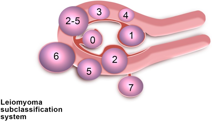
Figure 2 FIGO classification of uterine fibroids according to Munro et al. (2011). Fibroid types range from 0 to 8. 0 = Pedunculated, intracavitary; 1 = Submucosal, <50% intramural; 2 = Submucosal, ≥50% intramural; 3 = Contact with endometrium, 100% intramural; 4 = Intramural; 5 = Subserosal, ≥50% intramural; 6 = Subserosal, <50% intramural; 7 = Subserosal, pedunculated; 8 = Other (e.g. cervical, parasitic). Where two numbers are given (e.g. 2–5), the first number refers to the relationship with the endometrium, while the second number refers to the relationship with the serosa; e.g. 2–5 = Submucosal and subserosal, each with less than half the diameter in the endometrial and peritoneal cavities respectively. Fibroid classification cartoon republished with permission from Munro et al. (2011).
Figure 2 FIGO classification of uterine fibroids according to Munro et al. (2011). Fibroid types range from 0 to 8. 0 = Pedunculated, intracavitary; 1 = Submucosal, <50% intramural; 2 = Submucosal, ≥50% intramural; 3 = Contact with endometrium, 100% intramural; 4 = Intramural; 5 = Subserosal, ≥50% intramural; 6 = Subserosal, <50% intramural; 7 = Subserosal, pedunculated; 8 = Other (e.g. cervical, parasitic). Where two numbers are given (e.g. 2–5), the first number refers to the relationship with the endometrium, while the second number refers to the relationship with the serosa; e.g. 2–5 = Submucosal and subserosal, each with less than half the diameter in the endometrial and peritoneal cavities respectively. Fibroid classification cartoon republished with permission from Munro et al. (2011).CC BY · PubMed Central
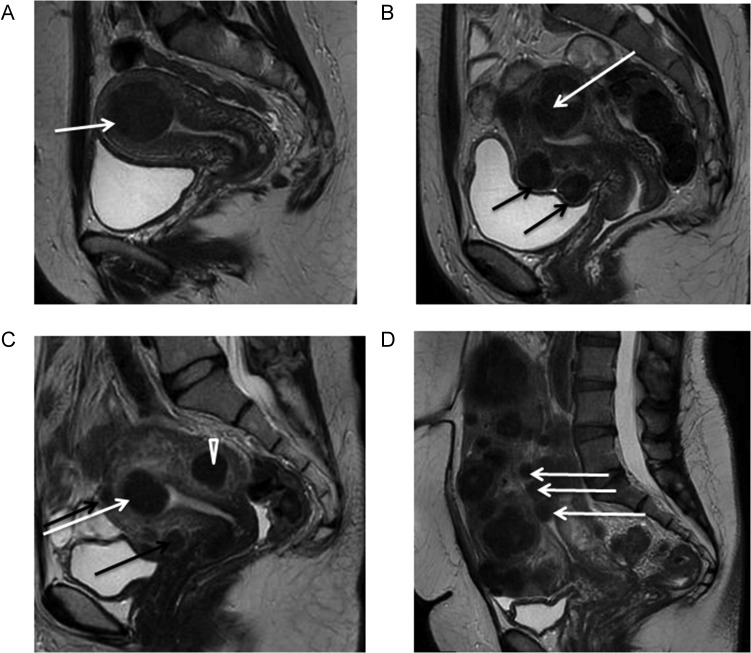
Figure 3 Magnetic resonance imaging (MRI) of fibroids. Midline sagittal T2-weighted images show different types of myomas according to the FIGO classification (Munro et al., 2011). Fibroids vary in size, number and site in the uterus. (A) Submucosal type 2 myoma. (B) Large type 2–5 myoma (white arrow): submucosal and subserosal, each with less than half the diameter in the endometrial and peritoneal cavities respectively. Subserosal type 5 myomas (subserosal, >50% intramural) (black arrows). (C) Submucosal type 2 myoma (>50% intramural) (white arrow). Intramural type 4 myoma (arrowhead). Small type 5 myomas (black arrows). (D) Multiple myomas, three of which are type 0 (intracavitary) (white arrows).
Figure 3 Magnetic resonance imaging (MRI) of fibroids. Midline sagittal T2-weighted images show different types of myomas according to the FIGO classification (Munro et al., 2011). Fibroids vary in size, number and site in the uterus. (A) Submucosal type 2 myoma. (B) Large type 2–5 myoma (white arrow): submucosal and subserosal, each with less than half the diameter in the endometrial and peritoneal cavities respectively. Subserosal type 5 myomas (subserosal, >50% intramural) (black arrows). (C) Submucosal type 2 myoma (>50% intramural) (white arrow). Intramural type 4 myoma (arrowhead). Small type 5 myomas (black arrows). (D) Multiple myomas, three of which are type 0 (intracavitary) (white arrows).CC BY · PubMed Central
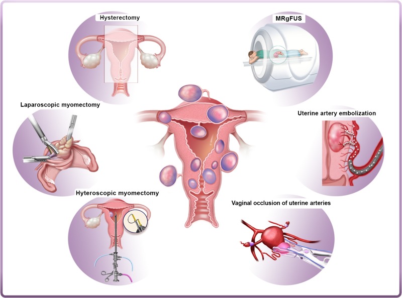
Figure 4 Current surgical and non-surgical management strategies of myomas. Left panel: hysterectomy, laparoscopic myomectomy and hysteroscopic myomectomy are the most widely used surgical interventions for myomas. Right panel: alternatives to surgical intervention include uterine artery embolization (UAE), high-frequency magnetic resonance-guided focused ultrasound surgery (MRgFUS) and vaginal occlusion of uterine arteries.
Figure 4 Current surgical and non-surgical management strategies of myomas. Left panel: hysterectomy, laparoscopic myomectomy and hysteroscopic myomectomy are the most widely used surgical interventions for myomas. Right panel: alternatives to surgical intervention include uterine artery embolization (UAE), high-frequency magnetic resonance-guided focused ultrasound surgery (MRgFUS) and vaginal occlusion of uterine arteries.CC BY · PubMed Central
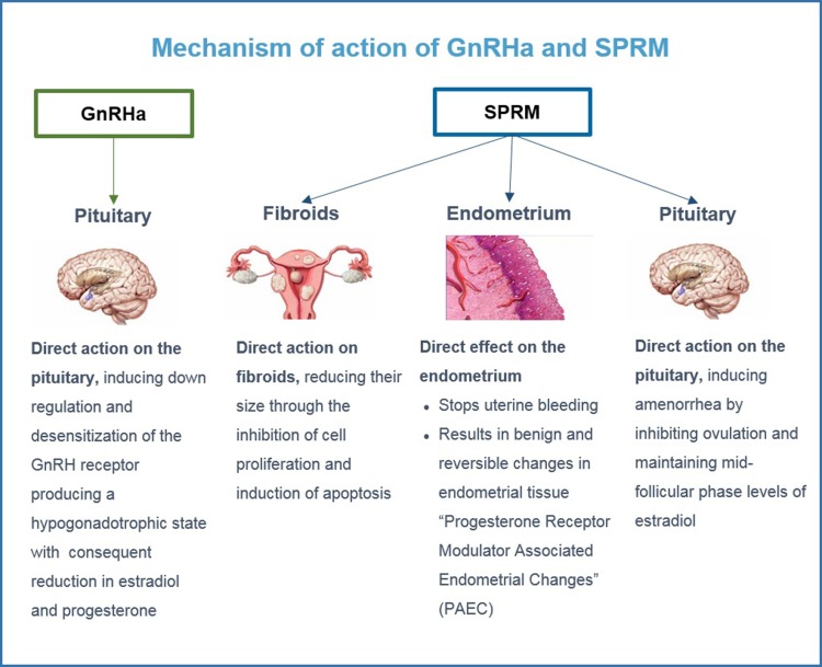
Figure 5 Mode of action of GnRH agonists and SPRMs (Selective Progesterone Receptor Modulators). GnRH agonists have a direct impact on the pituitary. SPRMs have a direct impact on fibroids, endometrium and the pituitary.
Figure 5 Mode of action of GnRH agonists and SPRMs (Selective Progesterone Receptor Modulators). GnRH agonists have a direct impact on the pituitary. SPRMs have a direct impact on fibroids, endometrium and the pituitary.CC BY · PubMed Central
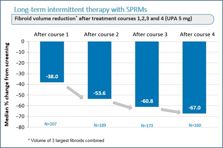
Figure 6 Effect on fibroid volume reduction after four courses of three months of ulipristal acetate (UPA) 5 mg daily. The off-period between two courses was two natural cycles.
Figure 6 Effect on fibroid volume reduction after four courses of three months of ulipristal acetate (UPA) 5 mg daily. The off-period between two courses was two natural cycles.CC BY · PubMed Central
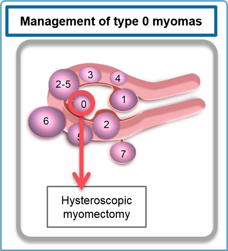
Figure 7 Management of type 0 myomas. Hysteroscopic myomectomy is the most appropriate approach. Fibroid classification cartoon republished with permission from Munro et al. (2011).
Figure 7 Management of type 0 myomas. Hysteroscopic myomectomy is the most appropriate approach. Fibroid classification cartoon republished with permission from Munro et al. (2011).CC BY · PubMed Central
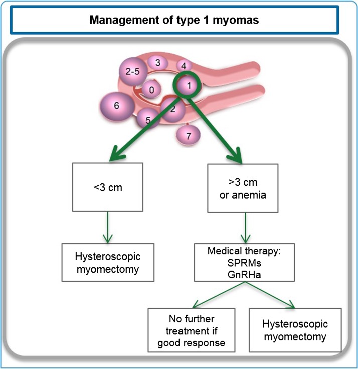
Figure 8 Management of type 1 myomas. Depending on the myoma size, presence of anemia and the surgeon's skill, hysteroscopic myomectomy combined or not with ulipristal acetate(UPA) should be proposed. Fibroid classification cartoon republished with permission from Munro et al.(2011).
Figure 8 Management of type 1 myomas. Depending on the myoma size, presence of anemia and the surgeon's skill, hysteroscopic myomectomy combined or not with ulipristal acetate(UPA) should be proposed. Fibroid classification cartoon republished with permission from Munro et al.(2011).CC BY · PubMed Central
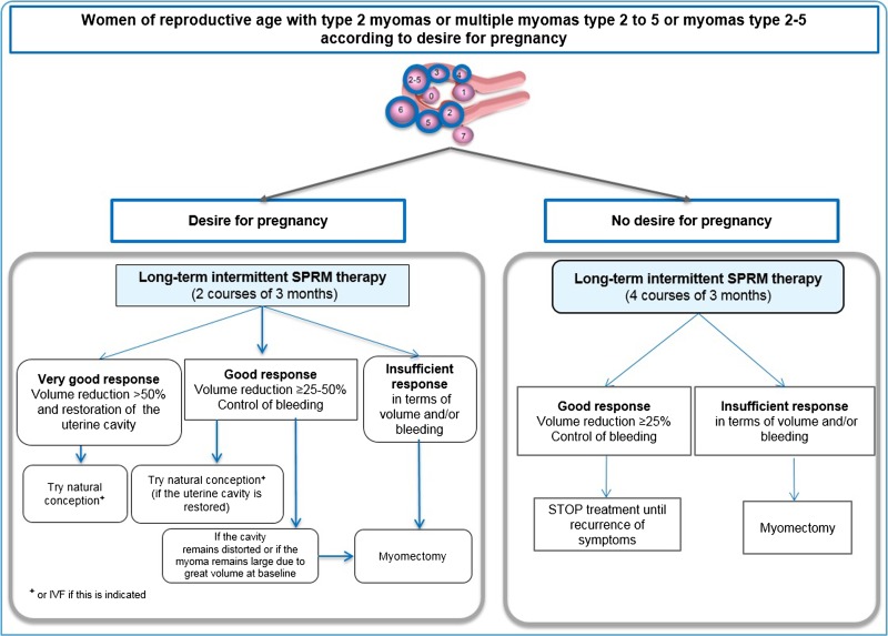
Figure 9 Management in case of myomas or multiple myomas (type 2–5) in women of reproductive age, according to desire for pregnancy. In cases of infertility, two courses of three months are recommended (left panel). Subsequent therapy is determined depending on the response to treatment and restoration of the uterine cavity. If there is no desire to conceive (right panel), long–term (four courses) intermittent therapy may be proposed. In case of a good response in terms of fibroid volume reduction and bleeding, treatment is stopped and only restarted if symptoms recur. Fibroid classification cartoon republished with permission from Munro et al. (2011).
Figure 9 Management in case of myomas or multiple myomas (type 2–5) in women of reproductive age, according to desire for pregnancy. In cases of infertility, two courses of three months are recommended (left panel). Subsequent therapy is determined depending on the response to treatment and restoration of the uterine cavity. If there is no desire to conceive (right panel), long–term (four courses) intermittent therapy may be proposed. In case of a good response in terms of fibroid volume reduction and bleeding, treatment is stopped and only restarted if symptoms recur. Fibroid classification cartoon republished with permission from Munro et al. (2011).CC BY · PubMed Central
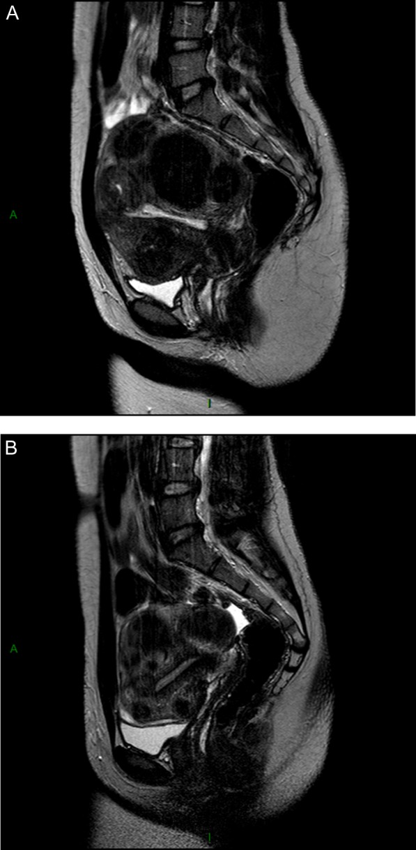
Figure 10 Considerable shrinkage of all myomas after four courses of intermittent ulipristal acetate (UPA) therapy. A patient aged 30 years presented with heavy menstrual bleedingand an unclear desire for pregnancy. (A) Before treatment, a midline sagittal T2-weighted magnetic resonance image (MRI) demonstrated the presence of multiple myomas: type 2, 3, 4 and 6. (B) Upon completion of treatment (intermittent UPA therapy (four courses of threemonths), the uterine cavity was no longer distorted. (C) One year after delivery of a healthy baby, no fibroid regrowth was observed after delivery.
Figure 10 Considerable shrinkage of all myomas after four courses of intermittent ulipristal acetate (UPA) therapy. A patient aged 30 years presented with heavy menstrual bleedingand an unclear desire for pregnancy. (A) Before treatment, a midline sagittal T2-weighted magnetic resonance image (MRI) demonstrated the presence of multiple myomas: type 2, 3, 4 and 6. (B) Upon completion of treatment (intermittent UPA therapy (four courses of threemonths), the uterine cavity was no longer distorted. (C) One year after delivery of a healthy baby, no fibroid regrowth was observed after delivery.CC BY · PubMed Central
Figure 11 Important shrinkage of the submucosal myoma was obtained after two courses of three months of intermittent ulipristal acetate (UPA) therapy. (A) Coronal T2-weighted magnetic resonance image (MRI) image illustrated the presence of multiple myomas (type 2, type 2-5) distorting the uterine cavity in a 19-year-old nulligravid patient, who presented to the emergency department, with heavy menstrual bleeding and anaemia (haemoglobin level of 7.4 g/l).The patient received two courses of UPA (5 mg) and iron. (B) At the end of therapy, MRI demonstrated a significant reduction in myoma volume (<50%) and restoration of the uterine cavity. Amenorrhea was achieved, with a haemoglobin level of 11.9 g/l. The patient was free of symptoms and did not wish to conceive; therefore, surgery was avoided.
Figure 11 Important shrinkage of the submucosal myoma was obtained after two courses of three months of intermittent ulipristal acetate (UPA) therapy. (A) Coronal T2-weighted magnetic resonance image (MRI) image illustrated the presence of multiple myomas (type 2, type 2-5) distorting the uterine cavity in a 19-year-old nulligravid patient, who presented to the emergency department, with heavy menstrual bleeding and anaemia (haemoglobin level of 7.4 g/l).The patient received two courses of UPA (5 mg) and iron. (B) At the end of therapy, MRI demonstrated a significant reduction in myoma volume (<50%) and restoration of the uterine cavity. Amenorrhea was achieved, with a haemoglobin level of 11.9 g/l. The patient was free of symptoms and did not wish to conceive; therefore, surgery was avoided.CC BY · PubMed Central
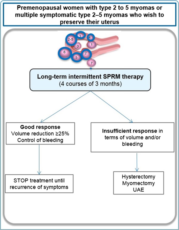
Figure 12 Management of type 2 to 5 myomas or multiple myomas (type 2–5) in premenopausal women wishing to preserve their uterus. In this case, long-term (four courses of three months) intermittent therapy with SPRMs is proposed. Fibroid classification cartoon republished with permission from Munro et al. (2011).
Figure 12 Management of type 2 to 5 myomas or multiple myomas (type 2–5) in premenopausal women wishing to preserve their uterus. In this case, long-term (four courses of three months) intermittent therapy with SPRMs is proposed. Fibroid classification cartoon republished with permission from Munro et al. (2011).CC BY · PubMed Central
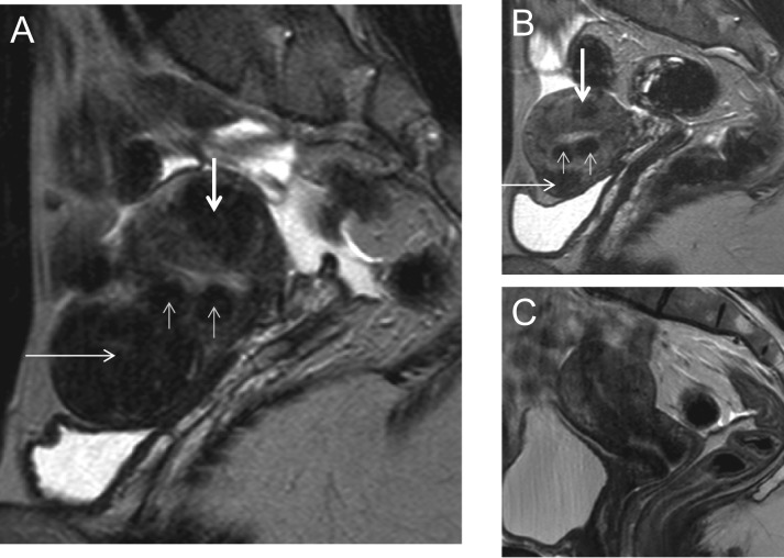
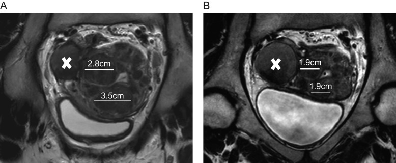
Figure 13 27 year-old women complaining of heavy menstrual bleeding and pelvic pain. A: Coronal T2-weighted MRI images illustrated the presence of type 2–5 and type 3 myomas distorting the uterine cavity and an endometrioma (indicated by X) of 4.3 cm in size. The white lines represent the diameter of the myomas. This patient received long-term intermittent therapy with 5 mg of UPA (2 courses of 3 months). B: At the end of therapy there was an important reduction in myoma volume, but not endometrioma volume.
Figure 13 27 year-old women complaining of heavy menstrual bleeding and pelvic pain. A: Coronal T2-weighted MRI images illustrated the presence of type 2–5 and type 3 myomas distorting the uterine cavity and an endometrioma (indicated by X) of 4.3 cm in size. The white lines represent the diameter of the myomas. This patient received long-term intermittent therapy with 5 mg of UPA (2 courses of 3 months). B: At the end of therapy there was an important reduction in myoma volume, but not endometrioma volume.CC BY · PubMed Central
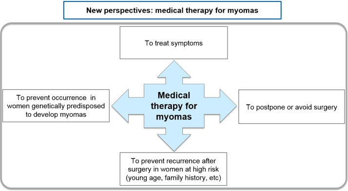
Figure 14 New avenues are emerging in medical fibroid therapy. The first goal of medical therapy is clearly to treat symptoms resulting from the presence of fibroids (heavy menstrual bleeding, pelvic pain, bulk symptoms, infertility, etc.), as well as to postpone or avoid surgery. Further avenues should be investigated by randomized trials, looking to avoid recurrence after surgery in women at high risk of recurrence, and to prevent occurrence of myomas in genetically predisposed women.
Figure 14 New avenues are emerging in medical fibroid therapy. The first goal of medical therapy is clearly to treat symptoms resulting from the presence of fibroids (heavy menstrual bleeding, pelvic pain, bulk symptoms, infertility, etc.), as well as to postpone or avoid surgery. Further avenues should be investigated by randomized trials, looking to avoid recurrence after surgery in women at high risk of recurrence, and to prevent occurrence of myomas in genetically predisposed women.CC BY · PubMed Central
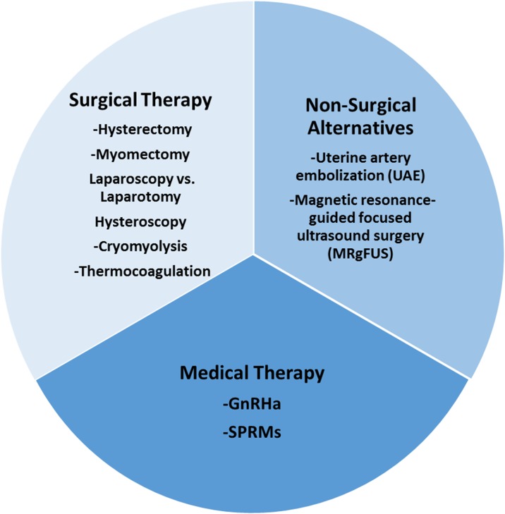
Figure 15 Surgical, non-surgical and medical therapy for the management of fibroids: the current armamentarium.
Figure 15 Surgical, non-surgical and medical therapy for the management of fibroids: the current armamentarium.CC BY · PubMed Central

Verification

Claims the paper makes

The claims and statistics of this paper haven’t been checked yet.

Run the check — free, no account needed. It reads the open-access text and checks the sources it cites.

A few free checks a day · papers already verified open instantly · results are public

Claims other papers make about this one

Each is checked against this paper’s own text: does it say what the citing paper says it does?

Self-check

Percentages, sums and reported differences re-computed from the paper’s own numbers.

The self-check needs the paper’s full text.

Citations

Cites

1 retracted

Show all 223 references

Research works printed but not linked: 3

  1. 10.1097/00001648-200303000-00021 · DOI
  2. 10.1097/00001703-200308000-00002 · DOI
  3. 10.1210/jc.76.5.1217 · DOI

Cited by

Sorted by citations

Metadata

Authors
Donnez, J., & Dolmans, M.-M.
Affiliations
  • UCLouvain (BE)
  • Cliniques Universitaires Saint-Luc (BE)
Corresponding author
Donnez, Jacques
Venue
Human Reproduction Update
Published
2016-07-27
Volume
22
Issue
6
Pages
665-686
ISSN
  • 1355-4786 (ISSN-L)
  • 1460-2369
OpenAlex
W2496662864
Cited by
780
References
223
Citations by year
2026: 76 · 2025: 86 · 2024: 77 · 2023: 109 · 2022: 89 · 2021: 92 · 2020: 94 · 2019: 48 · 2018: 54 · 2017: 45 · 2016: 9
FWCI
9.19
Type
journal article
Language
en
Open access
Licence
cc-by
Locations
  • Human Reproduction Update (publishedVersion)
  • PubMed Central (publishedVersion, cc-by)
  • PubMed (publishedVersion)
  • Digital Access to Libraries (Université catholique de Louvain (UCL), l'Université de Namur (UNamur) and the Université Saint-Louis (USL-B)) (submittedVersion)
Funders
Gedeon Richter
Topics
  • Health Sciences › Medicine › Obstetrics and Gynecology › Uterine Myomas and Treatments
  • Health Sciences › Medicine › Public Health, Environmental and Occupational Health › Reproductive Health and Contraception
  • Health Sciences › Medicine › Obstetrics and Gynecology › Gynecological conditions and treatments
Keywords
uterine fibroids, leiomyoma, fibroid management, myomectomy, uterine artery embolization, thermal ablation, selective progesterone receptor modulators, ulipristal acetate, fertility preservation, hysterectomy alternative
MeSH terms
Contraceptive Agents, Female, Humans, Hysterectomy, Leiomyoma, Magnetic Resonance Imaging, Norpregnadienes, Progesterone, Risk Factors, Uterine Neoplasms, Randomized Controlled Trials as Topic, Pelvic Pain, Uterine Artery

About these checks

Registry, retraction, Cites and statistics results are computed, not judged, with one exception: a printed reference that no index lists is read once by a model before it counts as a citation error. Claims are extracted by a model and kept only when their quote appears verbatim in the text; each cited source is then checked, with a model’s help, for whether it says what the claim relies on. Those verdicts can be wrong — read the claim’s quote and the cited source before relying on one.

Statistics and arithmetic are recomputed, not judged.

Confidence bands are shown only for fields calibrated on our annotated test set. None is calibrated yet, so no band is shown.

What counts as a claim ›How we measure confidence ›How we check this ›

Are you an author, or do you see a problem with this page? Request a review