

The DOI matches its Crossref record.
Effect of magnesium ions/Type I collagen promote the biological behavior of osteoblasts and its mechanism
Open accessAbstract
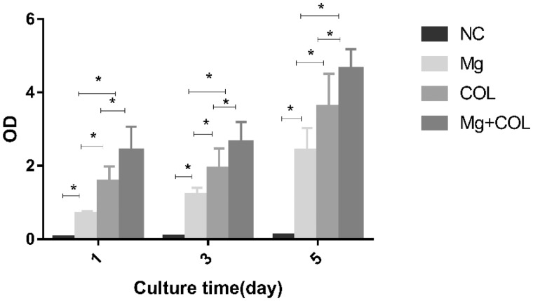
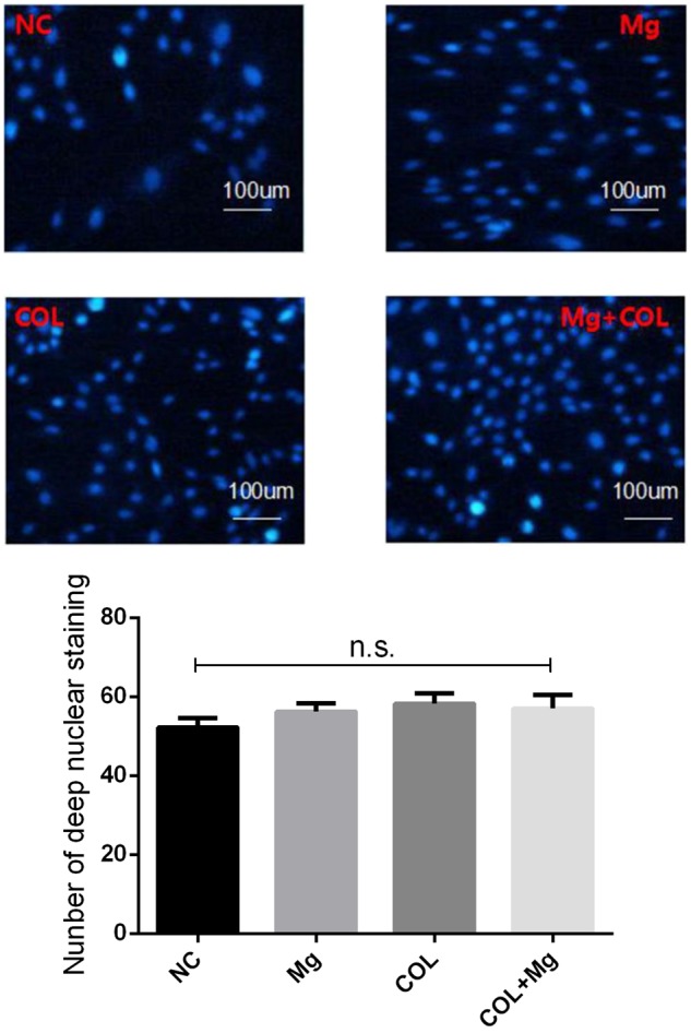
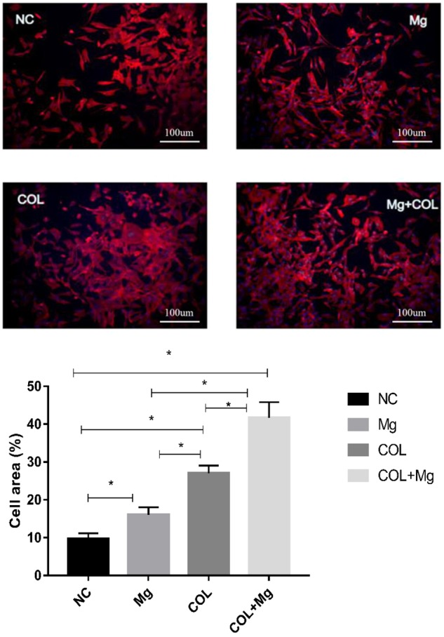
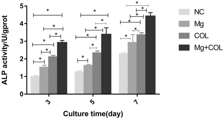
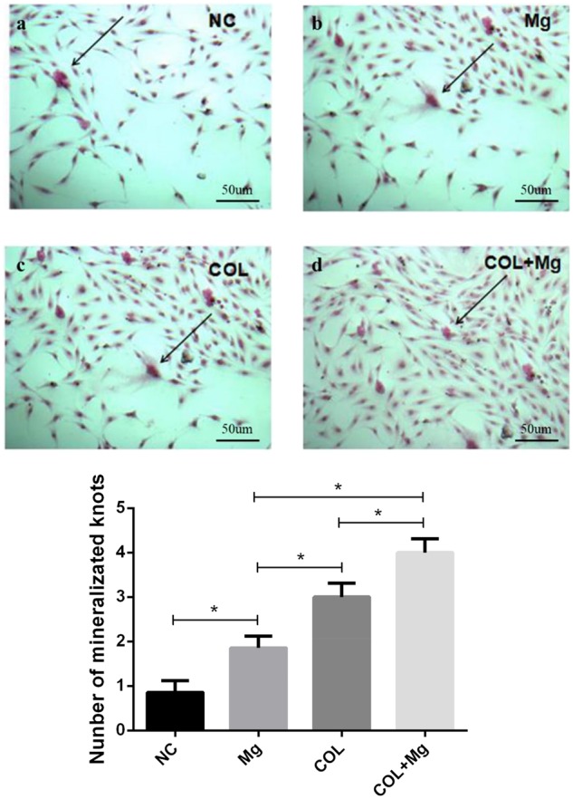
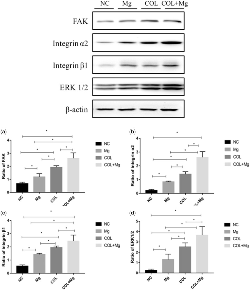
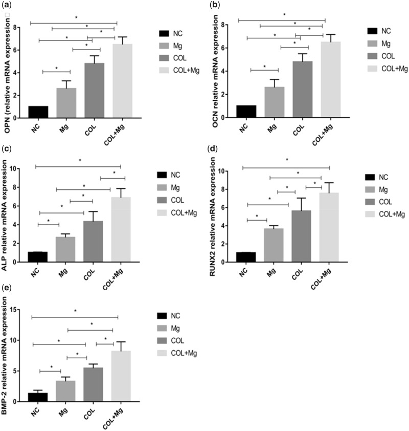
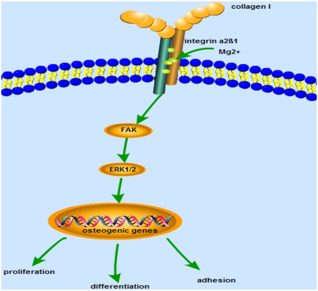
Type I collagen (Col I) is a main component of extracellular matrix (ECM). Its safety, biocompatibility, hydrophilicity and pyrogen immunogenicity make it suitable for tissues engineering applications. Mg2+ also control a myriad of cellular processes, including the bone development by enhancing the attachment and differentiation of osteoblasts and accelerating mineralization to enhance bone healing. In our studies, Mg2+ bind collagen to promote the proliferation and differentiation of osteoblasts through the expression of integrins and downstream signaling pathways. In order to clarify the biological behavior effect of 10 mM Mg2+/Col I coating, we performed 3-(4,5-Dimethylthiazol-2-yl)-2,5-diphenyltetrazolium bromide (MTT), alkaline phosphatase (ALP), 4′6-diamidino-2-phenylindole (DAPI), Alizarin red staining and Rhodamine B-isothiocyanate (RITC)-labeled phalloidin experiments and found that 10 mM Mg2+ group, Col I-coating group, 10 mM Mg2+/Col I-coating group, respectively, promoted the proliferation and differentiation of osteoblasts, especially 10 mM Mg2+/Col I-coating group. We detected the mRNA expression of osteogenic-related genes (Runx2, ALP and OCN, OPN and BMP-2) and the protein expression of signaling pathway (integrin α2, integrin β1, FAK and ERK1/2), these results indicated that 10 mM Mg2+/Col I coating play an critical role in up-regulating the MC3T3-E1 cells activity. The potential mechanisms of this specific performance may be through activating via integrin α2β1-FAK-ERK1/2 protein-coupled receptor pathway.
Overview
The claims and statistics of this paper haven’t been checked yet.
Run the check — free, no account needed. It reads the open-access text and checks the sources it cites.
Integrity signals
Registry
Cites
Statistics
Figures
Verification
Claims the paper makes
The claims and statistics of this paper haven’t been checked yet.
Run the check — free, no account needed. It reads the open-access text and checks the sources it cites.
Claims other papers make about this one
Each is checked against this paper’s own text: does it say what the citing paper says it does?
The cited source says what the claim attributes to it.
In the citing paper
“This can be interpreted as Mg 2+ released from PM under daily medium exchange conditions acting within a concentration range that promotes osteoblast proliferation and metabolic activity in the several mM range, as reported in the literature, rather than accumulating to toxic levels [45,46].”
From Functional Surface Modification of Magnesium Implant by Drug-Loaded Biodegradable Polymer Coating (2026) · cites this paper as [46]
Self-check
Percentages, sums and reported differences re-computed from the paper’s own numbers.
The self-check needs the paper’s full text.
Citations
Cites
No references could be resolved yet.
Research works printed but not linked: 37
- Krishnamoorthy, 2017, A novel strategy to enhance secretion of ECM components by stem cells: relevance to tissue engineering, Tissue Eng Part A, 24, 145, 10.1089/ten.tea.2017.0060 · DOI
- Davidenko, 2018, Selecting the correct cellular model for assessing of the biological response of collagen-based biomaterials, Acta Biomater, 65, 88, 10.1016/j.actbio.2017.10.035 · DOI
- Ling-Yu, 2018, Advances in functionalized polymer coatings on biodegradable magnesium alloys—a review, Acta Biomater, 79, 23, 10.1016/j.actbio.2018.08.030 · DOI · Paper page
- Hayrapetyan, 2016, Effect of nano-HA/collagen composite hydrogels on osteogenic behavior of mesenchymal stromal cells, Stem Cell Rev Rep, 12, 352, 10.1007/s12015-016-9644-x · DOI
- Peng, 2013, Enhancement of bone healing in rabbit ulnar critical bone defect by injectable nano-hydroxyapatite/collagen/(calcium sulfate hemihydrate) mixed with autogenous bone marrow, J Biomat Tissue Eng, 3, 534, 10.1166/jbt.2013.1117 · DOI
- Qing, 2018, Mineralized collagen/Mg-Ca alloy combined scaffolds with improved biocompatibility for enhanced bone response following tooth extraction, Biomed Mater, 13, 065008, 10.1088/1748-605X/aadb47 · DOI
- Wang, 2016, Comparison of bone regeneration in alveolar bone of dogs on mineralized collagen grafts with two composition ratios of nano-hydroxyapatite and collagen, Regen Biomater, 3, 33, 10.1093/rb/rbv025 · DOI
- Jaiswal, 2018, Mechanical, corrosion and biocompatibility behaviour of Mg-3Zn-HA biodegradable composites for orthopaedic fixture accessories, J Mech Behav Biomed Mater, 78, 442, 10.1016/j.jmbbm.2017.11.030 · DOI
- Ding, 2016, Opportunities and challenges for the biodegradable magnesium alloys as next-generation biomaterials, Regener Biomater, 3, 79, 10.1093/rb/rbw003 · DOI
- Zhang, 2017, Nanocomposite hydrogels stabilized by self-assembled multivalent bisphosphonate-magnesium nanoparticles mediate sustained release of magnesium ion and promote in-situ bone regeneration, Acta Biomater, 64, 389, 10.1016/j.actbio.2017.09.039 · DOI
- Federica, 2016, Magnesium modifies the structural features of enzymatically mineralized collagen gels affecting the retraction capabilities of human dermal fibroblasts embedded within this 3D system, Materials, 9, 477, 10.3390/ma9060477 · DOI
- Zuoying, 2019, Injectable PLGA microspheres with tunable magnesium ion release for promoting bone regeneration, Acta Biomater, 85, 294, 10.1016/j.actbio.2018.12.017 · DOI
- Wang, 2017, Magnesium ions promote the biological behaviour of rat calvarial osteoblasts by activating the PI3K/Akt signalling pathway, Biol Trace Elem Res, 179, 284, 10.1007/s12011-017-0948-8 · DOI
- Shimaya, 2010, Magnesium enhances adherence and cartilage formation of synovial mesenchymal stem cells through integrins, Osteoarthr Cartilage, 18, 1300, 10.1016/j.joca.2010.06.005 · DOI
- Leem, 2016, Magnesium ions facilitate integrin alpha 2‐ and alpha 3‐mediated proliferation and enhance alkaline phosphatase expression and activity in hBMSCs, J Tissue Eng Regen Med, 10, E527, 10.1002/term.1861 · DOI
- Brown, 2018, Salt-bridge modulates differential calcium-mediated ligand binding to integrin α1- and α2-I domains, Sci Rep, 8, 2916, 10.1038/s41598-018-21231-1 · DOI
- Kozlova, 2018, Implication of Integrin α2β1 in proliferation and invasion of human breast carcinoma and melanoma cells: noncanonical Function of Akt protein kinase, Biochemistry (Mosc), 83, 738, 10.1134/S0006297918060111 · DOI
- Mizuno, 2000, Type I collagen-induced osteoblastic differentiation of bone-marrow cells mediated by collagen-α2β1 integrin interaction, J Cell Physiol, 184, 207, 10.1002/1097-4652(200008)184:2<207::AID-JCP8>3.0.CO;2-U · DOI
- Arous, 2017, Role and impact of the extracellular matrix on integrin-mediated pancreatic β-cell functions, Biol Cell, 109, 223, 10.1111/boc.201600076 · DOI
- Eble, 2017, Dramatic and concerted conformational changes enable rhodocetin to block α2β1 integrin selectively, PLOS Biol, 15, e2001492, 10.1371/journal.pbio.2001492 · DOI
- Onley, 2000, Micromolar Ca2+ concentrations are essential for Mg2+-dependent binding of collagen by the integrin alpha 2beta 1 in human platelets, J Biol Chem, 275, 24560, 10.1074/jbc.M004111200 · DOI
- 0X.Zreiqat, 2002, Mechanisms of magnesium-stimulated adhesion of osteoblastic cells to commonly used orthopaedic implants, J Biomed Mater Res, 62, 175, 10.1002/jbm.10270 · DOI
- Witkowska-Zimny, 2015, α2β1 integrin-mediated mechanical signals during osteodifferentiation of stem cells from the Wharton’s jelly of the umbilical cord, Folia Histochem Cytobiol, 52, 297, 10.5603/FHC.a2014.0031 · DOI
- Mcatee, 2019, Prostate tumor cell exosomes containing hyaluronidase Hyal1 stimulate prostate stromal cell motility by engagement of FAK-mediated integrin signaling, Matrix Biol, 78-79, 165, 10.1016/j.matbio.2018.05.002 · DOI
- Jahan, 2018, Axed MUC4 (MUC4/X) aggravates pancreatic malignant phenotype by activating integrin-β1/FAK/ERK pathway, Biochim Biophys Acta Mol Basis Dis, 1864, 2538, 10.1016/j.bbadis.2018.05.008 · DOI
- Saleem, 2009, beta1 integrin/FAK/ERK signalling pathway is essential for human fetal islet cell differentiation and survival, J Pathol, 219, 182, 10.1002/path.2577 · DOI
- Gao, 2018, Glutathione homeostasis is significantly altered by quercetin via the Keap1/Nrf2 and MAPK signaling pathways in rats, J Clin Biochem Nutr, 62, 56, 10.3164/jcbn.17-40 · DOI
- Zhang, 2018, Panaxadiol saponins induce activation of MAPK/ERK signaling pathway in bone marrow cells of aplastic anemia mice, Chin J Pathophysiol, 34, 686
- Zhang, 2017, Ion channel functional protein kinase TRPM7 regulates Mg ions to promote the osteoinduction of human osteoblast via PI3K pathway: in vitro simulation of the bone-repairing effect of Mg-based alloy implant, Acta Biomater, 63, 369, 10.1016/j.actbio.2017.08.051 · DOI
- Mushahary, 2014, Collagen type-I leads to in vivo matrix mineralization and secondary stabilization of Mg–Zr–Ca alloy implants, Colloids Surf B, 122, 719, 10.1016/j.colsurfb.2014.08.005 · DOI
- Serre, 1998, Influence of magnesium substitution on a collagen-apatite biomaterial on the production of a calcifying matrix by human osteoblasts, J Biomed Mater Res, 42, 626, 10.1002/(SICI)1097-4636(19981215)42:4<626::AID-JBM20>3.0.CO;2-S · DOI
- Chin, 2014, Assignments of human integrin α1I domain in theapoand Mg2+bound states, Biomol NMR Assign, 8, 117, 10.1007/s12104-013-9465-7 · DOI
- Hamaia, 2014, Integrin recognition motifs in the human collagens, Adv Exper Med Biol, 819, 127, 10.1007/978-94-017-9153-3_9 · DOI
- Zhang, 2015, Magnesium modification of a calcium phosphate cement alters bone marrow stromal cell behavior via an integrin-mediated mechanism, Biomaterials, 53, 251, 10.1016/j.biomaterials.2015.02.097 · DOI
- Humtsoe, 2005, A streptococcal collagen-like protein interacts with the α2β1 integrin and induces intracellular signaling, J Biol Chem, 280, 13848, 10.1074/jbc.M410605200 · DOI
- Huang, 2008, Midkine promotes tetraspanin-integrin interaction and induces FAK-Stat1alpha pathway contributing to migration/invasiveness of human head and neck squamous cell carcinoma cells, Biochem Biophys Res Commun, 377, 474, 10.1016/j.bbrc.2008.09.138 · DOI
- Witkowskazimny, 2014, α2β1 integrin-mediated mechanical signals during osteodifferentiation of stem cells from the Wharton’s jelly of the umbilical cord, Folia Histochem Cytobiol, 52, 297, 10.5603/FHC.a2014.0031 · DOI
Cited by
Sorted by citations
Osteogenic Effects of Ion Released from Biodegradable Metallic Magnesium and Calcium Coating
Miyake, R., Shimabukuro, M., Terauchi, M., Marukawa, E., & Kawashita, M. (2025) · Advanced Biology · doi:10.1002/adbi.202500401 · Cited by 5
Functional Surface Modification of Magnesium Implant by Drug-Loaded Biodegradable Polymer Coating
Park, J.-E., Jang, Y.-S., Ko, S.-O., & Lee, M.-H. (2026) · Applied Sciences · doi:10.3390/app16031542 · Cited by 3
Metadata
- Authors
- Nie, X., Sun, X., Wang, C., & Yang, J.
- Affiliations
- Jinzhou Medical University (CN)
- Beijing Union University (CN)
- Corresponding author
- Wang, Chengyue
- Yang, Jingxin
- Venue
- Regenerative Biomaterials
- Publisher
- Oxford University Press (OUP)
- Published
- 2019-10-30
- Volume
- 7
- Issue
- 1
- Pages
- 53-61
- ISSN
- 2056-3418 (ISSN-L)
- 2056-3426
- PMC
- PMC7233620
- OpenAlex
- W2982515127
- Cited by
- 63
- References
- 0
- Citations by year
- 2026: 7 · 2025: 14 · 2024: 15 · 2023: 12 · 2022: 6 · 2021: 8 · 2020: 1
- FWCI
- 1.32
- Type
- journal article
- Language
- en
- Open access
- gold
- doi.org
- Licence
- Locations
- Regenerative Biomaterials (publishedVersion, cc-by)
- PubMed Central (publishedVersion, cc-by)
- PubMed (publishedVersion)
- Funders
- Science and Technology Fund of Liaoning Province (20180530071, 2019-MS-141)
- Topics
- Physical Sciences › Engineering › Biomedical Engineering › Bone Tissue Engineering Materials
- Physical Sciences › Materials Science › Biomaterials › Magnesium Alloys: Properties and Applications
- Life Sciences › Biochemistry, Genetics and Molecular Biology › Molecular Biology › Bone Metabolism and Diseases
- Keywords
- magnesium ions, type I collagen, osteoblast proliferation, osteogenic differentiation, alpha2beta1 integrin, focal adhesion kinase, ERK signaling, Runx2, alkaline phosphatase, MC3T3-E1 cells, bone tissue engineering, collagen coating
About these checks
Registry, retraction, Cites and statistics results are computed, not judged, with one exception: a printed reference that no index lists is read once by a model before it counts as a citation error. Claims are extracted by a model and kept only when their quote appears verbatim in the text; each cited source is then checked, with a model’s help, for whether it says what the claim relies on. Those verdicts can be wrong — read the claim’s quote and the cited source before relying on one.
Statistics and arithmetic are recomputed, not judged.
Confidence bands are shown only for fields calibrated on our annotated test set. None is calibrated yet, so no band is shown.
What counts as a claim ›How we measure confidence ›How we check this ›
Are you an author, or do you see a problem with this page? Request a review
