

The DOI matches its Crossref record; 61 of the 62 references it cites were found in a registry or an index.
Evidence of endoplasmic reticulum stress and protein synthesis inhibition in the placenta of non‐native women at high altitude
Open accessAbstract
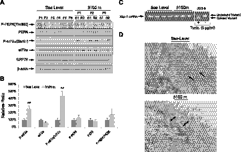
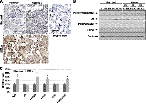
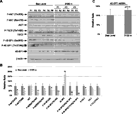
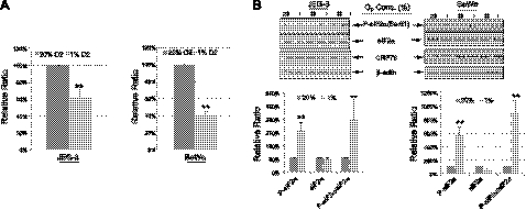
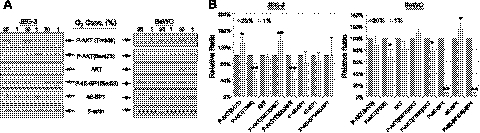
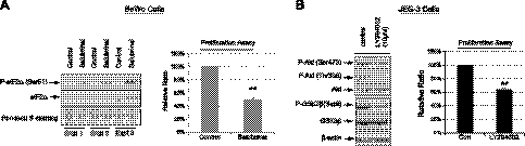
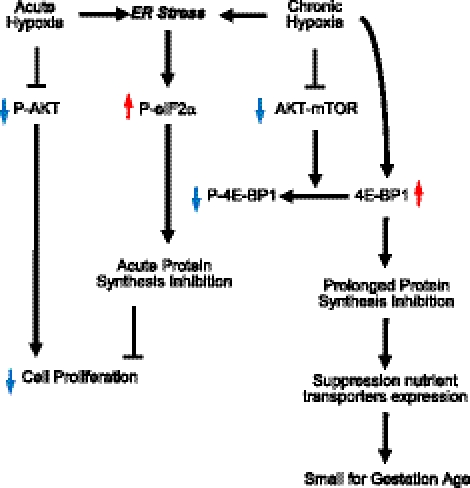
Pregnancy at high altitude is associated with a reduction in birth weight of ~100 g/1000 m of ascent. The underlying mechanisms are unclear but may involve alteration in energy‐demanding activities, such as protein synthesis. To test this hypothesis, both in vivo and in vitro approaches were used. Placental tissues from pregnant women residing at 3100 m were studied, and placental cells were incubated under hypoxia. In the 3100‐m placentas, we observed dilation of endoplasmic reticulum (ER) cisternae, increased phosphorylation of eukaryotic initiation factor 2 subunit α (P‐eIF2α), reduced AKT phosphorylation, and reduced P‐4E‐BP1 but increased 4E‐BP1 protein compared to sea level controls. These findings suggest the presence of ER stress and protein synthesis inhibition. Hypoxia (1% O 2 ) reduced proliferation of trophoblast‐like JEG‐3 cells, BeWo cells, and placental fibroblasts by ~40, ~60, and ~18%, respectively. Sublethal dosage of salubrinal, an eIF2α phosphatase inhibitor, increased P‐eIF2α and reduced BeWo cell and placental fibroblast proliferation by ~50%. Administration of the PI‐3K inhibitor LY294002 also reduced JEG‐3 proliferation. Our results demonstrate that exposure to chronic hypobaric hypoxia causes mild placental ER stress, which, in turn, modulates protein synthesis and slows proliferation. These effects may account for the reduced placental villous volume, and contribute to the low birth weight that typifies high‐altitude populations.—Yung, H. W., Cox, M., Tissot van Patot, M., Burton, G. J. Evidence of endoplasmic reticulum stress and protein synthesis inhibition in the placenta of non‐native women at high altitude. FASEB J. 26, 1970‐1981 (2012). www.fasebj.org
Overview
The claims and statistics of this paper haven’t been checked yet.
Run the check — free, no account needed. It reads the open-access text and checks the sources it cites.
Integrity signals
Registry
Cites
Statistics
Figures
Verification
Claims the paper makes
The claims and statistics of this paper haven’t been checked yet.
Run the check — free, no account needed. It reads the open-access text and checks the sources it cites.
Claims other papers make about this one
Each is checked against this paper’s own text: does it say what the citing paper says it does?
In the citing paper
“Mild activation of the UPR, with phosphorylation of eIF2α alone, has been reported in the high-altitude human placenta, where it may mediate homeostatic adaptations to the hypobaric hypoxia experienced (598).”
From Placental Origins of Chronic Disease (2016) · cites this paper as [598]
In the citing paper
“Equally, evidence of translational arrest has been reported in human placentas from normal healthy pregnancies at high altitude (3,100 m) where growth of the villus tree is impaired (598).”
From Placental Origins of Chronic Disease (2016) · cites this paper as [598]
In the citing paper
“These changes can be recapitulated by exposing trophoblast cell lines to hypoxia (1% O2), when they are associated with reduced cell proliferation.”
From Placental Origins of Chronic Disease (2016) · cites this paper as [598]
In the citing paper
“These placentas displayed increased phosphorylation of eIF2α, reduced phosphorylation of AKT and 4E-BP1, and an increase in total 4E-BP1 that will favor sequestration of eIF4E.”
From Placental Origins of Chronic Disease (2016) · cites this paper as [598]
In the citing paper
“However, there is evidence for a decrease in protein synthesis in the placenta of non-native women at high altitude (598).”
From Placental Origins of Chronic Disease (2016) · cites this paper as [598]
In the citing paper
“Most of the data relating to regulation of protein synthesis in response to hypoxia come from the cancer field (542, 585, 586), but it appears that the same pathways are operative in the placenta under physiological and pathological conditions (596, 598).”
From Placental Origins of Chronic Disease (2016) · cites this paper as [598]
Self-check
Percentages, sums and reported differences re-computed from the paper’s own numbers.
The self-check needs the paper’s full text.
Citations
Cites
Phosphorylation and regulation of Akt/PKB by the rictor-mTOR complex
Sarbassov, D. D., Guertin, D. A., Ali, S. M., & Sabatini, D. M. (2005) · Science · doi:10.1126/science.1106148 · Cited by 6,216
TSC2 is phosphorylated and inhibited by Akt and suppresses mTOR signalling
Inoki, K., Li, Y., Zhu, T., Wu, J., & Guan, K.-L. (2002) · Nature Cell Biology · doi:10.1038/ncb839 · Cited by 3,083
Mechanism of activation of protein kinase B by insulin and IGF‐1.
Alessi, D. R., Andjelković, M., Caudwell, B., Cron, P. D., MORRICE, N. A., Cohen, P., & Hemmings, B. A. (1996) · The EMBO Journal · doi:10.1002/j.1460-2075.1996.tb01045.x · Cited by 3,014
eIF4 Initiation Factors: Effectors of mRNA Recruitment to Ribosomes and Regulators of Translation
Gingras, A., Raught, B., & Sonenberg, N. (1999) · Annual Review of Biochemistry · doi:10.1146/annurev.biochem.68.1.913 · Cited by 2,103
Endoplasmic Reticulum Stress and Oxidative Stress: A Vicious Cycle or a Double-Edged Sword?
Malhotra, J. D., & Kaufman, R. J. (2007) · Antioxidants and Redox Signaling · doi:10.1089/ars.2007.1782 · Cited by 1,588
Regulation of translation initiation by FRAP/mTOR
Gingras, A.-C., Raught, B., & Sonenberg, N. (2001) · Genes & Development · doi:10.1101/gad.887201 · Cited by 1,545
Jacinto, E., Facchinetti, V., Liu, D., Soto, N., Wei, S., Jung, S. Y., Huang, Q., Qin, J., & Su, B. (2006) · Cell · doi:10.1016/j.cell.2006.08.033 · Cited by 1,407
A Selective Inhibitor of eIF2α Dephosphorylation Protects Cells from ER Stress
Boyce, M., Bryant, K. F., Jousse, C., Long, K., Harding, H. P., Scheuner, D. L., Kaufman, R. J., Ma, D., Coen, D. M., Ron, D., et al. (2005) · Science · doi:10.1126/science.1101902 · Cited by 1,403
Translational Control Is Required for the Unfolded Protein Response and In Vivo Glucose Homeostasis
Scheuner, D. L., Song, B., McEwen, E. L., Liu, C., Laybutt, D. R., Gillespie, P. J., Saunders, T. L., Bonner‐Weir, S., & Kaufman, R. J. (2001) · Molecular Cell · doi:10.1016/s1097-2765(01)00265-9 · Cited by 1,355
Coping with stress: eIF2 kinases and translational control
Wek, R. C., Jiang, H.-Y., & Anthony, T. G. (2005) · Biochemical Society Transactions · doi:10.1042/bst0340007 · Cited by 1,353
Research works printed but not linked: 1
- 10.1002/(SICI)1096-8644(1998)107:27 <25::AID-AJPA3>3.0.CO;2-L · DOI
Cited by
Sorted by citations
Pathophysiology of placental-derived fetal growth restriction
Burton, G. J., & Jauniaux, E. (2018) · American Journal of Obstetrics and Gynecology · doi:10.1016/j.ajog.2017.11.577 · Cited by 1,070
Self-citationPlacental Origins of Chronic Disease
Burton, G. J., Fowden, A. L., & Thornburg, K. L. (2016) · Physiological Reviews · doi:10.1152/physrev.00029.2015 · Cited by 717
Self-citationGDF15: a hormone conveying somatic distress to the brain
Lockhart, S. M., Saudek, V., & O’Rahilly, S. (2020) · Endocrine Reviews · doi:10.1210/endrev/bnaa007 · Cited by 256
Gestational Hypoxia and Developmental Plasticity
Ducsay, C. A., Goyal, R., Pearce, W. J., Wilson, S. M., Hu, X., & Zhang, L. (2018) · Physiological Reviews · doi:10.1152/physrev.00043.2017 · Cited by 208
Measuring high-altitude adaptation
MOORE, L. G. (2017) · Journal of Applied Physiology · doi:10.1152/japplphysiol.00321.2017 · Cited by 205
Metadata
- Authors
- Yung, H. W., Cox, M., van Patot, M. T., & Burton, G. J.
- Affiliations
- University of Cambridge (GB)
- Centre for Trophoblast Research University of Cambridge Cambridge UK
- Corresponding author
- Burton, Graham J.
- Venue
- The FASEB Journal
- Publisher
- Wiley
- Published
- 2012-01-20
- Volume
- 26
- Issue
- 5
- Pages
- 1970-1981
- ISSN
- 0892-6638 (ISSN-L)
- 1530-6860
- PMC
- PMC3336782
- OpenAlex
- W2115115666
- Cited by
- 112
- References
- 62
- Citations by year
- 2026: 2 · 2025: 9 · 2024: 5 · 2023: 6 · 2022: 8 · 2021: 9 · 2020: 8 · 2019: 10 · 2018: 9 · 2017: 7 · 2016: 8 · 2015: 12 · 2014: 5 · 2013: 10 · 2012: 4
- FWCI
- 5.97
- Type
- journal article
- Language
- en
- Open access
- hybrid
- doi.org
- Licence
- Locations
- The FASEB Journal (publishedVersion, cc-by-nc)
- Europe PMC (PubMed Central) (publishedVersion, cc-by-nc)
- PubMed Central (publishedVersion, cc-by-nc)
- PubMed (publishedVersion)
- Funders
- Wellcome Trust (084804/2/08/Z)
- Topics
- Health Sciences › Medicine › Physiology (medical) › High Altitude and Hypoxia
- Health Sciences › Medicine › Obstetrics and Gynecology › Pregnancy and preeclampsia studies
- Life Sciences › Biochemistry, Genetics and Molecular Biology › Cell Biology › Endoplasmic Reticulum Stress and Disease
- Keywords
- endoplasmic reticulum stress, hypoxia, high altitude, placenta, protein synthesis inhibition, eIF2α phosphorylation, 4E-BP1, Akt phosphorylation, PI3K signaling, salubrinal, cell proliferation, low birth weight, trophoblast
- MeSH terms
- Altitude, Endoplasmic Reticulum, Female, Humans, Phosphoproteins, Phosphorylation, Placenta, Pregnancy, Protein Biosynthesis, Blotting, Western, Eukaryotic Initiation Factor-2, Oxidative Stress, Cell Cycle Proteins, Cell Line, Tumor, Adaptor Proteins, Signal Transducing, Proto-Oncogene Proteins c-akt, Real-Time Polymerase Chain Reaction
About these checks
Registry, retraction, Cites and statistics results are computed, not judged, with one exception: a printed reference that no index lists is read once by a model before it counts as a citation error. Claims are extracted by a model and kept only when their quote appears verbatim in the text; each cited source is then checked, with a model’s help, for whether it says what the claim relies on. Those verdicts can be wrong — read the claim’s quote and the cited source before relying on one.
Statistics and arithmetic are recomputed, not judged.
Confidence bands are shown only for fields calibrated on our annotated test set. None is calibrated yet, so no band is shown.
What counts as a claim ›How we measure confidence ›How we check this ›
Are you an author, or do you see a problem with this page? Request a review
