

The DOI matches its Crossref record; 2 of the 2 references it cites were found in a registry or an index.
Tissue tropism in parasitic diseases
Abstract
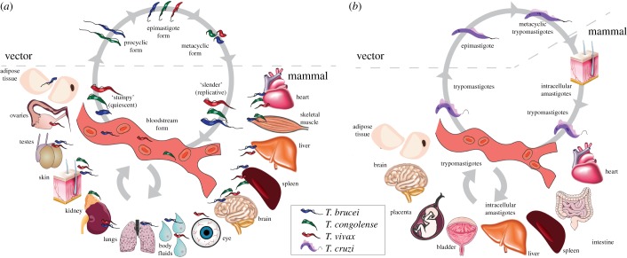
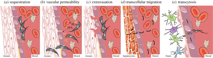
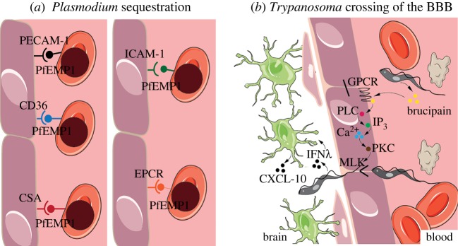
Parasitic diseases, such as sleeping sickness, Chagas disease and malaria, remain a major cause of morbidity and mortality worldwide, but particularly in tropical, developing countries. Controlling these diseases requires a better understanding of host–parasite interactions, including a deep appreciation of parasite distribution in the host. The preferred accumulation of parasites in some tissues of the host has been known for many years, but recent technical advances have allowed a more systematic analysis and quantifications of such tissue tropisms. The functional consequences of tissue tropism remain poorly studied, although it has been associated with important aspects of disease, including transmission enhancement, treatment failure, relapse and clinical outcome. Here, we discuss current knowledge of tissue tropism in Trypanosoma infections in mammals, describe potential mechanisms of tissue entry, comparatively discuss relevant findings from other parasitology fields where tissue tropism has been extensively investigated, and reflect on new questions raised by recent discoveries and their potential impact on clinical treatment and disease control strategies.
Overview
The claims and statistics of this paper haven’t been checked yet.
Run the check — free, no account needed. It reads the open-access text and checks the sources it cites.
Integrity signals
Registry
Cites
Statistics
Figures
Verification
Claims the paper makes
The claims and statistics of this paper haven’t been checked yet.
Run the check — free, no account needed. It reads the open-access text and checks the sources it cites.
Claims other papers make about this one
Each is checked against this paper’s own text: does it say what the citing paper says it does?
In the citing paper
“The tissue changes in parasitic diseases closely resemble those found in the skin of patients with atopic dermatitis, in the bronchial walls of asthmatics, and in the nasal mucosa of patients with allergic rhinitis.”
From Parasites and allergies: a complex bidirectional relationship from evolutionary origins to modern therapeutics (2026) · cites this paper as Silva Pereira et al. 2019
In the citing paper
“These similarities may arise from the molecular resemblance between atopic allergens and parasitic antigens (Silva Pereira et al. 2019; Wu et al. 2017).”
From Parasites and allergies: a complex bidirectional relationship from evolutionary origins to modern therapeutics (2026) · cites this paper as Silva Pereira et al. 2019
Self-check
Percentages, sums and reported differences re-computed from the paper’s own numbers.
The self-check needs the paper’s full text.
Citations
Cites
On the Cytoadhesion of Plasmodium vivax –Infected Erythrocytes
Carvalho, B. O., Lopes, S. C. P., Nogueira, P. A., Orlandi, P. P., Bargieri, D. Y., Blanco, Y. C., Mamoni, R., Leite, J. A., Rodrigues, M. M., Soares, I. S., et al. (2010) · The Journal of Infectious Diseases · doi:10.1086/654815 · Cited by 299
White, N. J. (2017) · Malaria Journal · doi:10.1186/s12936-017-1731-1 · Cited by 230
Research works printed but not linked: 182
- 10.1105/tpc.113.119727 · DOI · Paper page
- 10.1038/nrmicro1529 · DOI
- 10.2174/1874421401004010030 · DOI
- 10.1016/j.gheart.2015.06.001 · DOI
- Desquesnes M, Holzmuller P, Lai D-H, Dargantes A, Lun Z-R, Jittaplapong S, Przyborski JM. 1941 Trypanosoma evansi and surra: a review and perspectives on origin, history, distribution, taxonomy, morphology, hosts, and pathogenic effects. Biomed Res. Int. 2013, 194176. (doi:10.1155/2013/194176) · DOI
- 10.1016/S0368-1742(44)80012-1 · DOI
- 10.1161/CIR.0000000000000599 · DOI
- 10.1017/S0031182000054354 · DOI
- 10.1093/oxfordjournals.bmb.a072045 · DOI
- 10.3389/fimmu.2016.00233 · DOI
- 10.1371/journal.ppat.1005744 · DOI
- 10.7554/eLife.17716 · DOI
- 10.1371/journal.pbio.3000105 · DOI
- 10.1016/S0140-6736(01)70338-8 · DOI
- 10.1016/S0035-9203(37)90074-9 · DOI
- Zwart D. 1989 Aspects of comparative pathology and pathogenesis of trypanosomal infections in Africa. Ann. Soc. Belg. Med. Trop. (1920) 69, 105-112.
- 10.1007/BF00432240 · DOI
- 10.1172/JCI200421052 · DOI
- 10.1007/s00018-002-8472-0 · DOI
- 10.1007/BF02236387 · DOI
- 10.1080/00034983.1989.11812418 · DOI
- Morrison WI, Murray M, Sayer PD, Preston JM. 1981 The pathogenesis of experimentally induced Trypanosoma brucei infection in the dog. I. Tissue and organ damage. Am. J. Pathol. 102, 168-181.
- Chirimwami B, Van Marck EA, Brucher JM, Mulumba P, Wery M, Gigase PL. 1988 Light microscopic neuropathology of long-term experimental Trypanosoma brucei gambiense infection in the rat. Ann. Soc. Belg. Med. Trop. (1920) 68, 195-203.
- 10.1016/0035-9203(80)90184-4 · DOI
- 10.1177/030098587200901s01 · DOI
- Abenga JN. 2014 A comparative pathology of Trypanosoma brucei infections. Glob. Adv. Res. J. Med. Med. Sci. 3, 390-399.
- 10.1371/journal.pntd.0000509 · DOI
- 10.1371/journal.pntd.0000486 · DOI
- 10.1371/journal.pntd.0004350 · DOI
- 10.1016/j.chom.2016.05.002 · DOI
- 10.1136/hrt.38.8.827 · DOI
- Poltera AA, Hochmann A, Lambert PH. 1980 A model for cardiopathy induced by Trypanosoma brucei brucei in mice. A histologic and immunopathologic study. Am. J. Pathol. 99, 325-351.
- Poltera AA, Sayer PD, Rudin W, Bovell D. 1985 Trypanosomal cardiac valvulitis in vervet monkeys. Trop. Med. Parasitol. 36, 77-80.
- Cohen C. 1973 Trypanosomiasis on the Witwatersrand. South African Med. J. 47, 485.
- 10.1111/j.1365-3156.2007.01948.x · DOI
- 10.1371/journal.pntd.0000383 · DOI
- 10.1177/030098588402100217 · DOI
- 10.1371/journal.pntd.0006690 · DOI
- 10.1023/B:TROP.0000009528.91525.01 · DOI
- 10.1016/S0034-5288(18)30647-7 · DOI
- 10.1080/00034983.1950.11685428 · DOI
- 10.1177/030098587901600307 · DOI
- 10.1038/272613a0 · DOI
- Emery DL, Moloo SK. 1981 The dynamics of the cellular reactions elicited in the skin of goats by Glossina morsitans morsitans infected with Trypanosoma (Nannomonas) congolense or T. (Duttonella) vivax. Acta Trop. 38, 15-28.
- 10.1177/030098587801500504 · DOI
- 10.1016/S0007-1935(17)51448-7 · DOI
- 10.1016/0035-9203(73)90193-4 · DOI
- Losos GJ, Gwamaka G. 1973 Histological examination of wild animals naturally infected with pathogenic African trypanosomes. Acta Trop. 30, 57-63.
- 10.1007/BF01320994 · DOI
- 10.1111/pim.12099 · DOI
- Bungener W, Mehlitz D. 1977 Extravascular localization of Trypanosoma vivax in cattle. Tropenmed. Parasitol. 28, 8-10.
- 10.1136/vr.107.24.551 · DOI
- 10.1017/S0031182000066737 · DOI
- 10.1016/0304-4017(90)90076-N · DOI
- 10.1016/j.vetpar.2006.08.017 · DOI
- 10.1371/journal.pntd.0000792 · DOI
- 10.1016/j.vetpar.2011.05.016 · DOI
- 10.1186/1297-9716-42-63 · DOI
- Isoun TT, Anosa VO. 1974 Lesions in the reproductive organs of sheep and goats experimentally infected with Trypanosoma vivax. Tropenmed. Parasitol. 25, 469-476.
- 10.1590/S0100-736X2008001200002 · DOI
- 10.1186/1297-9716-44-1 · DOI
- 10.1016/j.vetpar.2012.08.001 · DOI
- Maxie MG, Losos GJ, Tabel H. 1979 Experimental bovine trypanosomiasis (Trypanosoma vivax and T. congolense). I. Sumptomatology and clinical pathology. Tropenmed. Parasitol. 30, 274-282.
- Kimeto BKA. 1989 Pathology and pathogenesis of Trypanosoma vivax infection in cattle. Nairobi, Kenya: University of Nairobi.
- van den Ingh T, de Neijs-Bakker M. 1979 Pancarditis in Trypanosoma vivax infection in cattle. Tropenmed. Parasitol. 30, 239-243.
- Emery DL, Barry JD, Moloo SK. 1980 The appearance of Trypanosoma (Duttonella) vivax in lymph following challenge of goats with infected Glossina morsitans morsitans. Acta Trop. 37, 375-379.
- 10.1007/s11250-017-1280-1 · DOI
- 10.1016/S0304-4017(97)00128-3 · DOI · Paper page
- 10.1016/S0304-4017(99)00164-8 · DOI
- 10.2307/3283007 · DOI
- 10.1354/vp.46-2-251 · DOI
- 10.3923/jp.2010.23.26 · DOI
- 10.1016/j.meegid.2015.10.018 · DOI
- 10.1017/S0031182000080756 · DOI
- 10.1080/00034983.1968.11686549 · DOI
- 10.1111/j.1574-6976.2012.00333.x · DOI
- 10.2307/3279787 · DOI
- 10.1590/S1807-59322009001200013 · DOI
- 10.1371/journal.pntd.0006388 · DOI
- 10.1371/journal.pone.0195879 · DOI
- 10.1590/S0074-02762010000600018 · DOI
- 10.1371/journal.pntd.0000864 · DOI
- 10.1074/jbc.M412802200 · DOI
- 10.1016/j.micinf.2011.06.002 · DOI
- 10.1016/j.pt.2019.01.002 · DOI
- 10.1016/S1286-4579(03)00162-X · DOI
- 10.1371/journal.ppat.1001032 · DOI
- 10.1002/1873-3468.12142 · DOI
- 10.1111/j.1550-7408.1978.tb04405.x · DOI
- 10.1017/S0031182000076514 · DOI
- 10.2307/3280584 · DOI
- 10.1007/BF00931503 · DOI
- 10.1016/S1471-4922(01)02079-7 · DOI
- 10.1182/blood-2013-09-521005 · DOI
- 10.3389/fimmu.2017.01148 · DOI
- 10.1186/1475-2875-11-33 · DOI
- 10.1073/pnas.0503386102 · DOI
- 10.1371/journal.ppat.1000068 · DOI
- 10.1084/jem.20110762 · DOI
- 10.1038/ncomms11659 · DOI
- 10.1007/BF01924436 · DOI
- Tizard IR, Holmes WL, York DA, Mellors A. 1977 The generation and identification of the hemolysin of Trypanosoma congolense. Specialia 660, 901-902. (doi:10.1007/bf01951271) · DOI
- 10.1016/0035-9203(78)90063-9 · DOI
- 10.1080/13550280802282934 · DOI
- 10.1016/j.physbeh.2007.05.045 · DOI
- 10.1016/j.pneurobio.2009.12.001 · DOI · Paper page
- 10.1155/2016/4503214 · DOI
- Mbaya A Kumshe H Nwosu CO. 2012 The mechanisms of anaemia in trypanosomosis: a review. In Anemia (ed. D Silverberg) pp. 269–282. Rijeka Croatia: InTech. See http://www.intechopen.com/books/anemia/the-mechanisms-of-anaemia-in-trypanosomosis-a-review. · DOI
- 10.1111/cmi.12123 · DOI
- 10.1126/science.291.5501.141 · DOI
- 10.1016/j.pt.2018.11.008 · DOI
- 10.1126/science.1129720 · DOI
- 10.1371/journal.ppat.0030171 · DOI
- 10.1016/j.micpath.2011.10.003 · DOI
- 10.1084/jem.20110560 · DOI
- 10.1111/j.1365-2958.2010.07230.x · DOI
- 10.1073/pnas.94.16.8807 · DOI
- 10.1046/j.1365-2958.2001.02286.x · DOI
- 10.1074/jbc.M305030200 · DOI
- 10.1371/journal.ppat.1005296 · DOI
- 10.1016/j.pt.2017.11.008 · DOI
- 10.1080/00034983.1950.11685444 · DOI
- 10.1590/0074-0276140362 · DOI
- 10.1186/1475-2875-10-297 · DOI
- 10.1186/1475-2875-13-144 · DOI
- 10.1016/j.pt.2016.08.014 · DOI
- 10.1046/j.0818-9641.2001.01048.x · DOI
- 10.1055/s-2007-979471 · DOI
- 10.1002/hep.27254 · DOI
- Bertolino P, Bowen DG. 2015 Malaria and the liver: immunological hide-and-seek or subversion of immunity from within? Front. Microbiol. 6, 41.
- 10.3410/B2-3 · DOI · Paper page
- 10.1186/1756-3305-4-41 · DOI · Paper page
- 10.1016/j.pt.2012.04.005 · DOI
- 10.1371/journal.ppat.1000122 · DOI
- 10.1017/S0031182000045200 · DOI
- 10.1017/S0031182000000834 · DOI
- 10.1111/j.1365-2958.2004.04256.x · DOI
- 10.1128/IAI.70.10.5412-5415.2002 · DOI
- 10.4269/ajtmh.2007.77.22 · DOI
- 10.1016/S1473-3099(07)70022-1 · DOI · Paper page
- 10.1016/0020-7519(79)90089-4 · DOI
- 10.1017/S0031182000050964 · DOI
- 10.1124/jpet.108.149872 · DOI
- 10.1016/S0035-9203(96)90132-7 · DOI
- 10.1111/eva.12338 · DOI
- 10.1038/s41467-018-04108-9 · DOI
- 10.1186/1475-2875-11-285 · DOI
- 10.1016/0035-9203(81)90019-5 · DOI
- 10.1111/j.1365-2141.1987.tb06913.x · DOI
- 10.1126/scitranslmed.3008882 · DOI
- 10.1007/s40588-016-0045-3 · DOI
- 10.1371/journal.ppat.1004414 · DOI
- 10.1371/journal.ppat.1005519 · DOI
- 10.1111/brv.12301 · DOI
- 10.1007/BF02239803 · DOI
- 10.1016/0304-4017(89)90070-8 · DOI
- 10.1016/0304-4017(89)90069-1 · DOI
- ILRAD. 1984 Anaemia in trypanosomiasls: haemorrhagic T. vivax may provide a clue. ILRAD Rep. 2, 1-9.
- 10.3390/ijms18102200 · DOI
- 10.1136/pgmj.2006.047357 · DOI
- Da Silva GB Jr, Pinto JR, Barros EJG, Farias GMN, Daher EDF. 2017 Kidney involvement in malaria: an update. Rev. Inst. Med. Trop. Sao Paulo 59, e53.
- 10.1038/s41467-017-02484-2 · DOI
- 10.1152/physiol.00006.2004 · DOI
- 10.1038/nmicrobiol.2017.32 · DOI
- 10.1590/S0074-02762008000100001 · DOI
- Desquesnes M, Dargantes A, Lai DH, Lun ZR, Holzmuller P, Jittapalapong S. 2013 Trypanosoma evansi and surra: a review and perspectives on transmission, epidemiology and control, impact, and zoonotic aspects. Biomed Res. Int. 2013, 1-20. · DOI
- 10.3389/fmicb.2016.02126 · DOI
- 10.1016/j.ijid.2011.03.018 · DOI
- 10.1007/s11250-012-0232-z · DOI
- 10.1079/BJN19970043 · DOI
- 10.1590/S0001-37652006000400004 · DOI
- 10.1038/s41598-018-27228-0 · DOI
- 10.1038/s41564-017-0014-5 · DOI
- 10.1038/nri2746 · DOI
- 10.1371/journal.pntd.0003714 · DOI
- 10.1016/j.meegid.2018.01.011 · DOI
- 10.1101/gr.115089.110 · DOI
- 10.1128/mBio.02097-14 · DOI
- 10.1128/mSphere.00340-16 · DOI
- 10.1017/S0031182014000894 · DOI
- 10.1016/j.molbiopara.2016.03.007 · DOI
- 10.2147/DDDT.S60973 · DOI
Cited by
Sorted by citations
Packi, K., Świerczewski, D., Klimczak, S., Ochab, D., Gałęziewska, J., Gromek, P., Kruczkowska, W., Płuciennik, E., Kałuzińska-Kołat, Ż., & Kołat, D. (2026) · Antonie van Leeuwenhoek · doi:10.1007/s10482-026-02425-4
Metadata
- Authors
- Silva Pereira, S., Trindade, S., De Niz, M., & Figueiredo, L. M.
- Venue
- Open Biology
- Publisher
- The Royal Society
- Published
- 2019-05
- Volume
- 9
- Issue
- 5
- ISSN
- 2046-2441
- PMC
- PMC6544988
- Cited by
- 85
- References
- 2
- Language
- en
- Update notices
- correction (2019-06-26)
- Funders
- European Molecular Biology Organization (ALTF 1048-2016)
- Fundação para a Ciência e a Tecnologia (IF/01050/2014, LISBOA-01-0145-FEDER-007391)
- H2020 European Research Council (771714)
About these checks
Registry, retraction, Cites and statistics results are computed, not judged, with one exception: a printed reference that no index lists is read once by a model before it counts as a citation error. Claims are extracted by a model and kept only when their quote appears verbatim in the text; each cited source is then checked, with a model’s help, for whether it says what the claim relies on. Those verdicts can be wrong — read the claim’s quote and the cited source before relying on one.
Statistics and arithmetic are recomputed, not judged.
Confidence bands are shown only for fields calibrated on our annotated test set. None is calibrated yet, so no band is shown.
What counts as a claim ›How we measure confidence ›How we check this ›
Are you an author, or do you see a problem with this page? Request a review
