CoChat

Cellular translational enhancer elements that recruit eukaryotic initiation factor 3

Open access

Preprint — published as 10.1261/rna.080310.124

The DOI matches its Crossref record; 1 of the 1 references it cites were found in a registry or an index.

Abstract

Translation initiation is a highly regulated process which broadly affects eukaryotic gene expression. Eukaryotic initiation factor 3 (eIF3) is a central player in canonical and alternative pathways for ribosome recruitment. Here we have investigated how direct binding of eIF3 contributes to the large and regulated differences in protein output conferred by different 5′-untranslated regions (5′-UTRs) of cellular mRNAs. Using an unbiased high-throughput approach to determine the affinity of budding yeast eIF3 for native 5′-UTRs from 4,252 genes, we demonstrate that eIF3 binds specifically to a subset of 5′-UTRs that contain a short unstructured binding motif, AMAYAA. eIF3 binding mRNAs have higher ribosome density in growing cells and are preferentially translated under certain stress conditions, supporting the functional relevance of this interaction. Our results reveal a new class of translational enhancer and suggest a mechanism by which changes in core initiation factor activity enact mRNA-specific translation programs.

Overview

The claims and statistics of this paper haven’t been checked yet.

Free: it reads the open-access text and checks the first 5 claims that cite a source we can check. Create a free account to check every claim.

A few free checks a day · papers already verified open instantly · results are public

Integrity signals

Registry

Crossref · 10.1101/2021.07.15.452589

Cites

1 of 1 references verified0 citation errors · 0 not checkable

Statistics

Figures

FIGURE 1. Hundreds of natural yeast 5′-UTRs bind to eIF3 specifically. (A) Scheme of RBNS assay. Designed DNA oligos encoding ∼10,000 yeast 5′-UTRs, plus at least 36 nt of endogenous coding sequence (CDS) are transcribed in vitro. The resulting RNA pool is incubated with various concentrations of purified eIF3, and protein-bound RNAs are captured on nitrocellulose and sequenced. Enrichment is the frequency of a 5′ UTR in the bound sample normalized to input RNA. (B) RBNS results are reproducible. Shown are bound reads in two replicates of 166 nM eIF3. (C) Enrichment between 55 and 500 nM eIF3 was well correlated. (D) Enrichment of the top 10 eIF3 binders from the 166 nM library traced across all libraries. Peak enrichment at 55 nM eIF3 is consistent with high-affinity binding. Less enrichment at higher (eIF3) is expected because low-affinity RNAs consume more of the sequencing lane. (E) Overlap of 5′-UTRs bound in 55, 166, and 500 nM libraries (red, green, and purple, respectively). See also Supplemental Figure S1.
FIGURE 1. Hundreds of natural yeast 5′-UTRs bind to eIF3 specifically. (A) Scheme of RBNS assay. Designed DNA oligos encoding ∼10,000 yeast 5′-UTRs, plus at least 36 nt of endogenous coding sequence (CDS) are transcribed in vitro. The resulting RNA pool is incubated with various concentrations of purified eIF3, and protein-bound RNAs are captured on nitrocellulose and sequenced. Enrichment is the frequency of a 5′ UTR in the bound sample normalized to input RNA. (B) RBNS results are reproducible. Shown are bound reads in two replicates of 166 nM eIF3. (C) Enrichment between 55 and 500 nM eIF3 was well correlated. (D) Enrichment of the top 10 eIF3 binders from the 166 nM library traced across all libraries. Peak enrichment at 55 nM eIF3 is consistent with high-affinity binding. Less enrichment at higher (eIF3) is expected because low-affinity RNAs consume more of the sequencing lane. (E) Overlap of 5′-UTRs bound in 55, 166, and 500 nM libraries (red, green, and purple, respectively). See also Supplemental Figure S1.CC BY-NC · PubMed Central
FIGURE 2. eIF3 binds to specific 5′ UTRs with nanomolar affinity. Purified eIF3 (15–1000 nM) was incubated with individual 32P-labeled 5′ UTRs from YEL070W (A), YOR052C (B), YKL054C (C), and YBR014C (D). Experimental data points (black dots) were fit (red lines) to determine dissociation constants (Kd). Error reflects standard error of the fit of nonlinear regression using Hill's equation. Enrichment score at 166 nM eIF3 is shown for comparison. See also Supplemental Figure S2.
FIGURE 2. eIF3 binds to specific 5′ UTRs with nanomolar affinity. Purified eIF3 (15–1000 nM) was incubated with individual 32P-labeled 5′ UTRs from YEL070W (A), YOR052C (B), YKL054C (C), and YBR014C (D). Experimental data points (black dots) were fit (red lines) to determine dissociation constants (Kd). Error reflects standard error of the fit of nonlinear regression using Hill's equation. Enrichment score at 166 nM eIF3 is shown for comparison. See also Supplemental Figure S2.CC BY-NC · PubMed Central
FIGURE 3. eIF3 recognizes AMAYAA motifs in unstructured regions. (A) eIF3-binding increases with increasing numbers of AMAYAA motifs. Distribution of observed enrichment (166 nM eIF3) for 5′ UTRs with 0, 1, 2, and 3 or more AMAYAA motifs, P-adjusted (Mann–Whitney) for selected pairwise comparisons. (B) eIF3 filter-binding assay of YOR092W_51_SL31s_7 which contains three consecutive AMAYAA motifs (red) and a 10 nt deletion mutant lacking AMAYAA motifs (gray). The best fit of experimental data points (dots) is shown as a line. (C) eIF3 preferentially binds unfolded 5′ UTRs. Enrichment correlates (Pearson) with 5′-UTR folding energies calculated in RNAfold. (D) Overview of RNA motif structure analysis. (E) AMAYAA motifs in tight-binding 5′ UTRs (top decile, red line) are more likely to be presented in unpaired contexts compared to motifs in weak binders (bottom decile, gray line). Median nucleotide pairing probability of 5′ UTRs binned based on their enrichment ranking in the 166 nM library. Position of the motif is indicated by the red rectangle. Benjamini–Hochberg adjusted P-values shown (top) for each nucleotide position. See also Supplemental Figure S3.
FIGURE 3. eIF3 recognizes AMAYAA motifs in unstructured regions. (A) eIF3-binding increases with increasing numbers of AMAYAA motifs. Distribution of observed enrichment (166 nM eIF3) for 5′ UTRs with 0, 1, 2, and 3 or more AMAYAA motifs, P-adjusted (Mann–Whitney) for selected pairwise comparisons. (B) eIF3 filter-binding assay of YOR092W_51_SL31s_7 which contains three consecutive AMAYAA motifs (red) and a 10 nt deletion mutant lacking AMAYAA motifs (gray). The best fit of experimental data points (dots) is shown as a line. (C) eIF3 preferentially binds unfolded 5′ UTRs. Enrichment correlates (Pearson) with 5′-UTR folding energies calculated in RNAfold. (D) Overview of RNA motif structure analysis. (E) AMAYAA motifs in tight-binding 5′ UTRs (top decile, red line) are more likely to be presented in unpaired contexts compared to motifs in weak binders (bottom decile, gray line). Median nucleotide pairing probability of 5′ UTRs binned based on their enrichment ranking in the 166 nM library. Position of the motif is indicated by the red rectangle. Benjamini–Hochberg adjusted P-values shown (top) for each nucleotide position. See also Supplemental Figure S3.CC BY-NC · PubMed Central
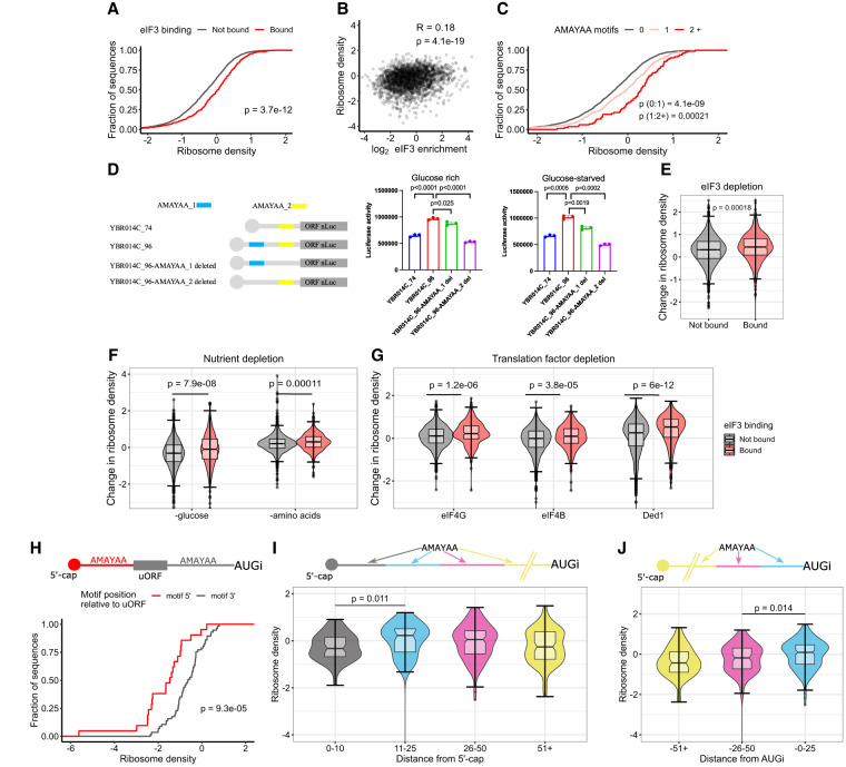
FIGURE 4. eIF3-binding and AMAYAA motifs enhance ribosome recruitment and start codon recognition in vivo. (A) mRNAs with 5′ UTRs that bound eIF3 in vitro show higher ribosome densities in growing yeast. Cumulative distributions of ribosome densities for bound (red) or not bound (gray) mRNAs. “Bound” includes the union of 55, 166, and 500 nM libraries. Ribosome density equals the average number of ribosome-protected mRNA fragments per mRNA for genes expressing a single dominant 5′-UTR isoform (see Materials and Methods). See Table 2 for ribosome profiling data sources. (B) eIF3 binding in vitro correlates with ribosome density in growing yeast (Pearson). (C) 5′-UTR AMAYAA motifs increase ribosome density in a dose-dependent manner. (D) 5′-UTR AMAYAA motifs promote higher translation of luciferase reporter mRNAs in unstarved conditions (left) and during glucose starvation (right). (E–G) eIF3-binding mRNAs (red) maintain higher ribosome densities than nonbinding (gray) under conditions of limiting eIF3 (E), following acute starvation for glucose or amino acids (F), and upon inactivation of various initiation factors (G). (H) mRNAs with a 5′-UTR AMAYAA motif 5′ to a uORF (red) show reduced ribosome density compared to genes with the motif 3′ (gray). mRNAs were grouped based on the position of the first AMAYAA motif relative to the last uORF. (I,J) Ribosome density varies by AMAYAA motif location for mRNAs without uORFs. Higher ribosome densities for mRNAs with motifs located 11–25 nt from the 5′ end of the mRNA (I) or 0–25 nt upstream of the start codon (J). Bonferroni corrected Mann–Whitney P-values shown for selected comparisons. (A–J) All values of ribosome densities and changes in ribosome densities have been log2-transformed. See also Supplemental Figure S4.
FIGURE 4. eIF3-binding and AMAYAA motifs enhance ribosome recruitment and start codon recognition in vivo. (A) mRNAs with 5′ UTRs that bound eIF3 in vitro show higher ribosome densities in growing yeast. Cumulative distributions of ribosome densities for bound (red) or not bound (gray) mRNAs. “Bound” includes the union of 55, 166, and 500 nM libraries. Ribosome density equals the average number of ribosome-protected mRNA fragments per mRNA for genes expressing a single dominant 5′-UTR isoform (see Materials and Methods). See Table 2 for ribosome profiling data sources. (B) eIF3 binding in vitro correlates with ribosome density in growing yeast (Pearson). (C) 5′-UTR AMAYAA motifs increase ribosome density in a dose-dependent manner. (D) 5′-UTR AMAYAA motifs promote higher translation of luciferase reporter mRNAs in unstarved conditions (left) and during glucose starvation (right). (E–G) eIF3-binding mRNAs (red) maintain higher ribosome densities than nonbinding (gray) under conditions of limiting eIF3 (E), following acute starvation for glucose or amino acids (F), and upon inactivation of various initiation factors (G). (H) mRNAs with a 5′-UTR AMAYAA motif 5′ to a uORF (red) show reduced ribosome density compared to genes with the motif 3′ (gray). mRNAs were grouped based on the position of the first AMAYAA motif relative to the last uORF. (I,J) Ribosome density varies by AMAYAA motif location for mRNAs without uORFs. Higher ribosome densities for mRNAs with motifs located 11–25 nt from the 5′ end of the mRNA (I) or 0–25 nt upstream of the start codon (J). Bonferroni corrected Mann–Whitney P-values shown for selected comparisons. (A–J) All values of ribosome densities and changes in ribosome densities have been log2-transformed. See also Supplemental Figure S4.CC BY-NC · PubMed Central
FIGURE 5. Model of location-dependent translational enhancement by direct binding of eIF3 to 5′-UTR motifs. eIF3 binding to AMAYAA motifs may enhance translation initiation by multiple mechanisms depending on the location of the motif. (Top left) AMAYAA motif binding to eIF3 increases initial recruitment of 43S PIC. (Top right) AMAYAA motif upstream of initiation codons (AUGi) increases their recognition during scanning. The presence of AMAYAA motifs increases ribosome loading during growth (left) and allows some mRNAs to maintain ribosome loading during starvation stress (right).
FIGURE 5. Model of location-dependent translational enhancement by direct binding of eIF3 to 5′-UTR motifs. eIF3 binding to AMAYAA motifs may enhance translation initiation by multiple mechanisms depending on the location of the motif. (Top left) AMAYAA motif binding to eIF3 increases initial recruitment of 43S PIC. (Top right) AMAYAA motif upstream of initiation codons (AUGi) increases their recognition during scanning. The presence of AMAYAA motifs increases ribosome loading during growth (left) and allows some mRNAs to maintain ribosome loading during starvation stress (right).CC BY-NC · PubMed Central
Jiří Koubek
Jiří Koubek CC BY-NC · PubMed Central

Verification

Claims the paper makes

The claims and statistics of this paper haven’t been checked yet.

Free: it reads the open-access text and checks the first 5 claims that cite a source we can check. Create a free account to check every claim.

A few free checks a day · papers already verified open instantly · results are public

Self-check

Percentages, sums and reported differences re-computed from the paper’s own numbers.

The self-check needs the paper’s full text.

Citations

Cites

Show all 1 references

Research works printed but not linked: 39

  1. Aitken, C.E. , Beznosková, P. , Vlčkova, V. , Chiu, W.-L. , Zhou, F. , Valášek, L.S. , Hinnebusch, A.G. , and Lorsch, J.R. (2016). Eukaryotic translation initiation factor 3 plays distinct roles at the mRNA entry and exit channels of the ribosomal preinitiation complex. Elife 5. · DOI
  2. 10.1074/mcp.M700468-MCP200 · DOI · Paper page
  3. 10.1093/nar/gkv416 · DOI · Paper page
  4. 10.1098/rstb.2016.0176 · DOI
  5. 10.1534/genetics.115.186221 · DOI
  6. 10.1016/j.sbi.2009.03.005 · DOI · Paper page
  7. Ghazalpour, A. , Bennett, B. , Petyuk, V.A. , Orozco, L. , Hagopian, R. , Mungrue, I.N. , Farber, C.R. , Sinsheimer, J. , Kang, H.M. , Furlotte, N. , et al. (2011). Comparative analysis of proteome and transcriptome variation in mouse. PLoS Genet. 7. · DOI · Paper page
  8. 10.1074/jbc.R110.150532 · DOI · Paper page
  9. 10.1046/j.1365-2958.2002.03172.x · DOI · Paper page
  10. 10.1093/nar/gkr765 · DOI
  11. 10.1126/science.aad9868 · DOI · Paper page
  12. 10.1128/MCB.26.4.1355-1372.2006 · DOI · Paper page
  13. 2017, Absolute Quantification of Protein and mRNA Abundances Demonstrate Variability in Gene-Specific Translation Efficiency in Yeast, Cell Syst., 4, 495, 10.1016/j.cels.2017.03.003 · DOI · Paper page
  14. 10.1016/j.molcel.2014.04.016 · DOI · Paper page
  15. 10.1038/nature14267 · DOI · Paper page
  16. 10.1016/j.molcel.2010.08.021 · DOI · Paper page
  17. Neupane, R. , Pisareva, V.P. , Rodriguez, C.F. , Pisarev, A. V , and Fernández, I.S. (2020). A complex IRES at the 5’-UTR of a viral mRNA assembles a functional 48S complex via an uAUG intermediate. Elife 9. · DOI
  18. Niederer, R.O. , Rojas-Duran, M.F. , Zinshteyn, B. , and Gilbert, W. V. (2020). Direct analysis of ribosome targeting illuminates thousand-fold regulation of translation initiation. BioRxiv 2020.04.28.066068. · DOI
  19. 10.1038/nprot.2014.121 · DOI · Paper page
  20. 10.1128/MCB.18.8.4935 · DOI · Paper page
  21. 10.1093/emboj/20.11.2954 · DOI
  22. 10.1038/emboj.2008.90 · DOI · Paper page
  23. 10.1261/rna.035865.112 · DOI · Paper page
  24. 2019, Cycloheximide can distort measurements of mRNA levels and translation efficiency, Nucleic Acids Res., 47, 4974, 10.1093/nar/gkz205 · DOI · Paper page
  25. 10.1038/nmeth.2089 · DOI · Paper page
  26. 2014, Roquin binding to target mRNAs involves a winged helix-turn-helix motif, Nat. Commun., 5, 5701, 10.1038/ncomms6701 · DOI
  27. Sen, N.D. , Zhou, F. , Ingolia, N.T. , and Hinnebusch, A.G. (2015). Genome-wide analysis of translational efficiency reveals distinct but overlapping functions of yeast DEAD-box RNA helicases Ded1 and eIF4A. Genome Res. 25. · DOI · Paper page
  28. 10.1073/pnas.1612398113 · DOI · Paper page
  29. 10.1016/j.cell.2013.05.049 · DOI · Paper page
  30. 2018, Cap-Independent Translation: What’s in a Name? Trends Biochem, Sci., 43, 882
  31. 10.1093/nar/gkt510 · DOI · Paper page
  32. 2016, RNA Sequence Context Effects Measured In Vitro Predict In Vivo Protein Binding and Regulation, Mol. Cell, 64, 294, 10.1016/j.molcel.2016.08.035 · DOI · Paper page
  33. 10.1093/nar/gkx805 · DOI · Paper page
  34. 10.1038/nrg3185 · DOI · Paper page
  35. 2020, Structure of the RNA Specialized Translation Initiation Element that Recruits eIF3 to the 5’-UTR of c-Jun, J. Mol. Biol., 432, 1841, 10.1016/j.jmb.2020.01.001 · DOI
  36. 10.1016/j.celrep.2016.01.043 · DOI · Paper page
  37. 10.1016/j.celrep.2013.07.024 · DOI · Paper page
  38. 10.1038/nature13578 · DOI · Paper page
  39. Zinshteyn, B. , Rojas Duran, M.F. , and Gilbert, W. V (2017). Translation initiation factor eIF4G1 preferentially binds yeast transcript leaders containing conserved oligo-uridine motifs. RNA rna.062059.117. · DOI

Metadata

Authors
Koubek, J., Niederer, R. O., Stanciu, A., Aitken, C. E., & Gilbert, W. V.
Affiliations
  • Yale University (US)
  • Vassar College (US)
Corresponding author
  • Aitken, Colin Echeverría
  • Gilbert, Wendy V.
Venue
RNA
Publisher
openRxiv
Published
2021-07-17
Volume
31
Issue
2
Pages
193-207
ISSN
1355-8382 (ISSN-L)
OpenAlex
W4404952642
Cited by
7
References
1
Citations by year
2026: 3 · 2025: 4
FWCI
0.95
Type
journal article
Language
en
Open access
Licence
cc-by-nc
Locations
  • RNA (publishedVersion, cc-by-nc)
  • bioRxiv (Cold Spring Harbor Laboratory) (acceptedVersion, cc-by)
  • Europe PMC (PubMed Central) (publishedVersion, cc-by-nc)
  • PubMed (publishedVersion)
  • PubMed Central (publishedVersion, cc-by-nc)
Funders
  • American Cancer Society
  • National Institutes of Health
  • National Institute of General Medical Sciences (R15GM140372, R01GM132358, K99GM135533)
Topics
  • Life Sciences › Biochemistry, Genetics and Molecular Biology › Molecular Biology › RNA and protein synthesis mechanisms
  • Life Sciences › Biochemistry, Genetics and Molecular Biology › Molecular Biology › RNA Research and Splicing
  • Life Sciences › Biochemistry, Genetics and Molecular Biology › Molecular Biology › Genomics and Chromatin Dynamics
Keywords
translation initiation, 5' untranslated region, translational enhancer, ribosome recruitment, ribosome density, Saccharomyces cerevisiae
MeSH terms
Binding Sites, Enhancer Elements, Genetic, Protein Binding, Ribosomes, RNA, Messenger, Saccharomyces cerevisiae, Protein Biosynthesis, Gene Expression Regulation, Fungal, 5' Untranslated Regions, Saccharomyces cerevisiae Proteins, Eukaryotic Initiation Factor-3

About these checks

Registry, retraction, Cites and statistics results are computed, not judged, with one exception: a printed reference that no index lists is read once by a model before it counts as a citation error. Claims are extracted by a model and kept only when their quote appears verbatim in the text; each cited source is then checked, with a model’s help, for whether it says what the claim relies on. Those verdicts can be wrong — read the claim’s quote and the cited source before relying on one.

Statistics and arithmetic are recomputed, not judged.

Confidence bands are shown only for fields calibrated on our annotated test set. None is calibrated yet, so no band is shown.

What counts as a claim ›How we measure confidence ›How we check this ›

Are you an author, or do you see a problem with this page? Request a review

How to cite

Cite CoChat for the checks on this page, and the paper for its own findings.

This page
CoChat. (2026). Citation check: Cellular translational enhancer elements that recruit eukaryotic initiation factor 3. https://app.cochat.ai/papers/10.1101/2021.07.15.452589
The paper
Koubek, J., Niederer, R. O., Stanciu, A., Aitken, C. E., & Gilbert, W. V. (2021). Cellular translational enhancer elements that recruit eukaryotic initiation factor 3. RNA. https://doi.org/10.1101/2021.07.15.452589

Data sources

Bibliographic data: Semantic Scholar, OpenAlex, Crossref.

Contains information from the Semantic Scholar Academic Graph Datasets, made available under the ODC Attribution License (ODC-BY 1.0).

Semantic Scholar’s data is described in: Kinney, R., et al. (2023). The Semantic Scholar Open Data Platform. arXiv:2301.10140.