

The DOI matches its Crossref record; 1 of the 1 references it cites were found in a registry or an index.
Structural and thermodynamic analyses of the β-to-α transformation in RfaH reveal principles of fold-switching proteins
Open accessPreprint — published as 10.7554/elife.76630 · Paper page
Abstract
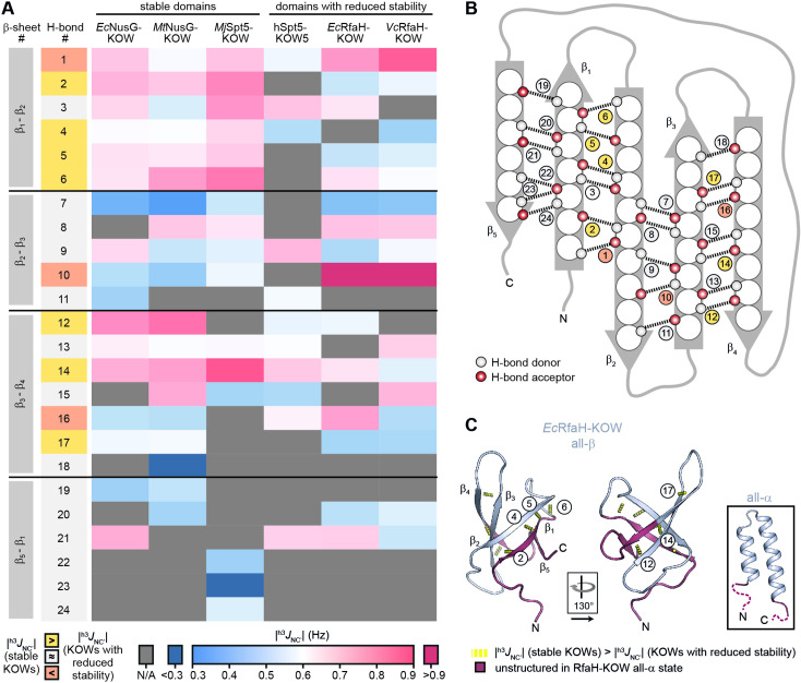
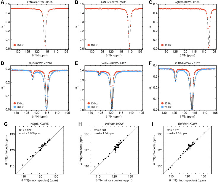
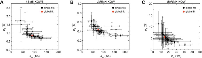
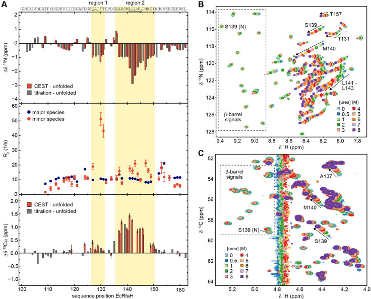
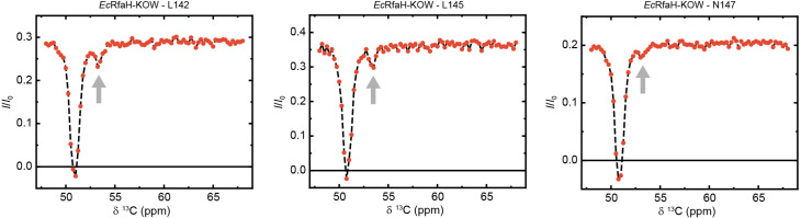
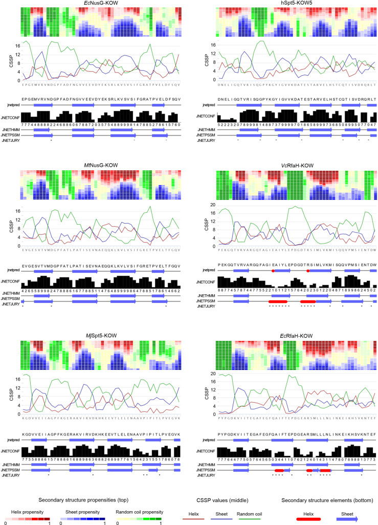
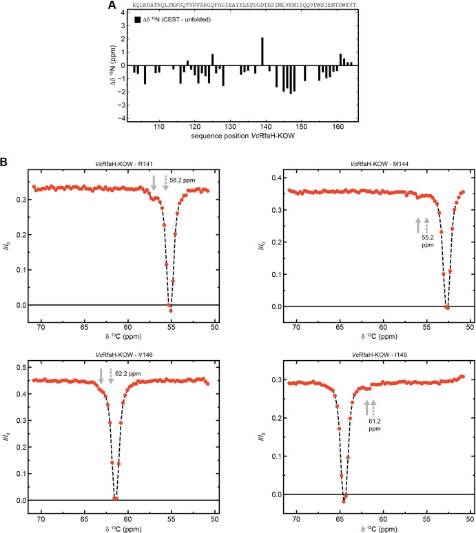
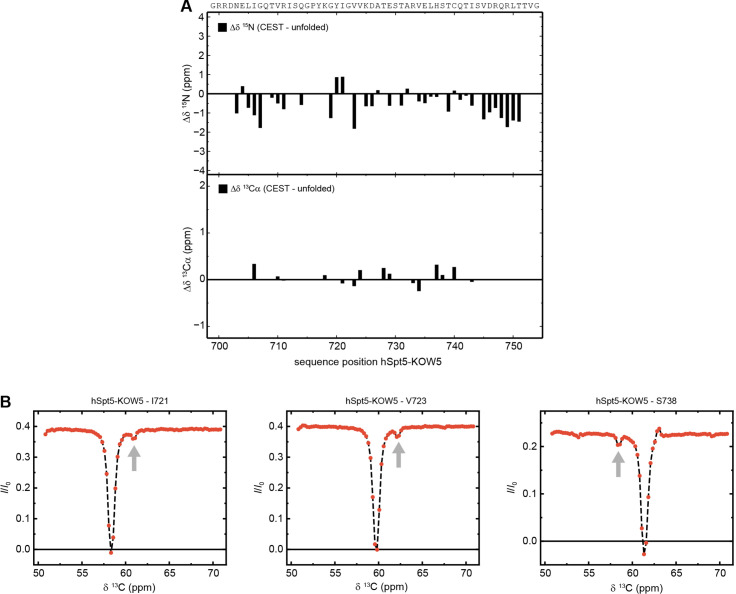
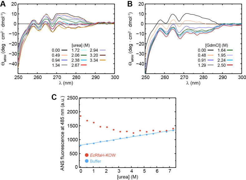
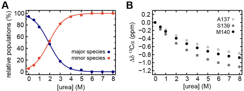
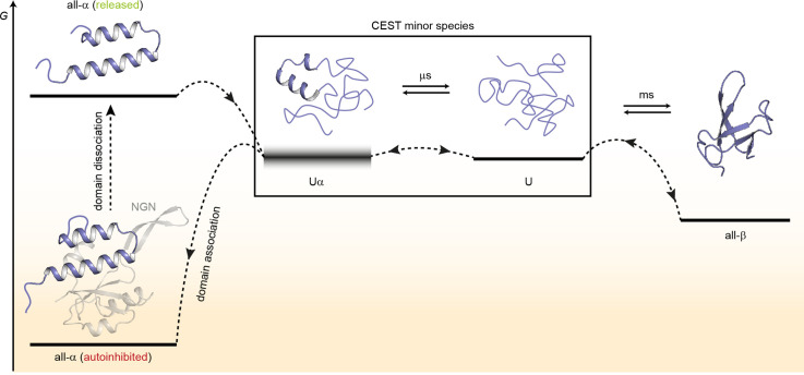
The two-domain protein RfaH, a paralog of the universally conserved NusG/Spt5 transcription factors, is regulated by autoinhibition coupled to the reversible conformational switch of its 60-residue C-terminal KOW domain between an α-hairpin and a β-barrel. In contrast, NusG/Spt5-KOW domains only occur in the β-barrel state. To understand the principles underlying the drastic fold switch in RfaH, we elucidated the thermodynamic stability and the structural dynamics of two RfaH- and four NusG/Spt5-KOW domains by combining biophysical and structural biology methods. We find that the RfaH-KOW β-barrel is thermodynamically less stable than that of most NusG/Spt5-KOWs and we show that it is in equilibrium with a globally unfolded species, which, strikingly, contains two helical regions that prime the transition towards the α-hairpin. Our results suggest that transiently structured elements in the unfolded conformation might drive the global folding transition in metamorphic proteins in general.
Overview
The claims and statistics of this paper haven’t been checked yet.
Run the check — free, no account needed. It reads the open-access text and checks the sources it cites.
Integrity signals
Registry
Cites
Statistics
Figures
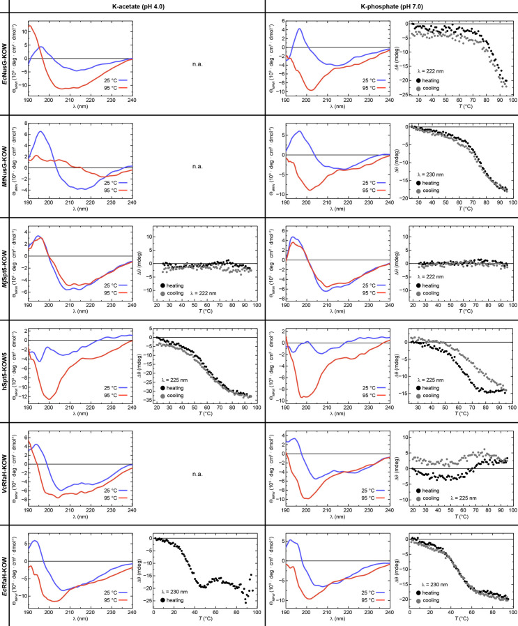
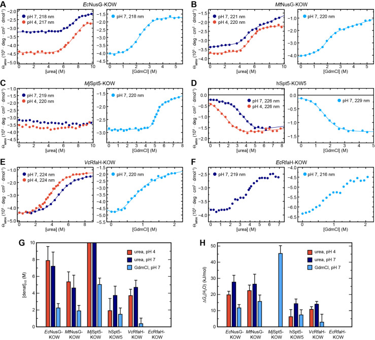
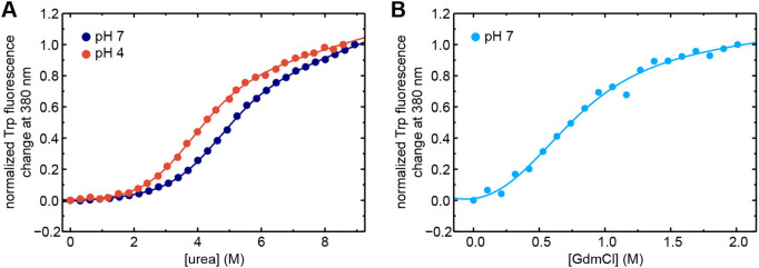
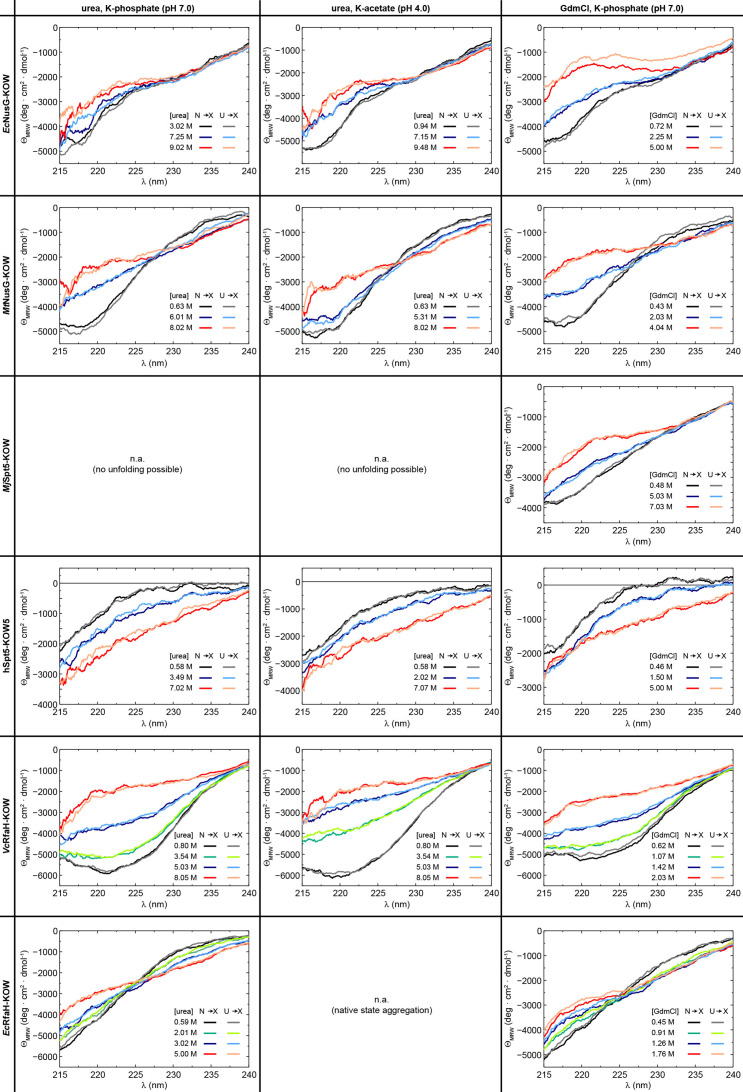
Verification
Claims the paper makes
The claims and statistics of this paper haven’t been checked yet.
Run the check — free, no account needed. It reads the open-access text and checks the sources it cites.
Self-check
Percentages, sums and reported differences re-computed from the paper’s own numbers.
The self-check needs the paper’s full text.
Citations
Cites
Highly accurate protein structure prediction with AlphaFold
Jumper, J., Evans, R., Pritzel, A., Green, T., Figurnov, M., Ronneberger, O., Tunyasuvunakool, K., Bates, R., Žídek, A., Potapenko, A., et al. (2021) · Nature · doi:10.1038/s41586-021-03819-2 · Cited by 43,764
Research works printed but not linked: 79
- 10.1002/pro.2253 · DOI
- 10.1128/mBio.01547-18 · DOI
- 10.1016/S0092-8674(02)00724-9 · DOI
- 10.1080/07391102.2014.994188 · DOI
- 10.1038/emboj.2008.268 · DOI
- 10.1016/j.molcel.2007.02.021 · DOI
- 10.1002/anie.200461794 · DOI
- 10.1038/nsmb.3465 · DOI
- 10.1021/acs.jpcb.7b11352 · DOI
- 10.1016/j.jmb.2013.10.041 · DOI
- 10.1016/j.sbi.2010.06.002 · DOI
- 10.1016/j.cell.2012.05.042 · DOI
- 10.1126/science.1184953 · DOI
- 10.1128/JB.186.9.2829-2840.2004 · DOI
- 10.1038/nprot.2007.498 · DOI
- 10.1023/A:1008392405740 · DOI · Paper page
- 10.1038/s41467-021-23167-z · DOI
- 10.1126/science.abd8700 · DOI
- 10.1021/acschembio.8b00276 · DOI
- 10.1093/nar/gkv332 · DOI
- 10.1101/SQB.1963.028.01.060 · DOI
- 10.1021/bi00003a021 · DOI
- 10.1016/j.bpj.2019.11.014 · DOI
- 10.1021/jp502193v · DOI
- Green MR , Sambrook J , Sambrook J . 2012. Molecular cloning: a laboratory manual, 4th ed. ed. Cold Spring Harbor, N.Y: Cold Spring Harbor Laboratory Press.
- 10.1016/j.pnmrs.2004.08.001 · DOI
- 10.1006/jmre.1998.1590 · DOI
- 10.1021/bi961079g · DOI
- 10.1093/nar/gkq135 · DOI
- 10.1007/s10858-012-9611-z · DOI
- 10.1042/BST20140226 · DOI
- 10.1016/S0968-0004(98)01335-8 · DOI · Paper page
- 10.1021/acs.jctc.8b00912 · DOI
- 10.1016/j.cell.2018.05.017 · DOI
- 10.1016/j.str.2020.10.006 · DOI
- 10.1093/nar/gkp351 · DOI
- 10.1073/pnas.1013828108 · DOI
- 10.4161/cc.22468 · DOI
- 10.1007/BF00228148 · DOI · Paper page
- 10.1021/acs.biochem.7b00375 · DOI
- 10.1016/j.jmr.2007.04.002 · DOI
- 10.1021/ct5002279 · DOI
- 10.1021/jp990413q · DOI
- 10.1016/S0022-2836(05)80197-4 · DOI
- 10.1110/ps.041205405 · DOI
- 10.1146/annurev.mi.37.100183.001425 · DOI
- 10.1128/MCB.00520-15 · DOI
- 10.1021/acs.jchemed.8b00649 · DOI
- 10.1016/j.jmb.2009.05.078 · DOI
- 10.1126/science.1158868 · DOI
- Pace CN , Shirley BA , Thomson JA , Creighton TE . 1990. Measuring the conformational stability of a proteinProtein Structure: A Practical Approach. pp. 311–330.
- 10.1073/pnas.94.23.12366 · DOI
- 10.1038/s41467-022-31532-9 · DOI
- 10.1073/pnas.1800168115 · DOI
- 10.1371/journal.pcbi.1004379 · DOI
- 10.1073/pnas.95.23.13585 · DOI
- 10.1021/bi00421a014 · DOI
- 10.1016/S0079-6565(98)00025-9 · DOI
- 10.1021/ja062025p · DOI
- 10.1016/S1090-7807(02)00014-9 · DOI
- 10.1002/bip.23420 · DOI
- 10.1023/A:1008398416292 · DOI
- 10.1093/nar/gkx523 · DOI
- 10.1080/07391102.2015.1031700 · DOI
- 10.1093/nar/gkt779 · DOI
- 10.1038/s41586-021-03828-1 · DOI · Paper page
- 10.1021/bi200750k · DOI
- 10.1021/ja3001419 · DOI
- 10.1021/cr400525m · DOI
- 10.1107/S174430910603658X · DOI
- 10.1006/jmbi.1996.0699 · DOI
- 10.1021/bi00174a022 · DOI
- 10.1016/j.jmb.2012.01.031 · DOI
- 10.1016/S0076-6879(94)39014-2 · DOI
- 10.1002/prot.24839 · DOI
- 10.1016/j.bmc.2016.08.012 · DOI
- 10.1002/cbic.201900446 · DOI
- 10.1038/s41598-018-30042-3 · DOI
- 10.1038/s41467-019-08567-6 · DOI
Metadata
- Authors
- Zuber, P. K., Daviter, T., Heißmann, R., Persau, U., Schweimer, K., & Knauer, S. H.
- Affiliations
- University of Bayreuth (DE)
- Birkbeck, University of London (GB)
- Corresponding author
- Knauer, Stefan H.
- Venue
- eLife
- Publisher
- openRxiv
- Published
- 2022-01-15
- Volume
- 11
- ISSN
- 2050-084X (ISSN-L)
- PMC
- PMC9683785
- OpenAlex
- W4306698552
- Cited by
- 29
- References
- 1
- Citations by year
- 2026: 5 · 2025: 8 · 2024: 7 · 2023: 7 · 2022: 2
- FWCI
- 1.96
- Type
- journal article
- Language
- en
- Open access
- gold
- doi.org
- Licence
- cc-by
- Locations
- eLife (publishedVersion, cc-by)
- bioRxiv (Cold Spring Harbor Laboratory) (acceptedVersion, cc-by)
- PubMed Central (publishedVersion, cc-by)
- PubMed (publishedVersion)
- DOAJ (DOAJ: Directory of Open Access Journals) (submittedVersion, cc-by-sa)
- ERef Bayreuth (University of Bayreuth) (submittedVersion)
- Funders
- European Cooperation in Science and Technology (CA15126)
- Deutsche Forschungsgemeinschaft (Ro617/21-1)
- Topics
- Life Sciences › Biochemistry, Genetics and Molecular Biology › Molecular Biology › Protein Structure and Dynamics
- Life Sciences › Biochemistry, Genetics and Molecular Biology › Molecular Biology › RNA and protein synthesis mechanisms
- Life Sciences › Biochemistry, Genetics and Molecular Biology › Molecular Biology › Heat shock proteins research
- Keywords
- RfaH, fold switching, autoinhibition, protein thermodynamic stability, protein folding dynamics, beta-barrel, metamorphic proteins, transcription factors, protein conformational switching
- MeSH terms
- Escherichia coli, Peptide Elongation Factors, Thermodynamics, Transcription Factors, Trans-Activators, Protein Folding, Escherichia coli Proteins
About these checks
Registry, retraction, Cites and statistics results are computed, not judged, with one exception: a printed reference that no index lists is read once by a model before it counts as a citation error. Claims are extracted by a model and kept only when their quote appears verbatim in the text; each cited source is then checked, with a model’s help, for whether it says what the claim relies on. Those verdicts can be wrong — read the claim’s quote and the cited source before relying on one.
Statistics and arithmetic are recomputed, not judged.
Confidence bands are shown only for fields calibrated on our annotated test set. None is calibrated yet, so no band is shown.
What counts as a claim ›How we measure confidence ›How we check this ›
Are you an author, or do you see a problem with this page? Request a review
