PreprintCited by 27Malaria Research and ControlBird parasitology and diseasesRNA Research and Splicing

The DOI matches its Crossref record; 1 of the 1 references it cites were found in a registry or an index.

The Plasmodium falciparum CCCH zinc finger protein ZNF4 plays an important role in gametocyte exflagellation through the regulation of male gametocyte enriched transcripts

Open access

Preprint — published as 10.3390/cells11101666 · Paper page

Abstract

CCCH zinc finger proteins (ZFPs) function mainly as RNA-binding proteins (RBPs) where they play a central role in the mRNA metabolism. Over 27 CCCH-ZFPs are encoded in the genome of the human malaria parasite Plasmodium falciparum , the causative agent of malaria tropica. However, little is known about their functions. In this study, we characterize one member of the PfCCCH-ZFP named ZNF4. We show that ZNF4 is highly expressed in mature gametocytes where it predominantly localizes to the cytoplasm. Targeted gene disruption of ZNF4 showed no significant effect in asexual blood stage replication and gametocyte development while male gametocyte exflagellation was significantly impaired leading to reduced malaria transmission in the mosquito. Comparative transcriptomics between wildtype (WT) and the ZNF4-deficient line (ZNF4-KO) demonstrated the de-regulation of about 473 genes (274-upregulated and 199 down-regulated) in mature gametocytes. Most of the down-regulated genes show peak expression in mature gametocyte with male enriched genes associated to the axonemal dynein complex formation and cell projection organization highly affected, pointing to the phenotype in male gametocyte exflagellation. Up-regulated genes are associated to ATP synthesis. Our combine data therefore indicate that ZNF4 is a CCCH zinc finger protein which plays an important role in male gametocyte exflagellation through the regulation of male gametocyte enriched genes. Author Summary: CCCH ZFPs have gained significant interest due to their ability to interact with RNA and control different RNA metabolic processes. The role of CCCH ZFPs in the malaria parasite Plasmodium falciparum has not been well studied. In this study we report the functional characterization of a PfCCCH-ZFP named ZNF4. Using mouse anti-ZNF4 antisera and a ZNF4-HA tag parasite line we show that the protein is mainly expressed in the cytosol of mature gametocytes. To determine the function of ZNF4, we generated a ZNF4 knockout parasite line and we found that the asexual blood stages and gametocytes developed normally while the ability of the male gametocytes to exflagellate as well as the further development of the parasite in the mosquito was significantly impaired. Interestingly, when the transcriptome of mature gametocytes of the ZNF4-KO was compared to the WT, down-regulated genes were mainly male gametocyte enriched genes associated to processes involved in gametocyte exflagellation such axonemal dynein complex formation and cell projection organization indicating that ZNF4 plays an important role in male gametocyte exflagellation through the regulation of male gametocyte enriched transcripts.

Overview

The claims and statistics of this paper haven’t been checked yet.

Run the check — free, no account needed. It reads the open-access text and checks the sources it cites.

A few free checks a day · papers already verified open instantly · results are public

Integrity signals

Registry

Crossref · 10.1101/2022.03.08.483571

Cites

1 of 1 references verified0 citation errors · 0 not checkable

Statistics

Figures

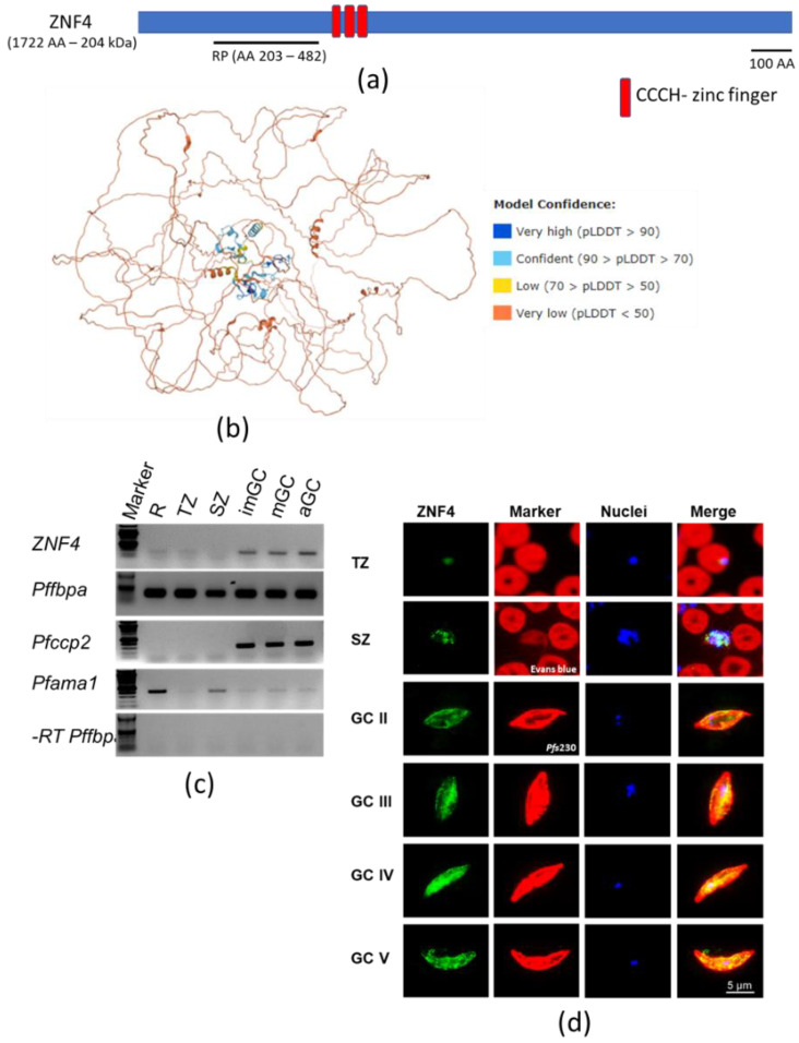
Figure 1 Domain architecture and protein expression of ZNF4: (a) Schematic of the ZNF4 domain structure. The 204 kDa protein contains three CCCH zinc finger domains indicated in red. The black line indicates the region used for the generation of the recombinant peptide. (b) Predicted 3D structure of ZNF4. The 3D structure of the protein was predicted using AlphaFold (https://alphafold.ebi.ac.uk/ accessed on 12 January 2022) [40,41]. Colours indicate the confidence level of prediction which ranged from blue (very high confidence) to orange (very low confidence). (c) Transcript expression of ZNF4 in the blood stages of P. falciparum. Complementary DNA was synthesized from rings (R), trophozoites (TZ), schizonts (SZ), immature (imGC), mature (mGC), and gametocytes at 30 min post-activation (aGC) and subjected to diagnostic PCR using ZNF4-specific primers. The expression levels of Pfama1 and Pfccp2 were used to verify asexual blood stage and gametocyte-specific expression. Samples lacking reverse transcriptase (-RT) were used as controls to check for any contamination with gDNA. Pffbpa was used as a loading control. (d) Localization of ZNF4 in different blood stages of P. falciparum. Anti-ZNF4 was used to immunolabel fixed samples of trophozoites (TZ), schizonts (SZ), and gametocytes (GC stage II to V) as well as of activated gametocytes (aGC) at 30 min post-activation (green). Asexual blood stages (trophozoites and schizonts) were visualized by labelling with Evans blue and gametocytes were visualized by using rabbit anti-Pfs230 (red); nuclei were highlighted by Hoechst nuclear stain 33342 (blue). Bar, 5 µm.
Figure 1 Domain architecture and protein expression of ZNF4: (a) Schematic of the ZNF4 domain structure. The 204 kDa protein contains three CCCH zinc finger domains indicated in red. The black line indicates the region used for the generation of the recombinant peptide. (b) Predicted 3D structure of ZNF4. The 3D structure of the protein was predicted using AlphaFold (https://alphafold.ebi.ac.uk/ accessed on 12 January 2022) [40,41]. Colours indicate the confidence level of prediction which ranged from blue (very high confidence) to orange (very low confidence). (c) Transcript expression of ZNF4 in the blood stages of P. falciparum. Complementary DNA was synthesized from rings (R), trophozoites (TZ), schizonts (SZ), immature (imGC), mature (mGC), and gametocytes at 30 min post-activation (aGC) and subjected to diagnostic PCR using ZNF4-specific primers. The expression levels of Pfama1 and Pfccp2 were used to verify asexual blood stage and gametocyte-specific expression. Samples lacking reverse transcriptase (-RT) were used as controls to check for any contamination with gDNA. Pffbpa was used as a loading control. (d) Localization of ZNF4 in different blood stages of P. falciparum. Anti-ZNF4 was used to immunolabel fixed samples of trophozoites (TZ), schizonts (SZ), and gametocytes (GC stage II to V) as well as of activated gametocytes (aGC) at 30 min post-activation (green). Asexual blood stages (trophozoites and schizonts) were visualized by labelling with Evans blue and gametocytes were visualized by using rabbit anti-Pfs230 (red); nuclei were highlighted by Hoechst nuclear stain 33342 (blue). Bar, 5 µm.CC BY · PubMed Central
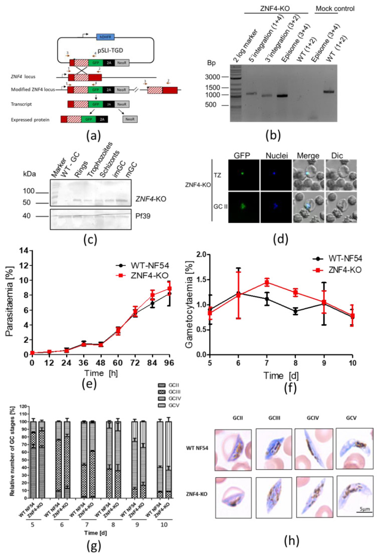
Figure 2 Targeted gene disruption of ZNF4 and its effect on asexual blood stage replication and gametocytogenesis: (a) Schematic depicting the generation of ZNF4-KO via single crossover recombination-based gene disruption using selective linked integration targeted gene disruption (SLI-TGD). The vector pSLI-TGD was modified to contain a 601 bp sequence block (white box with red stripes) from near the 5′ end of the ZNF4 coding region (red box). The coding region was maintained in frame with a GFP protein coding region (green box), a 2A “skip” peptide (black box), and the Neo-R gene (grey) that provides resistance to the antibiotic G418. Arrows indicate the position of primers 1–4, used to detect integration of the pSLI-TGD vector. Asterisks indicate a stop codon. GFP, green fluorescent protein; hDHFR; human dehydrofolate reductase for resistance to WR99210; NeoR, neomycin-resistance; 2A, skip peptide. (b) Confirmation of vector integration for the ZNF4-KO parasites by diagnostic PCR using gDNA obtained from ZNF4-KO and WT-NF54. 5′-integration was detected using primers 1 and 4 (1164 bp) and 3′-integration using primers 3 and 2 (955 bp). Primers 3 and 4 were used to detect the presence of episome (965 bp), and primers 1 and 2 were used for WT control (1194 bp). (c) Confirmation of truncated ZNF4 tagged with GFP. Parasite lysates obtained from different stages of the ZNF4-KO parasite line were subjected to Western blotting using polyclonal mouse anti-GFP (estimated size 53 kDa). Lysate from WT mature gametocyte was used as negative control. Immunoblotting with mouse anti-Pf39 antisera (39 kDa) served as a loading control. (d) Verification of GFP expression in the ZNF4-KO parasites by live imaging. Live images of trophozoites (TZ) and gametocyte stage II (GCII) of the ZNF4-KO line detected GFP (green) in the parasite. Dic, differential interference contrast. Nuclei were counterstained with Hoechst 33342 (blue). Bar, 5 µm. (e) Asexual blood stage replication of the ZNF4-KO. Synchronized ring stage cultures of WT and ZNF4-KO with an initial parasitaemia of 0.25% were maintained in cell culture medium, and the parasitaemia was followed over a time-period of 0 to 96 h via Giemsa stained smears. The data are a representation of one of two experiments performed in triplicate (mean ± SD). For the second experiment, see Figure S2a. (f) Disruption of ZNF4 shows no effect in gametocytaemia. Following two rounds of synchronization, a culture of 5% of the ring stage parasites of the WT and ZNF4-KO was induced for gametocytogenesis and, the next day, the parasites were washed and grown with medium supplemented with 50 mM GlcNac to kill asexual blood stages for 5 days, then with normal medium until day 10 post-induction. The gametocytaemia was monitored by Giemsa-stained blood smears from day 5 post-induction. The result is a representative of one of two experiments (see Figure S2b for second experiment). (g) Gametocyte maturation in the ZNF4-KO. The development of gametocyte was compared between the WT and the ZNF4-KO by counting the gametocyte stages of 50 gametocytes at each time point in triplicate (See Figure S2c for second experiment). (h) Gametocyte morphology in the ZNF4-KO line. Giemsa-stained pictures of gametocyte stages of the ZNF4-KO and WT.
Figure 2 Targeted gene disruption of ZNF4 and its effect on asexual blood stage replication and gametocytogenesis: (a) Schematic depicting the generation of ZNF4-KO via single crossover recombination-based gene disruption using selective linked integration targeted gene disruption (SLI-TGD). The vector pSLI-TGD was modified to contain a 601 bp sequence block (white box with red stripes) from near the 5′ end of the ZNF4 coding region (red box). The coding region was maintained in frame with a GFP protein coding region (green box), a 2A “skip” peptide (black box), and the Neo-R gene (grey) that provides resistance to the antibiotic G418. Arrows indicate the position of primers 1–4, used to detect integration of the pSLI-TGD vector. Asterisks indicate a stop codon. GFP, green fluorescent protein; hDHFR; human dehydrofolate reductase for resistance to WR99210; NeoR, neomycin-resistance; 2A, skip peptide. (b) Confirmation of vector integration for the ZNF4-KO parasites by diagnostic PCR using gDNA obtained from ZNF4-KO and WT-NF54. 5′-integration was detected using primers 1 and 4 (1164 bp) and 3′-integration using primers 3 and 2 (955 bp). Primers 3 and 4 were used to detect the presence of episome (965 bp), and primers 1 and 2 were used for WT control (1194 bp). (c) Confirmation of truncated ZNF4 tagged with GFP. Parasite lysates obtained from different stages of the ZNF4-KO parasite line were subjected to Western blotting using polyclonal mouse anti-GFP (estimated size 53 kDa). Lysate from WT mature gametocyte was used as negative control. Immunoblotting with mouse anti-Pf39 antisera (39 kDa) served as a loading control. (d) Verification of GFP expression in the ZNF4-KO parasites by live imaging. Live images of trophozoites (TZ) and gametocyte stage II (GCII) of the ZNF4-KO line detected GFP (green) in the parasite. Dic, differential interference contrast. Nuclei were counterstained with Hoechst 33342 (blue). Bar, 5 µm. (e) Asexual blood stage replication of the ZNF4-KO. Synchronized ring stage cultures of WT and ZNF4-KO with an initial parasitaemia of 0.25% were maintained in cell culture medium, and the parasitaemia was followed over a time-period of 0 to 96 h via Giemsa stained smears. The data are a representation of one of two experiments performed in triplicate (mean ± SD). For the second experiment, see Figure S2a. (f) Disruption of ZNF4 shows no effect in gametocytaemia. Following two rounds of synchronization, a culture of 5% of the ring stage parasites of the WT and ZNF4-KO was induced for gametocytogenesis and, the next day, the parasites were washed and grown with medium supplemented with 50 mM GlcNac to kill asexual blood stages for 5 days, then with normal medium until day 10 post-induction. The gametocytaemia was monitored by Giemsa-stained blood smears from day 5 post-induction. The result is a representative of one of two experiments (see Figure S2b for second experiment). (g) Gametocyte maturation in the ZNF4-KO. The development of gametocyte was compared between the WT and the ZNF4-KO by counting the gametocyte stages of 50 gametocytes at each time point in triplicate (See Figure S2c for second experiment). (h) Gametocyte morphology in the ZNF4-KO line. Giemsa-stained pictures of gametocyte stages of the ZNF4-KO and WT.CC BY · PubMed Central
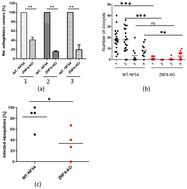
Figure 3 ZNF4-KO impairs male gametocyte exflagellation and parasite transmission in the mosquito: (a) Disruption of ZNF4 impairs male gametocyte exflagellation. Mature WT and ZNF4-KO gametocytes were activated in vitro and the number of exflagellation centres was counted in 30 fields in triplicate using the light microscope. Three independent experiments were performed, indicated in numbers 1 to 3. **, p < 0.01, Student’s t test. (b) Mosquito infectivity of ZNF4-KO. Enriched mature gametocytes of WT or the ZNF4-KO were fed to An. stephensi mosquitoes via standard membrane feeding assays. The number of oocysts per midgut were counted at day 7 post-infection in four independent experiments; ***, p < 0.001, **, p < 0.01; ns, not significant, Student’s t test. (c) Percentage of infected mosquitoes in WT versus ZNF4-KO in each standard membrane-feeding experiment; * p < 0.05, Student’s t test.
Figure 3 ZNF4-KO impairs male gametocyte exflagellation and parasite transmission in the mosquito: (a) Disruption of ZNF4 impairs male gametocyte exflagellation. Mature WT and ZNF4-KO gametocytes were activated in vitro and the number of exflagellation centres was counted in 30 fields in triplicate using the light microscope. Three independent experiments were performed, indicated in numbers 1 to 3. **, p < 0.01, Student’s t test. (b) Mosquito infectivity of ZNF4-KO. Enriched mature gametocytes of WT or the ZNF4-KO were fed to An. stephensi mosquitoes via standard membrane feeding assays. The number of oocysts per midgut were counted at day 7 post-infection in four independent experiments; ***, p < 0.001, **, p < 0.01; ns, not significant, Student’s t test. (c) Percentage of infected mosquitoes in WT versus ZNF4-KO in each standard membrane-feeding experiment; * p < 0.05, Student’s t test.CC BY · PubMed Central
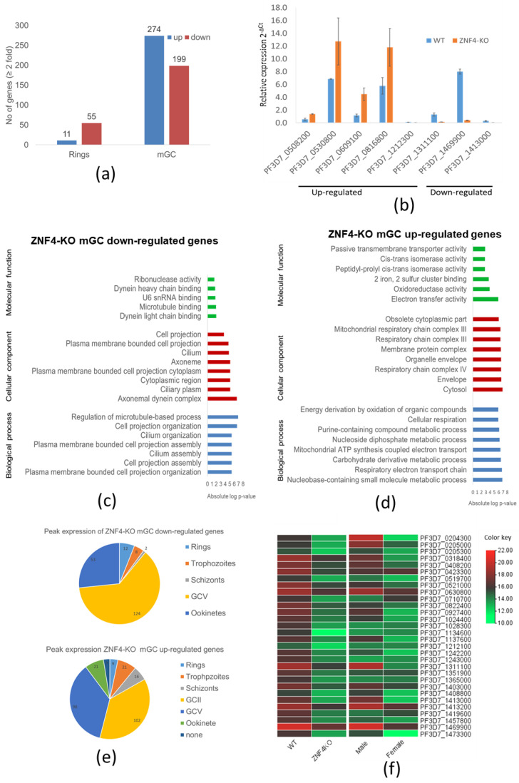
Figure 4 Deregulation of gene expression following disruption of ZNF4: (a) Comparative transcriptomics of deregulated genes in the ZNF4-KO parasite line in rings and mature gametocytes. Total RNA was extracted from ring stage parasites as well as the mature gametocytes of the ZNF4-KO and the WT and were subjected to comparative transcriptomics by RNA sequencing. Deregulated genes with relative gene expression greater than 2-fold were considered significant. (b) Validation of RNA-Seq data by qRT. Transcript analysis for 5 upregulated genes and 3 downregulated in mature gametocyte, as identified by RNA-Seq, were validated by real-time RT-PCR. Transcript expression levels were calculated by the 2−ΔCt method; the threshold cycle number (Ct) was normalized with the Ct of the gene encoding seryl tRNA-ligase (PF3D7_0717700) as reference. Genes were considered upregulated when the changes between ZNF4-KO and WT sample were greater than 2-fold. (c,d) Summary of Gene Ontology functional analysis of mature gametocyte differentially expressed genes following ZNF4 disruption. GO enrichment analysis of deregulated genes was determined using PlasmoDB (https://plasmodb.org/plasmo/app, accessed on 1 January 2022). The most significantly (p < 0.05) enriched GO terms in the biological process, the cellular component, and the molecular function are presented. For the complete list, see Table S2. All adjusted statistically significant values of the terms were the absolute log10 values. GO, gene ontology. (e) Pie chart showing peak expression stage of deregulated genes. The peak expression of the downregulated as well as the upregulated genes were determined using the 7 stage RNA-Seq data [44]. (f) Heat map of top 30 downregulated genes and their sex-specific expression. Heat map representing patterns of the top 30 downregulated genes and their sex-specific expression pattern [45]. Heat map was constructed using TB tools [42].
Figure 4 Deregulation of gene expression following disruption of ZNF4: (a) Comparative transcriptomics of deregulated genes in the ZNF4-KO parasite line in rings and mature gametocytes. Total RNA was extracted from ring stage parasites as well as the mature gametocytes of the ZNF4-KO and the WT and were subjected to comparative transcriptomics by RNA sequencing. Deregulated genes with relative gene expression greater than 2-fold were considered significant. (b) Validation of RNA-Seq data by qRT. Transcript analysis for 5 upregulated genes and 3 downregulated in mature gametocyte, as identified by RNA-Seq, were validated by real-time RT-PCR. Transcript expression levels were calculated by the 2−ΔCt method; the threshold cycle number (Ct) was normalized with the Ct of the gene encoding seryl tRNA-ligase (PF3D7_0717700) as reference. Genes were considered upregulated when the changes between ZNF4-KO and WT sample were greater than 2-fold. (c,d) Summary of Gene Ontology functional analysis of mature gametocyte differentially expressed genes following ZNF4 disruption. GO enrichment analysis of deregulated genes was determined using PlasmoDB (https://plasmodb.org/plasmo/app, accessed on 1 January 2022). The most significantly (p < 0.05) enriched GO terms in the biological process, the cellular component, and the molecular function are presented. For the complete list, see Table S2. All adjusted statistically significant values of the terms were the absolute log10 values. GO, gene ontology. (e) Pie chart showing peak expression stage of deregulated genes. The peak expression of the downregulated as well as the upregulated genes were determined using the 7 stage RNA-Seq data [44]. (f) Heat map of top 30 downregulated genes and their sex-specific expression. Heat map representing patterns of the top 30 downregulated genes and their sex-specific expression pattern [45]. Heat map was constructed using TB tools [42].CC BY · PubMed Central

Verification

Claims the paper makes

The claims and statistics of this paper haven’t been checked yet.

Run the check — free, no account needed. It reads the open-access text and checks the sources it cites.

A few free checks a day · papers already verified open instantly · results are public

Self-check

Percentages, sums and reported differences re-computed from the paper’s own numbers.

The self-check needs the paper’s full text.

Citations

Cites

Show all 1 references

Research works printed but not linked: 58

  1. 10.1111/cmi.12604 · DOI
  2. 10.1155/2010/976827 · DOI
  3. 10.1038/nrmicro3519 · DOI
  4. 10.1038/nature12920 · DOI
  5. 10.1038/nature12970 · DOI
  6. 10.1093/NAR/GKAB683 · DOI
  7. 10.1111/mmi.14334 · DOI
  8. 10.15252/EMBR.202051660 · DOI
  9. 10.1371/journal.ppat.1004905 · DOI
  10. 10.1016/j.chom.2016.12.003 · DOI
  11. 10.1111/mmi.14388 · DOI
  12. 10.1371/journal.ppat.1007249 · DOI
  13. 10.1371/journal.ppat.1000767 · DOI
  14. 10.1126/science.1125129 · DOI
  15. 10.1242/jcs.059824 · DOI
  16. 10.1242/jcs.186908 · DOI
  17. 10.3389/fcimb.2017.00320 · DOI
  18. 10.3390/ijms20205087 · DOI
  19. 10.1111/CMI.13387 · DOI
  20. 10.3390/v12040404 · DOI
  21. 10.1210/me.10.2.140 · DOI
  22. 10.1104/pp.109.145656 · DOI
  23. 10.4161/psb.6.5.15105 · DOI
  24. 10.1093/NAR/GKAB1061 · DOI · Paper page
  25. 10.1038/nmeth.4223 · DOI
  26. 10.1186/1471-2164-12-587 · DOI
  27. 10.1093/nar/gkw536 · DOI
  28. 10.1016/J.MOLP.2020.06.009 · DOI
  29. 10.1101/2021.09.02.458722 · DOI
  30. 10.1038/nature12311 · DOI · Paper page
  31. 10.1126/SCIENCE.AAP7847 · DOI
  32. 10.3389/fcimb.2021.624945 · DOI
  33. 10.1371/journal.pgen.1009306 · DOI
  34. 10.1242/jcs.051326 · DOI
  35. 10.1093/infdis/jix237 · DOI
  36. 10.7554/ELIFE.34463 · DOI
  37. 10.1038/s41431-019-0489-z · DOI
  38. 10.1371/JOURNAL.PGEN.1007602 · DOI
  39. 10.1126/science.8178152 · DOI
  40. 10.1101/gad.8.17.2072 · DOI
  41. 10.1016/j.celrep.2015.06.008 · DOI
  42. 10.1074/jbc.M110053200 · DOI
  43. 10.1128/MCB.01683-13 · DOI
  44. 10.1371/journal.ppat.1003268 · DOI
  45. 10.3390/pathogens10121536 · DOI
  46. 10.1038/nri.2016.129 · DOI
  47. 10.1186/1471-2164-14-256 · DOI
  48. 10.1038/294364a0 · DOI
  49. 10.2307/3280287 · DOI
  50. 10.1186/S12864-015-1695-X · DOI
  51. 10.1093/bioinformatics/bts635 · DOI · Paper page
  52. 10.1038/nbt.3122 · DOI
  53. 10.1093/bioinformatics/btt656 · DOI · Paper page
  54. 10.1006/meth.2001.1262 · DOI · Paper page
  55. 10.1046/j.1365-2958.2003.03570.x · DOI · Paper page
  56. 10.1186/1475-2875-9-147 · DOI
  57. 1998, Plasmodium falciparum: Purification of the various gametocyte developmental stages from in vitro-cultivated parasites, Am J Trop Med Hyg, 59, 505, 10.4269/ajtmh.1998.59.505 · DOI
  58. 10.1016/j.molbiopara.2007.04.008 · DOI

Metadata

Authors
Hanhsen, B., Farrukh, A., Pradel, G., & Ngwa, C. J.
Affiliations
RWTH Aachen University (DE)
Corresponding author
Ngwa, Che Julius
Venue
Cells
Publisher
openRxiv
Published
2022-03-10
Volume
11
Issue
10
Pages
1666-1666
ISSN
2073-4409 (ISSN-L)
OpenAlex
W4280578208
Cited by
27
References
1
Citations by year
2026: 4 · 2025: 4 · 2024: 5 · 2023: 11 · 2022: 2
FWCI
4.86
Type
journal article
Language
en
Open access
Licence
cc-by
Locations
  • Cells (publishedVersion, cc-by)
  • bioRxiv (Cold Spring Harbor Laboratory) (acceptedVersion, cc-by)
  • PubMed Central (publishedVersion, cc-by)
  • PubMed (publishedVersion)
  • DOAJ (DOAJ: Directory of Open Access Journals) (submittedVersion, cc-by-sa)
  • RWTH Publications (RWTH Aachen) (submittedVersion, other-oa)
Funders
Deutsche Forschungsgemeinschaft (PR905/20-1, No 731060, NG170/1‐1)
Topics
  • Health Sciences › Medicine › Public Health, Environmental and Occupational Health › Malaria Research and Control
  • Life Sciences › Immunology and Microbiology › Parasitology › Bird parasitology and diseases
  • Life Sciences › Biochemistry, Genetics and Molecular Biology › Molecular Biology › RNA Research and Splicing
Keywords
Plasmodium falciparum, CCCH zinc finger proteins, male gametocytes, ATP production, mosquito-borne transmission, gene knockout, transcriptomics, RNA-binding proteins
MeSH terms
Animals, Cytoplasm, Malaria, Male, Culicidae, Plasmodium falciparum, Zinc Fingers

About these checks

Registry, retraction, Cites and statistics results are computed, not judged, with one exception: a printed reference that no index lists is read once by a model before it counts as a citation error. Claims are extracted by a model and kept only when their quote appears verbatim in the text; each cited source is then checked, with a model’s help, for whether it says what the claim relies on. Those verdicts can be wrong — read the claim’s quote and the cited source before relying on one.

Statistics and arithmetic are recomputed, not judged.

Confidence bands are shown only for fields calibrated on our annotated test set. None is calibrated yet, so no band is shown.

What counts as a claim ›How we measure confidence ›How we check this ›

Are you an author, or do you see a problem with this page? Request a review

How to cite

Cite CoChat for the checks on this page, and the paper for its own findings.

This page
CoChat. (2026). Citation check: The Plasmodium falciparum CCCH zinc finger protein ZNF4 plays an important role in gametocyte exflagellation through the regulation of male gametocyte enriched transcripts. https://app.cochat.ai/papers/10.1101/2022.03.08.483571
The paper
Hanhsen, B., Farrukh, A., Pradel, G., & Ngwa, C. J. (2022). The Plasmodium falciparum CCCH zinc finger protein ZNF4 plays an important role in gametocyte exflagellation through the regulation of male gametocyte enriched transcripts. Cells. https://doi.org/10.1101/2022.03.08.483571