PreprintCited by 15Evolution and Genetic DynamicsFungal and yeast genetics researchGlycosylation and Glycoproteins Research

The DOI matches its Crossref record; 1 of the 1 references it cites were found in a registry or an index.

Experimental evolution of phosphomannomutase-deficient yeast reveals compensatory mutations in a phosphoglucomutase

Open access

Preprint — published as 10.7554/elife.79346 · Paper page

Abstract

The most common cause of human congenital disorders of glycosylation (CDG) are mutations in the phosphomannomutase gene PMM2 , which affect protein N -linked glycosylation. The yeast gene SEC53 encodes a nearly-identical homolog of human PMM2 . We evolved 384 populations of yeast harboring one of two human-disease-associated alleles, sec53 -V238M and sec53 -F126L, or wild-type SEC53 . We find that after 1,000 generations, most populations compensate for the slow-growth phenotype associated with the sec53 human-disease-associated alleles. Through whole-genome sequencing we identify compensatory mutations, including known SEC53 genetic interactors. We observe an enrichment of compensatory mutations in other genes whose human homologs are associated with Type 1 CDG, including PGM1 , which encodes the minor isoform of phosphoglucomutase in yeast. By genetic reconstruction, we show that evolved pgm1 mutations are dominant and allele-specific genetic interactors that restore both protein glycosylation and growth of yeast harboring the sec53 -V238M allele. Finally, we characterize the enzymatic activity of purified Pgm1 mutant proteins. We find that reduction, but not elimination, of Pgm1 activity best compensates for the deleterious phenotypes associated with the sec53 -V238M allele. Broadly, our results demonstrate the power of experimental evolution as a tool for identifying genes and pathways that compensate for human-disease associated alleles.

Overview

The claims and statistics of this paper haven’t been checked yet.

Run the check — free, no account needed. It reads the open-access text and checks the sources it cites.

A few free checks a day · papers already verified open instantly · results are public

Integrity signals

Registry

Crossref · 10.1101/2022.04.06.487342

Cites

1 of 1 references verified0 citation errors · 0 not checkable

Statistics

Figures

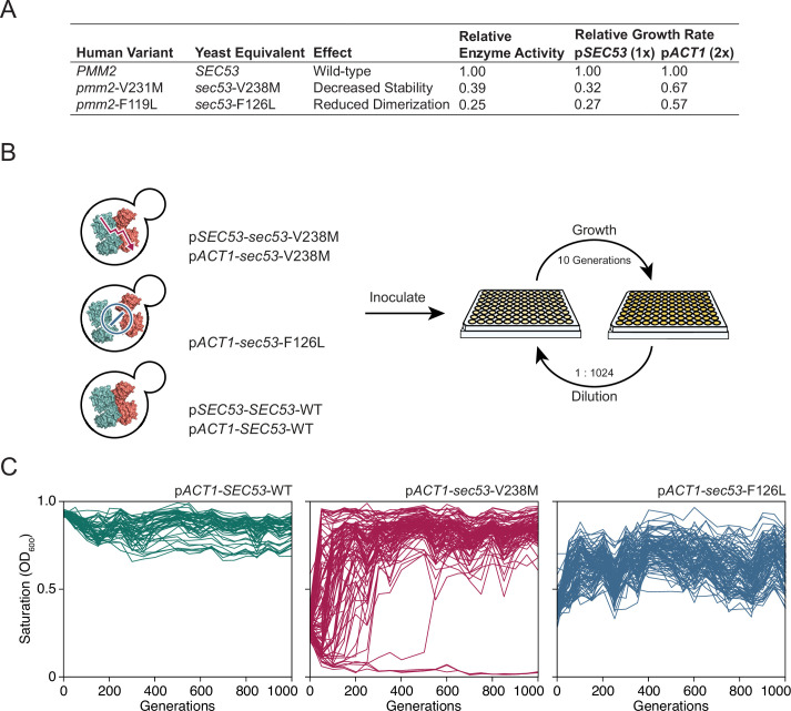
Figure 1. Experimental evolution of yeast models of congenital disorders of glycosylation. (A) Table of the various sec53 alleles used in this study. In vitro enzymatic activities of Pmm2 are relative to wild-type Pmm2 and were previously reported (Pirard et al., 1999). Relative growth rates of yeast carrying various SEC53 alleles were previously reported (Lao et al., 2019). (B) Diagram of the evolution experiment. Yeast carrying sec53 mutations implicated in human disease were used to initiate replicate populations in 96-well plates: 96 populations of each mutant genotype and 48 populations of each wild-type genotype. Populations were propagated in rich glucose media, unshaken, for 1000 generations. (C) Single time-point OD600 readings of populations were taken every 50 generations during the evolution experiment as a measure of growth rate. Each line represents one population.
Figure 1. Experimental evolution of yeast models of congenital disorders of glycosylation. (A) Table of the various sec53 alleles used in this study. In vitro enzymatic activities of Pmm2 are relative to wild-type Pmm2 and were previously reported (Pirard et al., 1999). Relative growth rates of yeast carrying various SEC53 alleles were previously reported (Lao et al., 2019). (B) Diagram of the evolution experiment. Yeast carrying sec53 mutations implicated in human disease were used to initiate replicate populations in 96-well plates: 96 populations of each mutant genotype and 48 populations of each wild-type genotype. Populations were propagated in rich glucose media, unshaken, for 1000 generations. (C) Single time-point OD600 readings of populations were taken every 50 generations during the evolution experiment as a measure of growth rate. Each line represents one population.CC BY · PubMed Central
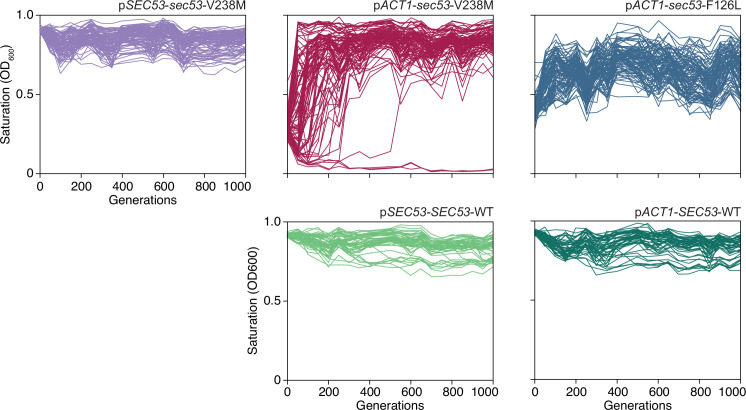
Figure 1—figure supplement 1. OD600 readings of each population. Single time-point OD600 readings of populations over the course of the evolution experiment. Every 50 generations, following daily transfer, populations were resuspended and OD was measured. Each line represents one population.
Figure 1—figure supplement 1. OD600 readings of each population. Single time-point OD600 readings of populations over the course of the evolution experiment. Every 50 generations, following daily transfer, populations were resuspended and OD was measured. Each line represents one population.CC BY · PubMed Central
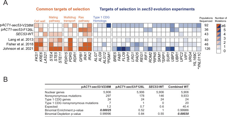
Figure 2. Mutations in Type 1 congenital disorders of glycosylation (CDG) homologs are enriched in sec53-V238M populations. (A) Heatmap showing the number of nonsynonymous mutations per gene that arose in the evolution experiment. Genes with two or more unique nonsynonymous mutations (and each Type 1 CDG homolog with at least one mutation) are shown. pSEC53-SEC53-WT and pACT1-SEC53-WT are grouped as ‘SEC53-WT’. For comparison, we show data from previously reported evolution experiments where the experimental conditions were identical to the conditions used here, aside from strain background and experiment duration (bottom three rows). Commonly mutated pathways are grouped for clarity. Asterisks (*) indicate previously known SEC53 genetic interactors (Costanzo et al., 2016; Kuzmin et al., 2018). (B) Binomial test for enrichment or depletion of mutations in Type 1 CDG homologs based on the number of nonsynonymous mutations observed in each experiment, the total number of yeast genes (5906), and the number of genes that are Type 1 CDG homologs (24). ‘Combined WT’ includes the SEC53-WT populations as well as three additional datasets: Lang et al., 2013, Fisher et al., 2018, and Johnson et al., 2021.
Figure 2. Mutations in Type 1 congenital disorders of glycosylation (CDG) homologs are enriched in sec53-V238M populations. (A) Heatmap showing the number of nonsynonymous mutations per gene that arose in the evolution experiment. Genes with two or more unique nonsynonymous mutations (and each Type 1 CDG homolog with at least one mutation) are shown. pSEC53-SEC53-WT and pACT1-SEC53-WT are grouped as ‘SEC53-WT’. For comparison, we show data from previously reported evolution experiments where the experimental conditions were identical to the conditions used here, aside from strain background and experiment duration (bottom three rows). Commonly mutated pathways are grouped for clarity. Asterisks (*) indicate previously known SEC53 genetic interactors (Costanzo et al., 2016; Kuzmin et al., 2018). (B) Binomial test for enrichment or depletion of mutations in Type 1 CDG homologs based on the number of nonsynonymous mutations observed in each experiment, the total number of yeast genes (5906), and the number of genes that are Type 1 CDG homologs (24). ‘Combined WT’ includes the SEC53-WT populations as well as three additional datasets: Lang et al., 2013, Fisher et al., 2018, and Johnson et al., 2021.CC BY · PubMed Central
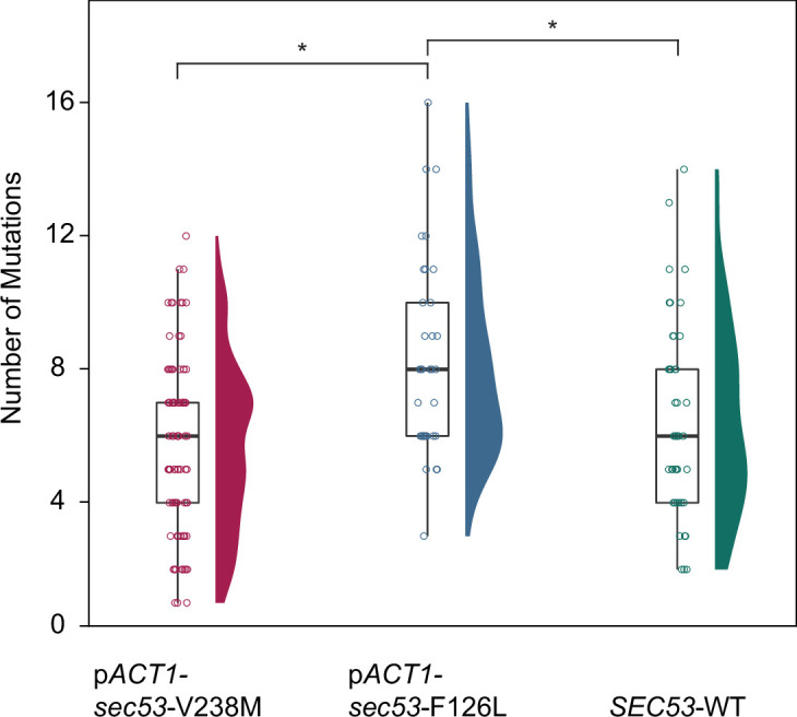
Figure 2—figure supplement 1. The number of de novo mutations per SEC53 genotype. Each circle represents the number of SNPs and small indels present in a sequenced clone (Supplementary file 1). pSEC53-SEC53-WT and pACT1-SEC53-WT are grouped as ‘SEC53-WT’. Distribution of points are shown as violin plots. Only statistically significant differences shown (p < 0.05) with bars and astersisks (*). pACT1-sec53-V238M versus SEC53-WT (p = 0.326), pACT1-sec53-V238M versus pACT1-sec53-F126L (p > 0.0001), pACT1-sec53-F126L versus SEC53-WT (p = 0.0234) (df = 167, F = 9.67, one-way analysis of variance [ANOVA] with Tukey post hoc test).
Figure 2—figure supplement 1. The number of de novo mutations per SEC53 genotype. Each circle represents the number of SNPs and small indels present in a sequenced clone (Supplementary file 1). pSEC53-SEC53-WT and pACT1-SEC53-WT are grouped as ‘SEC53-WT’. Distribution of points are shown as violin plots. Only statistically significant differences shown (p < 0.05) with bars and astersisks (*). pACT1-sec53-V238M versus SEC53-WT (p = 0.326), pACT1-sec53-V238M versus pACT1-sec53-F126L (p > 0.0001), pACT1-sec53-F126L versus SEC53-WT (p = 0.0234) (df = 167, F = 9.67, one-way analysis of variance [ANOVA] with Tukey post hoc test).CC BY · PubMed Central
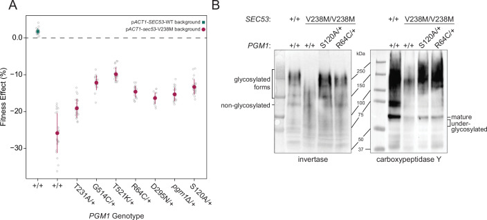
Figure 3. Evolved mutations rescue fitness and protein glycosylation defects of pACT1-sec53-V238M. (A) Average fitness effects and standard deviations of reconstructed heterozygous pgm1 mutations. Fitness effects were determined by competitive fitness assays against a fluorescently labeled version of the diploid pACT1-SEC53-WT ancestor. Replicate measurements are plotted as gray circles. Pairs of pgm1 fitness effects with non-statistically significant differences: R64C-D295N, R64C-pgm1Δ, R64C-S120A, D295N-pgm1Δ, G514C-T521K, G514C-S120A, and pgm1Δ-S120A (df = 194, F = 208.1, each p > 0.05, one-way analysis of variance [ANOVA] with Tukey post hoc test). (B) Western blots of invertase (left) and carboxypeptidase Y (right) from ancestral and reconstructed strains. In panels A and B, plus signs (+) indicate wild-type alleles. Genotypes are either homozygous wild-type (+/+), homozygous mutant (mutation/mutation), or heterozygous (mutation/+).
Figure 3. Evolved mutations rescue fitness and protein glycosylation defects of pACT1-sec53-V238M. (A) Average fitness effects and standard deviations of reconstructed heterozygous pgm1 mutations. Fitness effects were determined by competitive fitness assays against a fluorescently labeled version of the diploid pACT1-SEC53-WT ancestor. Replicate measurements are plotted as gray circles. Pairs of pgm1 fitness effects with non-statistically significant differences: R64C-D295N, R64C-pgm1Δ, R64C-S120A, D295N-pgm1Δ, G514C-T521K, G514C-S120A, and pgm1Δ-S120A (df = 194, F = 208.1, each p > 0.05, one-way analysis of variance [ANOVA] with Tukey post hoc test). (B) Western blots of invertase (left) and carboxypeptidase Y (right) from ancestral and reconstructed strains. In panels A and B, plus signs (+) indicate wild-type alleles. Genotypes are either homozygous wild-type (+/+), homozygous mutant (mutation/mutation), or heterozygous (mutation/+).CC BY · PubMed Central
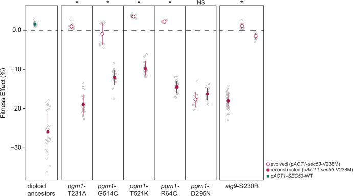
Figure 3—figure supplement 1. Fitness effects of reconstructed mutations and evolved clones containing those same mutations. Average fitness effects and standard deviations of reconstructed heterozygous pgm1 and alg9 mutations (closed circles) compared to evolved clones containing those mutations plus three to nine additional mutations (open circles). Replicate measurements are plotted as gray circles. Note there are two clones from different populations with an alg9-S230R mutation. Asterisks (*) represent statically significant differences between fitness effects of evolved clones and fitness effects of reconstructed clones (Welch’s modified t-test): pgm1-T231A (p < 0.0001, t = 36.1, df = 28.9); pgm1-G514C (p < 0.0001, t = 11.8, df = 18.4); pgm1-T521K (p < 0.0001, t = 33.8, df = 27.3); pgm1-R64C (p < 0.0001, t = 52.9, df = 24.3); pgm1-D295N (p = 0.05, t = 2.10, df = 19.9); alg9-S230R (left) (p < 0.0001, t = 53.7, df = 49.7); alg9-S230R (right) (p < 0.0001, t = 44.1, df = 46.1).
Figure 3—figure supplement 1. Fitness effects of reconstructed mutations and evolved clones containing those same mutations. Average fitness effects and standard deviations of reconstructed heterozygous pgm1 and alg9 mutations (closed circles) compared to evolved clones containing those mutations plus three to nine additional mutations (open circles). Replicate measurements are plotted as gray circles. Note there are two clones from different populations with an alg9-S230R mutation. Asterisks (*) represent statically significant differences between fitness effects of evolved clones and fitness effects of reconstructed clones (Welch’s modified t-test): pgm1-T231A (p < 0.0001, t = 36.1, df = 28.9); pgm1-G514C (p < 0.0001, t = 11.8, df = 18.4); pgm1-T521K (p < 0.0001, t = 33.8, df = 27.3); pgm1-R64C (p < 0.0001, t = 52.9, df = 24.3); pgm1-D295N (p = 0.05, t = 2.10, df = 19.9); alg9-S230R (left) (p < 0.0001, t = 53.7, df = 49.7); alg9-S230R (right) (p < 0.0001, t = 44.1, df = 46.1).CC BY · PubMed Central
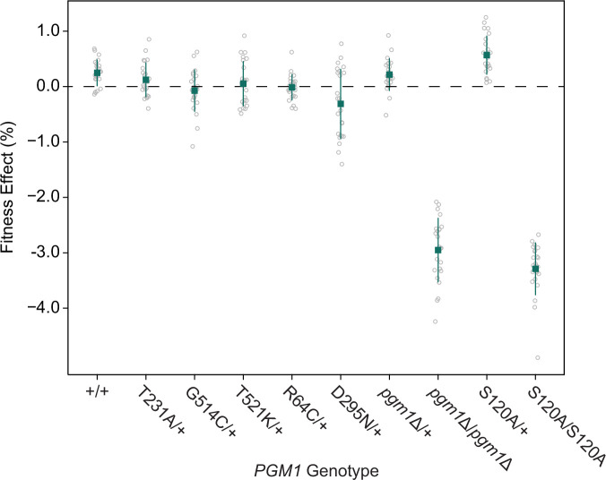
Figure 3—figure supplement 2. Fitness effects of pgm1 mutations in the SEC53-WT background. Average fitness effects and standard deviations of reconstructed pgm1 mutations in the diploid pACT1-SEC53-WT background. Replicate measurements are plotted as gray circles. Plus signs (+) indicate wild-type alleles.
Figure 3—figure supplement 2. Fitness effects of pgm1 mutations in the SEC53-WT background. Average fitness effects and standard deviations of reconstructed pgm1 mutations in the diploid pACT1-SEC53-WT background. Replicate measurements are plotted as gray circles. Plus signs (+) indicate wild-type alleles.CC BY · PubMed Central
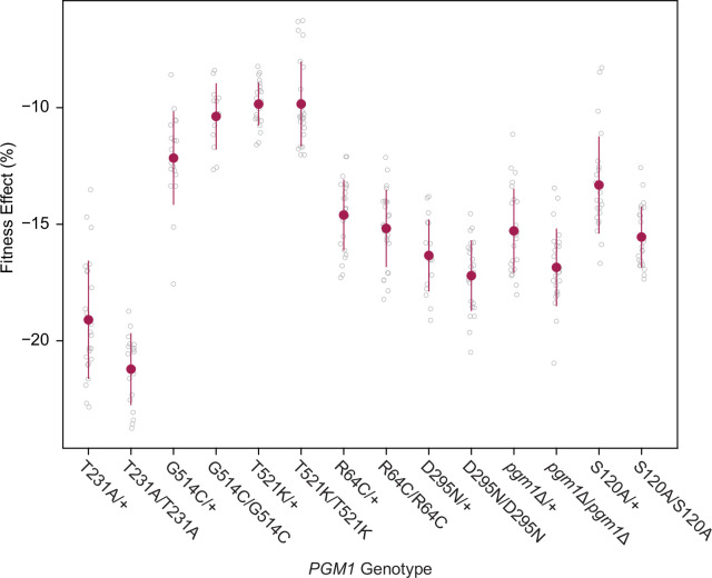
Figure 3—figure supplement 3. Fitness effects of homozygous pgm1 mutations. Average fitness effects and standard deviations of reconstructed pgm1 mutations in the diploid pACT1-sec53-V238M background as heterozygous (mutation/+) or homozygous (mutation/mutation) alleles. Replicate measurements are plotted as gray circles. Comparison of heterozygous and homozygous allele fitness effects (Welch’s modified t-test): pgm1-T231A (p = 0.002, t = 3.35, df = 38.6); pgm1-G514C (p = 0.008, t = 2.85, df = 27.8); pgm1-T521K (p = 0.996, t = 0.005, df = 34.0); pgm1-R64C (p = 0.219, t = 1.24, df = 45.7); pgm1-T231A (p = 0.077, t = 1.84, df = 36.4); pgm1Δ (p = 0.003, t = 3.12, df = 45.7); pgm1-S120A (p < 0.0001, t = 4.45, df = 38.6).
Figure 3—figure supplement 3. Fitness effects of homozygous pgm1 mutations. Average fitness effects and standard deviations of reconstructed pgm1 mutations in the diploid pACT1-sec53-V238M background as heterozygous (mutation/+) or homozygous (mutation/mutation) alleles. Replicate measurements are plotted as gray circles. Comparison of heterozygous and homozygous allele fitness effects (Welch’s modified t-test): pgm1-T231A (p = 0.002, t = 3.35, df = 38.6); pgm1-G514C (p = 0.008, t = 2.85, df = 27.8); pgm1-T521K (p = 0.996, t = 0.005, df = 34.0); pgm1-R64C (p = 0.219, t = 1.24, df = 45.7); pgm1-T231A (p = 0.077, t = 1.84, df = 36.4); pgm1Δ (p = 0.003, t = 3.12, df = 45.7); pgm1-S120A (p < 0.0001, t = 4.45, df = 38.6).CC BY · PubMed Central
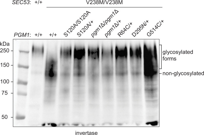
Figure 3—figure supplement 4. Effects of pgm1 mutations on invertase glycosylation in the pACT1-sec53-V238M background. Western blot of invertase from reconstructed sec53/pgm1 strains.
Figure 3—figure supplement 4. Effects of pgm1 mutations on invertase glycosylation in the pACT1-sec53-V238M background. Western blot of invertase from reconstructed sec53/pgm1 strains.CC BY · PubMed Central
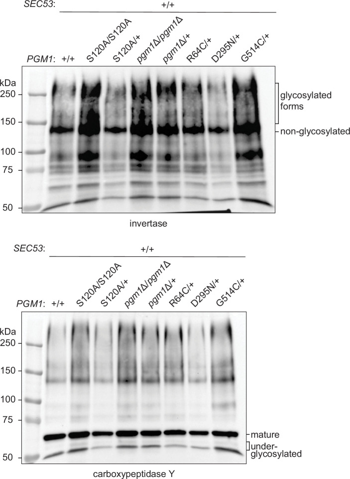
Figure 3—figure supplement 5. Effects of pgm1 mutations on invertase and carboxypeptidase Y (CPY) glycosylation in the pACT1-SEC53-WT background. Western blots of invertase and CPY from constructed SEC53/pgm1 strains.
Figure 3—figure supplement 5. Effects of pgm1 mutations on invertase and carboxypeptidase Y (CPY) glycosylation in the pACT1-SEC53-WT background. Western blots of invertase and CPY from constructed SEC53/pgm1 strains.CC BY · PubMed Central
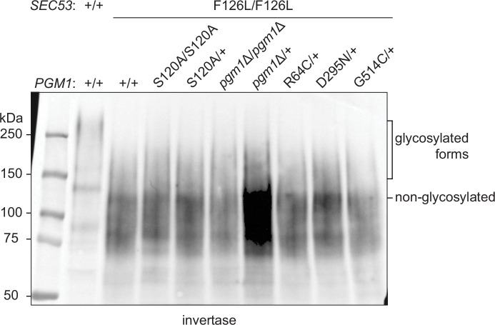
Figure 3—figure supplement 6. Effects of pgm1 mutations on invertase glycosylation in the pACT1-sec53-F126L background. Western blot of invertase from reconstructed sec53/pgm1 strains.
Figure 3—figure supplement 6. Effects of pgm1 mutations on invertase glycosylation in the pACT1-sec53-F126L background. Western blot of invertase from reconstructed sec53/pgm1 strains.CC BY · PubMed Central
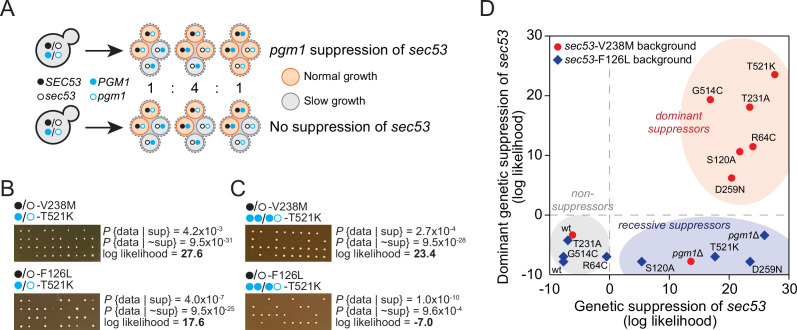
Figure 4. pgm1 mutations are dominant suppressors of sec53-V238M. (A) Diagram of possible spore genotypes in the tetrad dissections. Heterozygous (e.g., SEC53/sec53 PGM1/pgm1) diploid strains could produce one of three tetrad genotypes based on allele segregation. (B) Example of pgm1-T521K dissections in a sec53-V238M background (top) and a sec53-F126L background (bottom). Listed are the probability and log likelihood of suppression, based on the ratio of normal growth (large colonies) to slow growth (no colonies or small colonies). (C) Example of PGM1::pgm1-T521K dissections in a sec53-V238M background (top) and a sec53-F126L background (bottom). (D) Plot of recessive suppression versus dominant suppression of the pgm1 mutations in the sec53-V238M background (red circles) and sec53-F126L background (blue diamonds), based on the tetrad dissections.
Figure 4. pgm1 mutations are dominant suppressors of sec53-V238M. (A) Diagram of possible spore genotypes in the tetrad dissections. Heterozygous (e.g., SEC53/sec53 PGM1/pgm1) diploid strains could produce one of three tetrad genotypes based on allele segregation. (B) Example of pgm1-T521K dissections in a sec53-V238M background (top) and a sec53-F126L background (bottom). Listed are the probability and log likelihood of suppression, based on the ratio of normal growth (large colonies) to slow growth (no colonies or small colonies). (C) Example of PGM1::pgm1-T521K dissections in a sec53-V238M background (top) and a sec53-F126L background (bottom). (D) Plot of recessive suppression versus dominant suppression of the pgm1 mutations in the sec53-V238M background (red circles) and sec53-F126L background (blue diamonds), based on the tetrad dissections.CC BY · PubMed Central
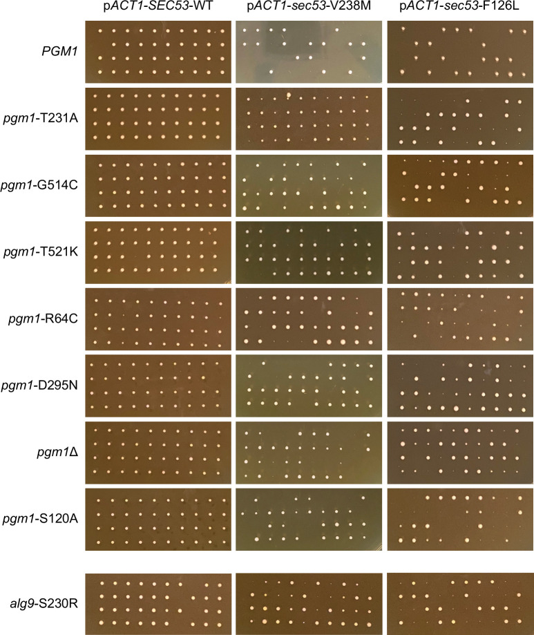
Figure 4—figure supplement 1. Tetrad dissections of PGM1/pgm1 and ALG9/alg9 strains. Ten tetrads were dissected for each strain. Spores from the same tetrad are grouped vertically. The parental diploid strains contained heterozygous SEC53/sec53 alleles and heterozygous PGM1/pgm1 or ALG9/alg9 alleles.
Figure 4—figure supplement 1. Tetrad dissections of PGM1/pgm1 and ALG9/alg9 strains. Ten tetrads were dissected for each strain. Spores from the same tetrad are grouped vertically. The parental diploid strains contained heterozygous SEC53/sec53 alleles and heterozygous PGM1/pgm1 or ALG9/alg9 alleles.CC BY · PubMed Central
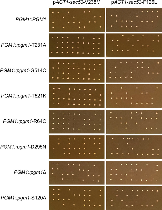
Figure 4—figure supplement 2. Tetrad dissections of PGM1-linked strains. Ten tetrads were dissected for each strain. Spores from the same tetrad are grouped vertically. The parental diploid strains contained heterozygous SEC53/sec53 alleles and four copies of PGM1; a wild-type copy of PGM1 was introduced next to the endogenous PGM1 ORF. Each haploid spore, then, contained either two wild-type copies (PGM1::PGM1) or a wild-type and mutant copy (PGM1::pgm1).
Figure 4—figure supplement 2. Tetrad dissections of PGM1-linked strains. Ten tetrads were dissected for each strain. Spores from the same tetrad are grouped vertically. The parental diploid strains contained heterozygous SEC53/sec53 alleles and four copies of PGM1; a wild-type copy of PGM1 was introduced next to the endogenous PGM1 ORF. Each haploid spore, then, contained either two wild-type copies (PGM1::PGM1) or a wild-type and mutant copy (PGM1::pgm1).CC BY · PubMed Central
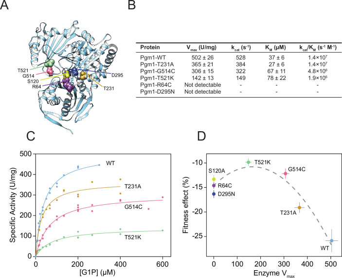
Figure 5. Complete loss of Pgm1 activity overshoots a fitness optimum. (A) AlphaFold structure of S. cerevisiae Pgm1 (Jumper et al., 2021). Mutated residues shown as spheres. (B) Kinetic parameters of recombinant Pgm1 enzymes as determined by a coupled enzymatic assay. Values ± 95% confidence intervals. (C) Michaelis–Menten curves of wild-type and mutant Pgm1. Replicate measurements are plotted as circles. (D) Pgm1 enzyme Vmax versus the corresponding allele’s fitness effect in the diploid pACT1-sec53-V238M background (as shown in Figure 3A). Horizontal and vertical error bars represent 95% confidence intervals. Best fit regression shown as a dashed curve (y = 14.583 + 0.04613x − 0.00014x2, R2 = 0.907, df = 4, F = 37.34, p = 0.0258, analysis of variance [ANOVA]). Note that we did not measure Pgm1-S120A activity and assume null activity.
Figure 5. Complete loss of Pgm1 activity overshoots a fitness optimum. (A) AlphaFold structure of S. cerevisiae Pgm1 (Jumper et al., 2021). Mutated residues shown as spheres. (B) Kinetic parameters of recombinant Pgm1 enzymes as determined by a coupled enzymatic assay. Values ± 95% confidence intervals. (C) Michaelis–Menten curves of wild-type and mutant Pgm1. Replicate measurements are plotted as circles. (D) Pgm1 enzyme Vmax versus the corresponding allele’s fitness effect in the diploid pACT1-sec53-V238M background (as shown in Figure 3A). Horizontal and vertical error bars represent 95% confidence intervals. Best fit regression shown as a dashed curve (y = 14.583 + 0.04613x − 0.00014x2, R2 = 0.907, df = 4, F = 37.34, p = 0.0258, analysis of variance [ANOVA]). Note that we did not measure Pgm1-S120A activity and assume null activity.CC BY · PubMed Central
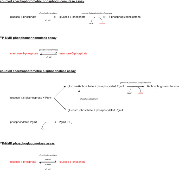
Figure 5—figure supplement 1. Enzymatic assays. Diagrams of the enzymatic reactions assayed in this study.
Figure 5—figure supplement 1. Enzymatic assays. Diagrams of the enzymatic reactions assayed in this study.CC BY · PubMed Central
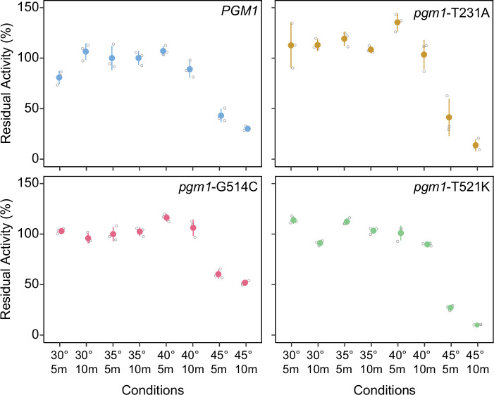
Figure 5—figure supplement 2. Thermostabilities of mutant Pgm1. Average residual activity and standard deviations of recombinant Pgm1 enzymes determined after incubation at the conditions described on the x-axis. Replicate measurements plotted as gray circles. Proteins were incubated in the presence bovine serum albumin (BSA; 0.1 mg/ml).
Figure 5—figure supplement 2. Thermostabilities of mutant Pgm1. Average residual activity and standard deviations of recombinant Pgm1 enzymes determined after incubation at the conditions described on the x-axis. Replicate measurements plotted as gray circles. Proteins were incubated in the presence bovine serum albumin (BSA; 0.1 mg/ml).CC BY · PubMed Central
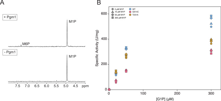
Figure 5—figure supplement 3. Phosphomannomutase activity of mutant Pgm1. (A) Representative phosphomannomutase assay by 31P-NMR spectroscopy. Pgm1 was incubated with 1 mM mannose-1-phosphate (M1P) and 20 μM glucose-1,6-bisphosphate (G16P) for 0 or 60 min. The amount of M1P or M6P was measured by integrating the area of the signals and comparing them to creatine phosphate, added as an internal standard. (B) Spectrophotometric phosphoglucomutase assays were conducted at different concentrations of glucose-1-phosphate (G1P) and M1P. Shapes indicate varying concentrations of M1P and colors indicate Pgm1 variants.
Figure 5—figure supplement 3. Phosphomannomutase activity of mutant Pgm1. (A) Representative phosphomannomutase assay by 31P-NMR spectroscopy. Pgm1 was incubated with 1 mM mannose-1-phosphate (M1P) and 20 μM glucose-1,6-bisphosphate (G16P) for 0 or 60 min. The amount of M1P or M6P was measured by integrating the area of the signals and comparing them to creatine phosphate, added as an internal standard. (B) Spectrophotometric phosphoglucomutase assays were conducted at different concentrations of glucose-1-phosphate (G1P) and M1P. Shapes indicate varying concentrations of M1P and colors indicate Pgm1 variants.CC BY · PubMed Central
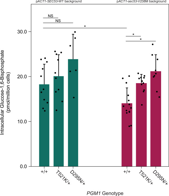
Figure 5—figure supplement 4. pgm1 mutations increase glucose-1,6-bisphosphate (G16P) levels in the pACT1-sec53-V238M background. Average abundance and standard deviations of G16P in SEC53-WT and sec53-V238M strains following metabolite extraction. Plus signs (+) indicate wild-type alleles. Replicate measurements plotted as circles. Asterisks (*) represent statistically significant differences (Mann–Whitney U-test, p < 0.01).
Figure 5—figure supplement 4. pgm1 mutations increase glucose-1,6-bisphosphate (G16P) levels in the pACT1-sec53-V238M background. Average abundance and standard deviations of G16P in SEC53-WT and sec53-V238M strains following metabolite extraction. Plus signs (+) indicate wild-type alleles. Replicate measurements plotted as circles. Asterisks (*) represent statistically significant differences (Mann–Whitney U-test, p < 0.01).CC BY · PubMed Central
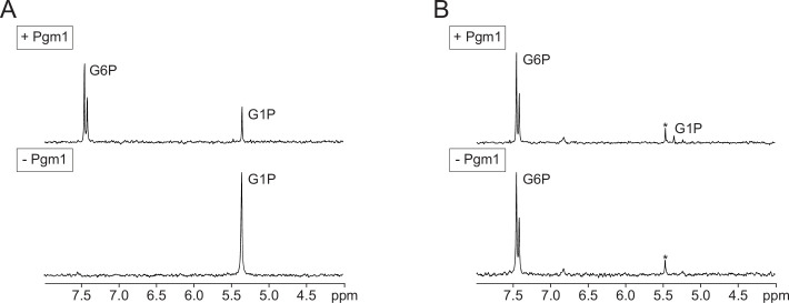
Figure 5—figure supplement 5. Representative forward/reverse phosphoglucomutase assay. Equal amounts of Pgm1 were incubated with glucose-1-phosphate (G1P) (A) or G6P (B) in the presence of glucose-1,6-bisphosphate (G16P) for 0, 5, 10, or 15 min. The amount of the substrates and products was measured by integrating the area of the signals and comparing them to creatine phosphate, added as an internal standard. Asterisks (*) indicate Pi contaminants.
Figure 5—figure supplement 5. Representative forward/reverse phosphoglucomutase assay. Equal amounts of Pgm1 were incubated with glucose-1-phosphate (G1P) (A) or G6P (B) in the presence of glucose-1,6-bisphosphate (G16P) for 0, 5, 10, or 15 min. The amount of the substrates and products was measured by integrating the area of the signals and comparing them to creatine phosphate, added as an internal standard. Asterisks (*) indicate Pi contaminants.CC BY · PubMed Central

Verification

Claims the paper makes

The claims and statistics of this paper haven’t been checked yet.

Run the check — free, no account needed. It reads the open-access text and checks the sources it cites.

A few free checks a day · papers already verified open instantly · results are public

Self-check

Percentages, sums and reported differences re-computed from the paper’s own numbers.

The self-check needs the paper’s full text.

Citations

Cites

Research works printed but not linked: 83

  1. 10.1023/A:1017249723165 · DOI
  2. 10.1371/journal.pone.0139882 · DOI
  3. 10.1073/pnas.1215510110 · DOI
  4. 10.1128/mcb.4.11.2381-2388.1984 · DOI
  5. 10.1007/s10545-014-9757-9 · DOI
  6. 1969, Genetic Control of Phosphoglucomutase Variants in Saccharomyces cerevisiae, J Bacteriol, 98, 532, 10.1128/jb.98.2.532-535.1969 · DOI
  7. 10.1093/bioinformatics/btr670 · DOI
  8. 10.1093/bioinformatics/btu170 · DOI · Paper page
  9. 10.1016/j.gene.2013.07.083 · DOI
  10. 10.1534/genetics.111.130765 · DOI
  11. 10.1002/jimd.12461 · DOI
  12. 10.1073/pnas.93.14.7160 · DOI
  13. 10.1093/glycob/9.6.617 · DOI
  14. 10.1073/pnas.1702314114 · DOI
  15. 10.21037/atm.2018.10.45 · DOI
  16. 10.1093/bioinformatics/btv195 · DOI
  17. 10.4161/fly.19695 · DOI · Paper page
  18. 10.1093/glycob/cwf082 · DOI
  19. 10.1371/journal.pone.0189629 · DOI
  20. 10.3390/ijms19082218 · DOI
  21. 10.1128/mSphere.00121-18 · DOI · Paper page
  22. 10.1126/science.1180823 · DOI · Paper page
  23. 10.1016/j.tibtech.2007.03.010 · DOI
  24. 10.1007/s10545-018-0156-5 · DOI
  25. 10.15252/msb.20156444 · DOI
  26. 10.1371/journal.pgen.1007396 · DOI
  27. 10.1093/glycob/cwj002 · DOI
  28. 10.1086/422367 · DOI
  29. 10.1155/2013/760629 · DOI
  30. 10.1016/S0300-9084(01)01292-5 · DOI
  31. 10.1534/genetics.107.071084 · DOI
  32. Garrison E , Marth G. 2012. Haplotype-based variant detection from short-read sequencing. arXiv: 12073907 [q-bio].
  33. 10.1016/S0021-9258(18)93580-5 · DOI
  34. 10.1002/(SICI)1097-0061(199910)15:14<1541::AID-YEA476>3.0.CO;2-K · DOI
  35. 10.1111/1574-6976.12082 · DOI · Paper page
  36. 10.1093/glycob/7.6.829 · DOI
  37. 10.1186/1471-2148-9-106 · DOI
  38. 10.1111/j.1432-1033.1978.tb12275.x · DOI
  39. 10.1093/molbev/msaa172 · DOI
  40. 10.1242/dmm.040584 · DOI
  41. 10.7554/eLife.63910 · DOI
  42. 10.1126/science.aaa0769 · DOI
  43. 10.1038/sj.ejhg.5200398 · DOI
  44. 10.1073/pnas.84.8.2145 · DOI
  45. 10.1126/science.aao1729 · DOI
  46. 10.1371/journal.pgen.1003972 · DOI
  47. 10.7554/eLife.09638 · DOI
  48. 10.1016/J.CUB.2020.03.046 · DOI
  49. 10.1371/journal.pone.0025290 · DOI
  50. 10.1534/genetics.107.071506 · DOI · Paper page
  51. 10.1038/nature12344 · DOI
  52. 10.1534/g3.118.200934 · DOI
  53. 10.1002/yea.3098 · DOI
  54. 10.1093/bfgp/elv041 · DOI
  55. 10.1093/bioinformatics/btp324 · DOI · Paper page
  56. 10.1107/S0907444997000875 · DOI
  57. 1969, Phosphoglucomutase kinetics with the phosphates of fructose, glucose, mannose, ribose, and galactose, Journal of Biological Chemistry, 244, 910, 10.1016/S0021-9258(18)91872-7 · DOI
  58. 10.1074/jbc.M706818200 · DOI
  59. 10.1038/s41559-018-0503-9 · DOI
  60. 10.1534/genetics.111.137471 · DOI
  61. 10.7554/eLife.23570 · DOI
  62. 10.3390/ijms20174164 · DOI
  63. 10.1186/s12864-017-3952-7 · DOI
  64. 10.1101/2021.12.02.470922 · DOI
  65. 10.1016/S0014-5793(99)00673-0 · DOI
  66. 10.1371/journal.pone.0146021 · DOI
  67. 10.1155/2012/640923 · DOI
  68. 10.1074/jbc.M601505200 · DOI
  69. 10.1016/j.str.2018.07.005 · DOI
  70. 10.1111/febs.14025 · DOI
  71. 10.1016/j.jmb.2016.02.032 · DOI
  72. 10.1016/bs.apcsb.2017.04.005 · DOI
  73. 10.1371/journal.pbio.1001935 · DOI
  74. 10.1093/bib/bbs017 · DOI · Paper page
  75. 10.1093/hmg/dds123 · DOI
  76. 10.1093/glycob/cww086 · DOI · Paper page
  77. 10.1074/jbc.M805224200 · DOI
  78. 10.1016/j.cell.2016.08.002 · DOI
  79. 10.1093/molbev/msab087 · DOI
  80. 10.1093/hmg/11.5.599 · DOI
  81. 10.1021/jo050278f · DOI
  82. 10.1039/D0GC03290E · DOI
  83. 10.1002/(SICI)1096-9888(199904)34:4<311::AID-JMS773>3.0.CO;2-F · DOI

Metadata

Authors
Vignogna, R. C., Allocca, M., Monticelli, M., Norris, J. W., Steet, R., Andreotti, G., Perlstein, E. O., & Lang, G. I.
Affiliations
  • Lehigh University (US)
  • National Academies of Sciences, Engineering, and Medicine (US)
  • Greenwood Genetic Center (US)
  • Istituto di Chimica Biomolecolare (IT)
Corresponding author
Lang, Gregory I.
Venue
eLife
Publisher
openRxiv
Published
2022-04-08
Volume
11
ISSN
2050-084X (ISSN-L)
OpenAlex
W4303986851
Cited by
15
References
1
Citations by year
2025: 3 · 2024: 7 · 2023: 3 · 2022: 2
FWCI
3.01
Type
journal article
Language
en
Open access
Licence
cc-by
Locations
  • eLife (publishedVersion, cc-by)
  • bioRxiv (Cold Spring Harbor Laboratory) (acceptedVersion, cc-by-nc-nd)
  • PubMed Central (publishedVersion, cc-by)
  • PubMed (publishedVersion)
  • DOAJ (DOAJ: Directory of Open Access Journals) (submittedVersion, cc-by-sa)
  • CNR ExploRA (submittedVersion)
Funders
  • National Science Foundation (2019035)
  • Lehigh University
  • National Institutes of Health
  • National Institute of General Medical Sciences (R01GM127420, P20GM139769)
Topics
  • Life Sciences › Biochemistry, Genetics and Molecular Biology › Genetics › Evolution and Genetic Dynamics
  • Life Sciences › Biochemistry, Genetics and Molecular Biology › Molecular Biology › Fungal and yeast genetics research
  • Life Sciences › Biochemistry, Genetics and Molecular Biology › Molecular Biology › Glycosylation and Glycoproteins Research
Keywords
phosphomannomutase deficiency, PMM2, congenital disorders of glycosylation, N-glycosylation, experimental evolution, compensatory mutations, PGM1, phosphoglucomutase
MeSH terms
Humans, Phosphoglucomutase, Saccharomyces cerevisiae, Phosphotransferases (Phosphomutases), Congenital Disorders of Glycosylation, Saccharomyces cerevisiae Proteins, Mutant Proteins

About these checks

Registry, retraction, Cites and statistics results are computed, not judged, with one exception: a printed reference that no index lists is read once by a model before it counts as a citation error. Claims are extracted by a model and kept only when their quote appears verbatim in the text; each cited source is then checked, with a model’s help, for whether it says what the claim relies on. Those verdicts can be wrong — read the claim’s quote and the cited source before relying on one.

Statistics and arithmetic are recomputed, not judged.

Confidence bands are shown only for fields calibrated on our annotated test set. None is calibrated yet, so no band is shown.

What counts as a claim ›How we measure confidence ›How we check this ›

Are you an author, or do you see a problem with this page? Request a review