PreprintCited by 45Bacteriophages and microbial interactionsVibrio bacteria research studiesBacterial Genetics and Biotechnology

The DOI matches its Crossref record; 1 of the 1 references it cites were found in a registry or an index.

The Vibrio cholerae Seventh Pandemic islands act in tandem to defend against a circulating phage

Open access

Preprint — published as 10.1371/journal.pgen.1010250 · Paper page

Abstract

The current circulating pandemic El Tor biotype of Vibrio cholerae has persisted for over sixty years and is characterized by its acquisition of two unique genomic islands called the Vibrio Seventh Pandemic Islands 1 and 2 (VSP-I and VSP-II). However, the functions of most of the genes on VSP-I and VSP-II are unknown and the advantages realized by El Tor through these two islands are not clear. Recent studies have broadly implicated these two mobile genetic elements with phage defense. Still, protection against phage infection through these islands has not been observed directly in any V. cholerae El Tor biotype. Here we report the isolation of a circulating phage from a cholera patient stool sample and demonstrate that propagation of this phage in its native host is inhibited by elements in both VSP-I and VSP-II, providing direct evidence for the role of these genomic islands in phage defense. Moreover, we show that these defense systems are regulated by quorum sensing and active only at certain cell density. Finally, we have isolated a naturally occurring phage variant that is resistant to the defense conferred by the VSP islands, illustrating the countermeasures used by phages to evade these defense mechanisms. Together, this work demonstrates a functional role for the VSPs in V. cholerae and highlights the key regulatory and mechanistic insights that can be gained by studying anti-phage systems in their native contexts. SIGNIFICANCE (AUTHOR SUMMARY): The current pandemic strain of Vibrio cholerae carries two unique genomic islands. How these two islands confer evolutionary advantage to the pathogen is unknown. We show here the identification of a circulating phage that is sensitive to the defense systems present on these two islands and demonstrate how phage variants can evade these defenses. Our studies provide the first direct evidence showing the importance of these genomic islands in defending against phage in their native environments; and in doing so provide novel insight into the mechanisms of these highly conserved defense elements.

Overview

The claims and statistics of this paper haven’t been checked yet.

Run the check — free, no account needed. It reads the open-access text and checks the sources it cites.

A few free checks a day · papers already verified open instantly · results are public

Integrity signals

Registry

Crossref · 10.1101/2022.05.16.492052

Cites

1 of 1 references verified0 citation errors · 0 not checkable

Statistics

Figures

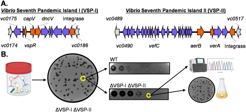
Fig 1 Isolation of VSP-sensitive phages from cholera patient stool samples. (A) Genomic organization of VSP-I &VSP-II, drawn to scale. Genes characterized before this study are marked in orange. Genes on either island with unknown function are blue. Non-VSP genes are grey. (B) Workflow for isolation of VSP sensitive phage. Filter sterilized supernatant from stool samples was plated on a lawn of the ΔVSP-I ΔVSP-II mutant of V. cholerae. Plaques were picked, diluted and spot plated on multiple hosts to identify changes in plaquing. Plaques were picked and plaque purified before sequencing and further downstream assays. This figure is created with Biorender.com.
Fig 1 Isolation of VSP-sensitive phages from cholera patient stool samples. (A) Genomic organization of VSP-I &VSP-II, drawn to scale. Genes characterized before this study are marked in orange. Genes on either island with unknown function are blue. Non-VSP genes are grey. (B) Workflow for isolation of VSP sensitive phage. Filter sterilized supernatant from stool samples was plated on a lawn of the ΔVSP-I ΔVSP-II mutant of V. cholerae. Plaques were picked, diluted and spot plated on multiple hosts to identify changes in plaquing. Plaques were picked and plaque purified before sequencing and further downstream assays. This figure is created with Biorender.com.CC BY · PubMed Central
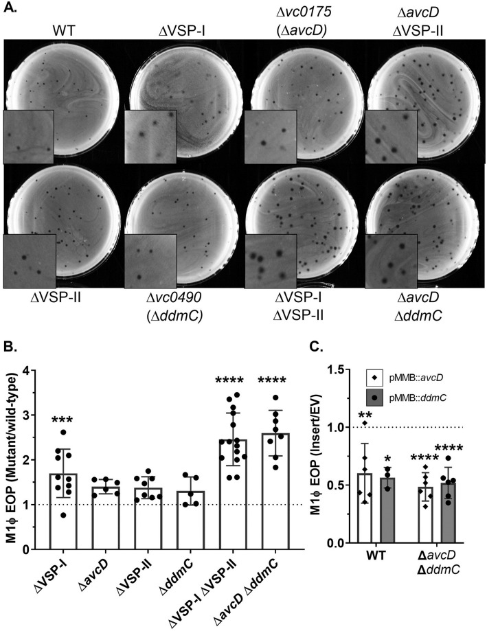
Fig 2 AvcD (VSP-I) and DdmC (VSP-II) inhibit phage plaque formation. (A) Representative images of M1Φ plaqued on 0.7% LB agar with various hosts. (B) Efficiency of plating (EOP) of M1Φ on the indicated hosts vs WT. EOP was calculated by dividing the number of plaques obtained from the indicated mutant strain by the plaques obtained from the parental VSP+ strain (WT). (C) EOP of different V. cholerae strains expressing vc0175 (avcD) or vc0490 (ddmC) from a plasmid. Strains containing plasmid expressing avcD (white bars/diamonds) or plasmid expressing ddmC (grey bars/circles), or plasmid with no insert (EV), on the low copy number plasmid pMMB67eh were infected with M1Φ under inducing conditions in either the WT or ΔavcD ΔddmC background as indicated in the figure. EOP was calculated by dividing the number of plaques from the strains ectopically expressing either avcD or ddmC by the number of plaques from the strains carrying the empty vector. Significance was determined by one-way ANOVA with Dunnett’s multiple comparison test comparing the EOP of each strain to the control of the WT strain. Signifiers for significance were displayed based on p-values as follows: 0.1234(ns), 0.0332(*), 0.0021(**), 0.0002(***), <0.0001(****).
Fig 2 AvcD (VSP-I) and DdmC (VSP-II) inhibit phage plaque formation. (A) Representative images of M1Φ plaqued on 0.7% LB agar with various hosts. (B) Efficiency of plating (EOP) of M1Φ on the indicated hosts vs WT. EOP was calculated by dividing the number of plaques obtained from the indicated mutant strain by the plaques obtained from the parental VSP+ strain (WT). (C) EOP of different V. cholerae strains expressing vc0175 (avcD) or vc0490 (ddmC) from a plasmid. Strains containing plasmid expressing avcD (white bars/diamonds) or plasmid expressing ddmC (grey bars/circles), or plasmid with no insert (EV), on the low copy number plasmid pMMB67eh were infected with M1Φ under inducing conditions in either the WT or ΔavcD ΔddmC background as indicated in the figure. EOP was calculated by dividing the number of plaques from the strains ectopically expressing either avcD or ddmC by the number of plaques from the strains carrying the empty vector. Significance was determined by one-way ANOVA with Dunnett’s multiple comparison test comparing the EOP of each strain to the control of the WT strain. Signifiers for significance were displayed based on p-values as follows: 0.1234(ns), 0.0332(*), 0.0021(**), 0.0002(***), <0.0001(****).CC BY · PubMed Central
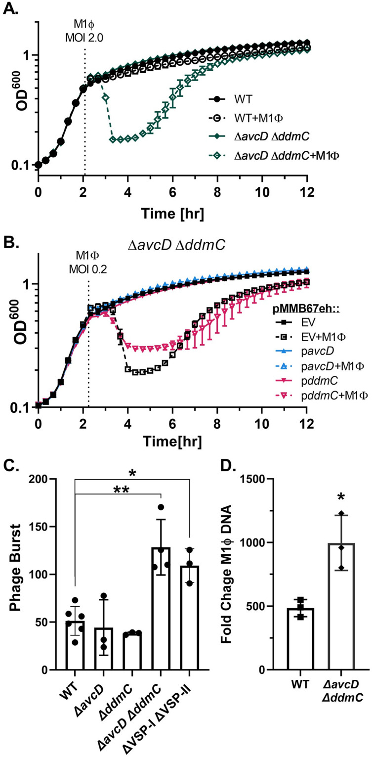
Fig 3 avcD and ddmC protect growing cultures of V. cholerae from M1Φ infection. (A) Growth and lysis curves of V. cholerae. Indicated strains were infected with M1Φ at an MOI of 2.0 at the time displayed by the dotted line. All strains were grown in LB shaking at 37°C and optical density was read at regular intervals with a plate reader. (B) V. cholerae ΔavcD ΔddmC strain containing the plasmid pMMB67eh or the same plasmid expressing avcD or ddmC was grown and infected with M1Φ at an MOI of 0.2. (C) Phage burst assays. Strains were grown to mid-log phase before being infected with phage at an MOI of 0.1. After a period of absorption cultures were further diluted 1/2500,1/25000, and 1/250000. Burst is calculated by dividing the total output phage by absorbed phage (T0-T10). Strains are indicated on the x-axis. P-values were determined by Unpaired T-tests with Welch’s correction and significance shown as *p<0.0332 and **p<0.0021. (D) Fold change in M1Φ genome copy through infection. The indicated strain was grown to mid-log phase before being infected with M1Φ at an MOI of 0.02 and grown in LB at 37°C on a roller. Immediately upon infection and at 20 minutes post infection a portion of the culture was removed and boiled. The boiled culture was then diluted 1:50 and used as template for qPCR with primers targeting the ICP3 DNA polymerase. Fold change was calculated by dividing the CT value at 20minutes by the value at 0 minutes for each culture.
Fig 3 avcD and ddmC protect growing cultures of V. cholerae from M1Φ infection. (A) Growth and lysis curves of V. cholerae. Indicated strains were infected with M1Φ at an MOI of 2.0 at the time displayed by the dotted line. All strains were grown in LB shaking at 37°C and optical density was read at regular intervals with a plate reader. (B) V. cholerae ΔavcD ΔddmC strain containing the plasmid pMMB67eh or the same plasmid expressing avcD or ddmC was grown and infected with M1Φ at an MOI of 0.2. (C) Phage burst assays. Strains were grown to mid-log phase before being infected with phage at an MOI of 0.1. After a period of absorption cultures were further diluted 1/2500,1/25000, and 1/250000. Burst is calculated by dividing the total output phage by absorbed phage (T0-T10). Strains are indicated on the x-axis. P-values were determined by Unpaired T-tests with Welch’s correction and significance shown as *p<0.0332 and **p<0.0021. (D) Fold change in M1Φ genome copy through infection. The indicated strain was grown to mid-log phase before being infected with M1Φ at an MOI of 0.02 and grown in LB at 37°C on a roller. Immediately upon infection and at 20 minutes post infection a portion of the culture was removed and boiled. The boiled culture was then diluted 1:50 and used as template for qPCR with primers targeting the ICP3 DNA polymerase. Fold change was calculated by dividing the CT value at 20minutes by the value at 0 minutes for each culture.CC BY · PubMed Central
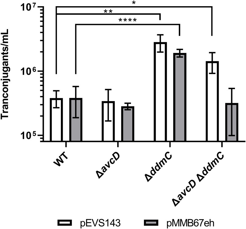
Fig 4 DdmC reduces plasmid conjugation. E. coli donors carrying either pEVS143(white bars) or pMMB67eh (grey bars) were mixed with different V. cholerae recipients as indicated and incubated together for 2.5 hours. Cells were then resuspended and an aliquot was plated on LB containing polymyxin B which is lethal to E. coli and either kanamycin (for pEVS143) or ampicillin (for pMMB67eh) and incubated overnight before the resultant colonies were counted. Significance was determined by Unpaired T tests with Welch’s correction with significance shown as *p<0.0332 and **p<0.0021 ****p<0.0001.
Fig 4 DdmC reduces plasmid conjugation. E. coli donors carrying either pEVS143(white bars) or pMMB67eh (grey bars) were mixed with different V. cholerae recipients as indicated and incubated together for 2.5 hours. Cells were then resuspended and an aliquot was plated on LB containing polymyxin B which is lethal to E. coli and either kanamycin (for pEVS143) or ampicillin (for pMMB67eh) and incubated overnight before the resultant colonies were counted. Significance was determined by Unpaired T tests with Welch’s correction with significance shown as *p<0.0332 and **p<0.0021 ****p<0.0001.CC BY · PubMed Central
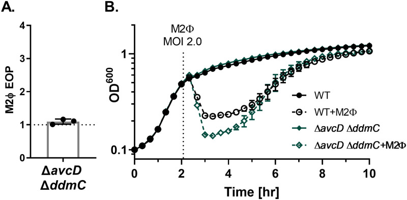
Fig 5 M2Φ is not susceptible to AvcD and DdmC. (A) EOP of M2Φ on ΔavcD ΔddmC compared to the parental VSP+ strain (WT). (B) Growth and lysis curves of the indicated strains with or without M2Φ added at an MOI of 2.0 at the time shown with a dotted line. Uninfected data is the same as shown in Fig 3A.
Fig 5 M2Φ is not susceptible to AvcD and DdmC. (A) EOP of M2Φ on ΔavcD ΔddmC compared to the parental VSP+ strain (WT). (B) Growth and lysis curves of the indicated strains with or without M2Φ added at an MOI of 2.0 at the time shown with a dotted line. Uninfected data is the same as shown in Fig 3A.CC BY · PubMed Central
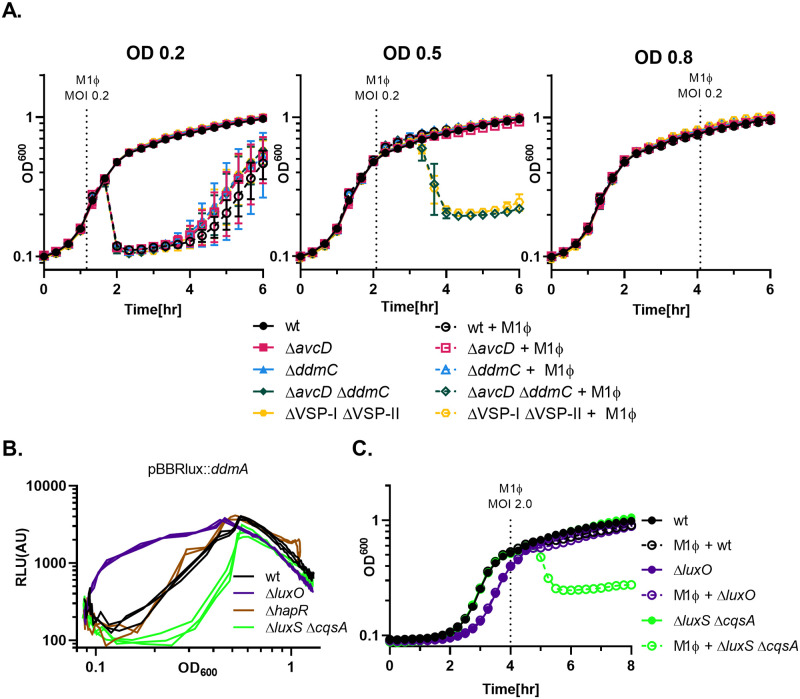
Fig 6 Defense by DdmC is controlled by quorum sensing. (A) Growth and lysis curves of V. cholerae infected by M1Φ at different cell densities. M1Φ was added at an MOI 0.2 at the time indicated with a dotted line that corresponds closely with the OD600 shown at the top of each graph. Strains used for all three graphs are indicated on the far right of the figure. (B) Quorum sensing mutant strains of V. cholerae carrying the reporter plasmid pBBRlux with the lux operon under the control of the region upstream of ddmA were grown at 37°C with shaking. Optical density (OD600) and bioluminescence were measured at regular intervals. Relative luminescence (RLU) was calculated by dividing the luminescence by optical density and was then plotted relative to the OD600 to show the normalized bioluminescence throughout growth. (C) Grow and lysis curves of C6706 (WT) and various quorum sensing mutants upon M1Φ infection. M1Φ was added to the indicated strains at the dotted line at an MOI of 2.0.
Fig 6 Defense by DdmC is controlled by quorum sensing. (A) Growth and lysis curves of V. cholerae infected by M1Φ at different cell densities. M1Φ was added at an MOI 0.2 at the time indicated with a dotted line that corresponds closely with the OD600 shown at the top of each graph. Strains used for all three graphs are indicated on the far right of the figure. (B) Quorum sensing mutant strains of V. cholerae carrying the reporter plasmid pBBRlux with the lux operon under the control of the region upstream of ddmA were grown at 37°C with shaking. Optical density (OD600) and bioluminescence were measured at regular intervals. Relative luminescence (RLU) was calculated by dividing the luminescence by optical density and was then plotted relative to the OD600 to show the normalized bioluminescence throughout growth. (C) Grow and lysis curves of C6706 (WT) and various quorum sensing mutants upon M1Φ infection. M1Φ was added to the indicated strains at the dotted line at an MOI of 2.0.CC BY · PubMed Central

Verification

Claims the paper makes

The claims and statistics of this paper haven’t been checked yet.

Run the check — free, no account needed. It reads the open-access text and checks the sources it cites.

A few free checks a day · papers already verified open instantly · results are public

Self-check

Percentages, sums and reported differences re-computed from the paper’s own numbers.

The self-check needs the paper’s full text.

Citations

Cites

Show all 1 references

Research works printed but not linked: 63

  1. 10.1073/pnas.1608732113 · DOI
  2. 10.1073/pnas.042667999 · DOI
  3. 10.1038/nrmicro2204 · DOI
  4. 10.1073/pnas.0502069102 · DOI
  5. 10.1128/mBio.00334-10 · DOI
  6. 10.1073/pnas.1810138116 · DOI
  7. 10.1038/s41467-016-0009-6 · DOI · Paper page
  8. 10.1371/journal.ppat.1004691 · DOI
  9. 10.1371/journal.pbio.3001514 · DOI
  10. 10.1371/journal.pgen.1010065 · DOI
  11. 10.1371/journal.pgen.1006563 · DOI
  12. 10.1126/science.abg2166 · DOI
  13. Colwell RR , Fidelma E . Crispr-Cas and Contact-Dependent Secretion Systems Present on Excisable Pathogenicity Islands with Conserved Recombination Modules. 2017;199: 1–20. doi:https://doi.org/10.1128/JB.00842-16. · DOI
  14. 10.1007/s11103-011-9767-z · DOI
  15. 10.1073/pnas.1801233115 · DOI
  16. 10.1038/s41586-019-1605-5 · DOI
  17. 10.1038/s41586-019-0953-5 · DOI
  18. Huiting E , Athukoralage J , Guan J , Silas S , Carion H , Bondy-Denomy J . Bacteriophages antagonize cGAS-like immunity in bacteria. bioRxiv. 2022;1: 2022.03.30.486325. doi:https://doi.org/10.1101/2022.03.30.486325 · DOI
  19. Severin GB , Hsueh BY , Elg CA , Dover JA , Rhoades CR , Wessel AJ , et al. A Broadly Conserved Deoxycytidine Deaminase Protects Bacteria from Phage Infection. BioRxiv. 2021. doi:https://doi.org/10.1101/2021.03.31.437871 · DOI
  20. Tal N , Millman A , Stokar-Avihail A , Fedorenko T , Leavitt A , Melamed S , et al. Antiviral defense via nucleotide depletion in bacteria. bioRxiv. 2021; 2021.04.26.441389. doi:https://doi.org/10.1101/2021.04.26.441389 · DOI
  21. 10.1111/j.1574-6968.2010.02008.x · DOI
  22. 10.1128/JB.00704-15 · DOI
  23. Murphy SG , Johnson BA , Ledoux CM , Dörr T . Vibrio cholerae’s mysterious Seventh Pandemic island (VSP-II) encodes novel Zur-regulated zinc starvation genes involved in chemotaxis and autoaggregation. PLoS Genet. 2021. doi:https://doi.org/10.1371/journal.pgen.1009624 June · DOI
  24. 10.1038/s41586-022-04546-y · DOI
  25. 10.1126/science.aba0372 · DOI
  26. 10.1128/JCM.42.10.4657-4671.2004 · DOI
  27. 10.1371/journal.pntd.0008275 · DOI
  28. 10.1126/science.272.5270.1910 · DOI
  29. 10.1111/j.1365-2958.1995.mmi_18040685.x · DOI
  30. 10.1016/j.cell.2012.01.053 · DOI
  31. 10.1007/978-1-60327-164-6_17 · DOI
  32. 10.1186/1471-2180-11-181 · DOI
  33. 10.1073/pnas.1718787115 · DOI
  34. 10.7554/eLife.42057 · DOI
  35. 10.1371/journal.ppat.1004837 · DOI
  36. 10.1016/S0092-8674(02)00829-2 · DOI
  37. 10.1016/j.cell.2004.06.009 · DOI
  38. 10.1073/pnas.0703860104 · DOI
  39. 10.3391/mbi.2016.7.1.01 · DOI
  40. 10.1038/s41598-018-23276-8 · DOI
  41. 10.1093/nar/gkaa726 · DOI
  42. 10.1016/j.chom.2022.03.001 · DOI
  43. 10.1128/mBio.00939-20 · DOI
  44. 10.1146/annurev-virology-011620-040628 · DOI
  45. 10.1038/s41598-016-0001-8 · DOI · Paper page
  46. 10.1073/pnas.1315647110 · DOI
  47. 10.1038/s41467-020-19415-3 · DOI
  48. 10.1126/science.aar4120 · DOI
  49. 10.1073/pnas.69.11.3365 · DOI
  50. 10.1126/science.1225829 · DOI · Paper page
  51. 10.1111/j.1462-2920.2012.02886.x · DOI
  52. 10.3389/fgene.2019.00735 · DOI
  53. 10.1093/infdis/145.3.296 · DOI
  54. 10.1371/journal.ppat.1002917 · DOI
  55. 10.1128/mBio.01028-13 · DOI
  56. 10.1073/pnas.1406478111 · DOI
  57. 10.1128/AEM.00066-06 · DOI
  58. 10.1007/978-1-60327-164-6_18 · DOI
  59. 10.1128/jb.92.3.787-788.1966 · DOI
  60. 10.1128/IAI.64.7.2853-2856.1996 · DOI · Paper page
  61. 10.1128/JB.01307-08 · DOI
  62. 10.1016/0378-1119(86)90358-6 · DOI
  63. 10.1128/AEM.72.1.802-810.2006 · DOI

Metadata

Authors
O’Hara, B. J., Alam, M., & Ng, W.-L.
Affiliations
  • Tufts University (US)
  • International Centre for Diarrhoeal Disease Research (BD)
Corresponding author
Ng, Wai-Leung
Venue
PLoS Genetics
Publisher
openRxiv
Published
2022-05-16
Volume
18
Issue
8
Pages
e1010250-e1010250
ISSN
1553-7390 (ISSN-L)
OpenAlex
W4293155768
Cited by
45
References
1
Citations by year
2026: 7 · 2025: 14 · 2024: 11 · 2023: 10 · 2022: 3
FWCI
5.06
Type
journal article
Language
en
Open access
Licence
cc-by
Locations
  • PLoS Genetics (publishedVersion, cc-by)
  • bioRxiv (Cold Spring Harbor Laboratory) (acceptedVersion, cc-by)
  • PubMed Central (publishedVersion, cc-by)
  • PubMed (publishedVersion)
  • DOAJ (DOAJ: Directory of Open Access Journals) (submittedVersion, cc-by-sa)
  • Greater South Information System
Funders
  • International Centre for Diarrhoeal Disease Research, Bangladesh
  • National Institutes of Health
  • National Institute of General Medical Sciences (T32GM007310)
  • National Institute of Allergy and Infectious Diseases (R01AI121337)
Topics
  • Life Sciences › Immunology and Microbiology › Virology › Bacteriophages and microbial interactions
  • Life Sciences › Immunology and Microbiology › Microbiology › Vibrio bacteria research studies
  • Life Sciences › Biochemistry, Genetics and Molecular Biology › Genetics › Bacterial Genetics and Biotechnology
Keywords
Vibrio cholerae, phage defense, bacteriophages, El Tor biotype, quorum sensing, cell density regulation, phage resistance, mobile genetic elements, genomic islands
MeSH terms
Bacteriophages, Cholera, Humans, Vibrio cholerae O1, Genomic Islands, Pandemics

About these checks

Registry, retraction, Cites and statistics results are computed, not judged, with one exception: a printed reference that no index lists is read once by a model before it counts as a citation error. Claims are extracted by a model and kept only when their quote appears verbatim in the text; each cited source is then checked, with a model’s help, for whether it says what the claim relies on. Those verdicts can be wrong — read the claim’s quote and the cited source before relying on one.

Statistics and arithmetic are recomputed, not judged.

Confidence bands are shown only for fields calibrated on our annotated test set. None is calibrated yet, so no band is shown.

What counts as a claim ›How we measure confidence ›How we check this ›

Are you an author, or do you see a problem with this page? Request a review

How to cite

Cite CoChat for the checks on this page, and the paper for its own findings.

This page
CoChat. (2026). Citation check: The Vibrio cholerae Seventh Pandemic islands act in tandem to defend against a circulating phage. https://app.cochat.ai/papers/10.1101/2022.05.16.492052
The paper
O’Hara, B. J., Alam, M., & Ng, W.-L. (2022). The Vibrio cholerae Seventh Pandemic islands act in tandem to defend against a circulating phage. PLoS Genetics. https://doi.org/10.1101/2022.05.16.492052