

The DOI matches its Crossref record; 1 of the 1 references it cites were found in a registry or an index.
Insight into the structural basis for dual nucleic acid - recognition by the Scaffold Attachment Factor B2 protein
Open accessPreprint — published as 10.3390/ijms24043286 · Paper page
Abstract
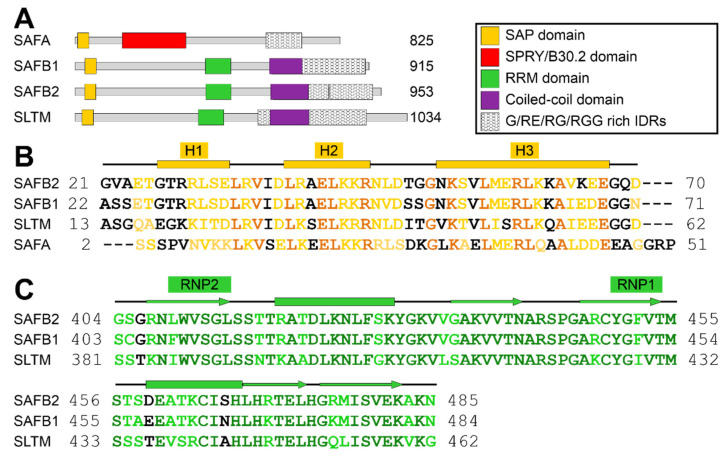
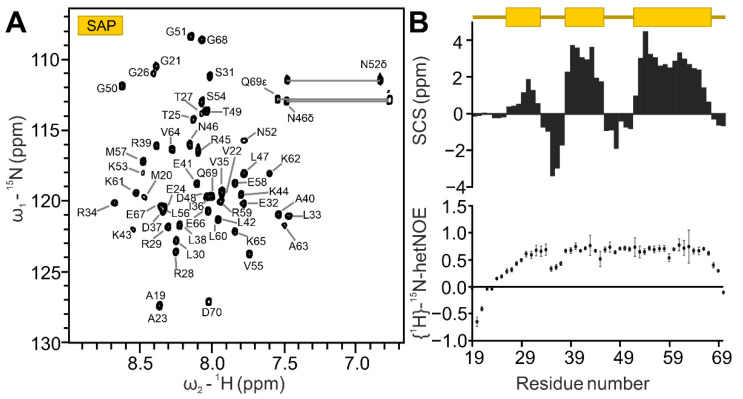
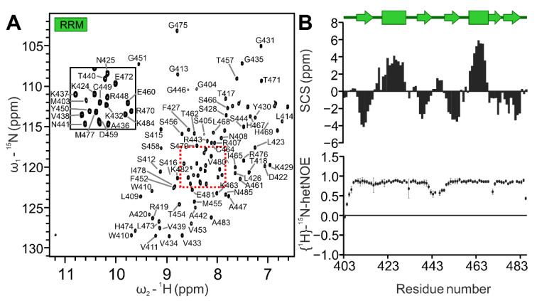
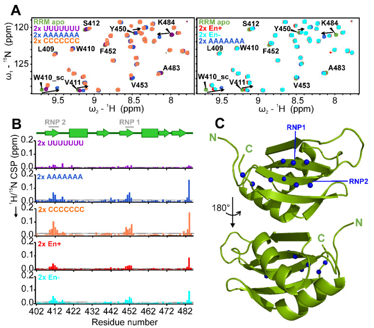
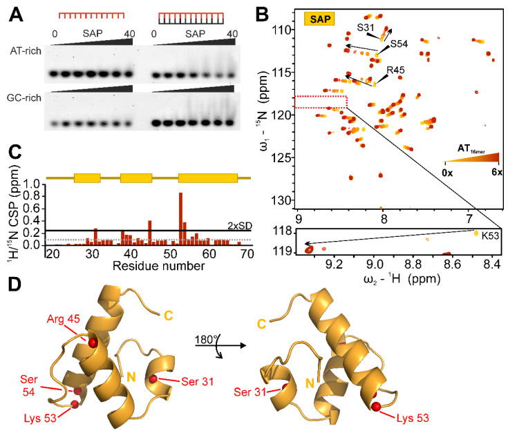
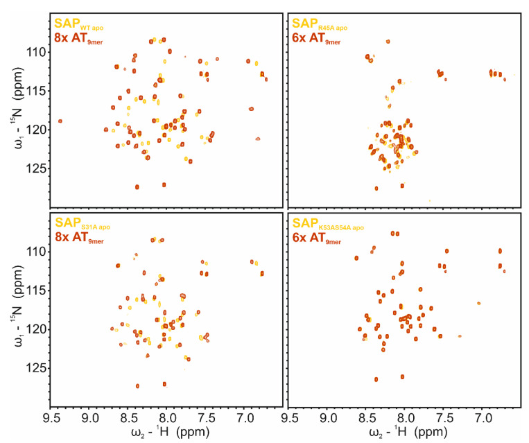
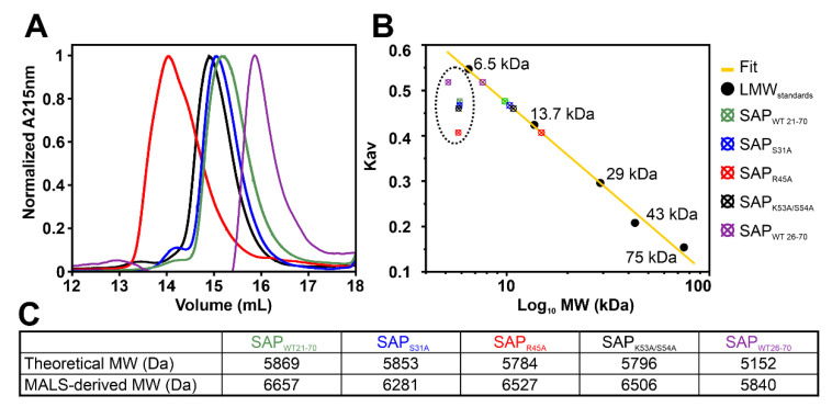
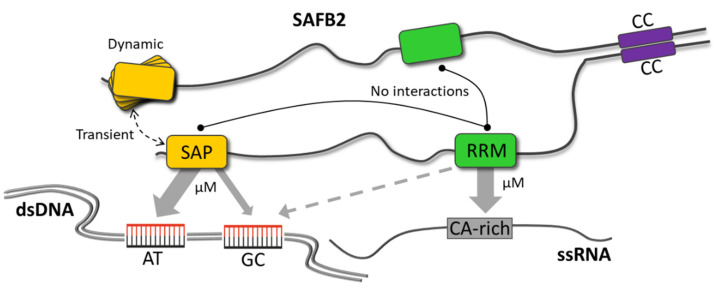
The family of scaffold attachment factor B (SAFB) proteins comprises three members and was first identified as binders of the nuclear matrix/scaffold. Over the past two decades, SAFBs were shown to act in DNA repair, mRNA/(l)ncRNA processing, and as part of protein complexes with chromatin-modifying enzymes. SAFB proteins are approximately-100-kDa-sized dual nucleic acid-binding proteins with dedicated domains in an otherwise largely unstructured context, but whether and how they discriminate DNA- and RNA-binding has remained enigmatic. We here provide the SAFB2 DNA- and RNA-binding SAP and RRM domains in their functional boundaries and use solution NMR spectroscopy to ascribe DNA- and RNA-binding functions. We give insight into their target nucleic acid preferences and map the interfaces with respective nucleic acids on sparse data-derived SAP and RRM domain structures. Further, we provide evidence that the SAP domain exhibits intra-domain dynamics and a potential tendency to dimerise, which may expand its specifically targeted DNA sequence range. Our data provide a first molecular basis of and a starting point towards deciphering DNA- and RNA-binding functions of SAFB2 on the molecular level and serve a basis for understanding its localization to specific regions of chromatin and its involvement in the processing of specific RNA species.
Overview
The claims and statistics of this paper haven’t been checked yet.
Run the check — free, no account needed. It reads the open-access text and checks the sources it cites.
Integrity signals
Registry
Cites
Statistics
Figures
Verification
Claims the paper makes
The claims and statistics of this paper haven’t been checked yet.
Run the check — free, no account needed. It reads the open-access text and checks the sources it cites.
Self-check
Percentages, sums and reported differences re-computed from the paper’s own numbers.
The self-check needs the paper’s full text.
Citations
Cites
Highly accurate protein structure prediction with AlphaFold
Jumper, J., Evans, R., Pritzel, A., Green, T., Figurnov, M., Ronneberger, O., Tunyasuvunakool, K., Bates, R., Žídek, A., Potapenko, A., et al. (2021) · Nature · doi:10.1038/s41586-021-03819-2 · Cited by 43,764
Research works printed but not linked: 82
- 10.1016/j.cell.2019.06.001 · DOI · Paper page
- 10.1038/nrm3884 · DOI · Paper page
- Davis, B.N. ; Hilyard, A.C. ; Nguyen, P.H. ; Lagna, G. ; Hata, A. SMAD proteins bind a conserved RNA sequence to promote microRNA maturation by Drosha. Mol. Cell 2010, 39. · DOI · Paper page
- Shi, L. ; Godfrey, W.R. ; Lin, J. ; Zhao, G. ; Kao, P.N. NF90 regulates inducible IL-2 gene expression in T cells. J. Exp. Med. 2007, 204. · DOI
- Shim, J. ; Lim, H. ; Yates Iii, J. ; Karin, M. Nuclear export of NF90 is required for interleukin-2 mRNA stabilization. Mol. Cell 2002, 10. · DOI
- Kuo, P.H. ; Doudeva, L.G. ; Wang, Y.T. ; Shen, C.K.J. ; Yuan, H.S. Structural insights into TDP43 in nucleic-acid binding and domain interactions. Nucleic Acids Res. 2009, 37. · DOI · Paper page
- Lukavsky, P.J. Molecular basis of UG-rich RNA recognition by the human splicing factor TDP43. Nature Struct. Mol. Biol. 2013, 20. · DOI · Paper page
- Huang, D.B. Crystal structure of NF-κB (p50)2 complexed to a high-affinity RNA aptamer. Proc. Natl Acad. Sci. USA 2003, 100. · DOI
- Müller, C.W. ; Rey, F.A. ; Sodeoka, M. ; Verdine, G.L. ; Harrison, S.C. Structure of the NF-κB p50 homodimer bound to DNA. Nature 1995, 373. · DOI · Paper page
- 10.1002/j.1460-2075.1992.tb05331.x · DOI · Paper page
- 10.1002/j.1460-2075.1992.tb05422.x · DOI · Paper page
- 10.1093/nar/24.5.843 · DOI
- 10.1242/jcs.112.10.1465 · DOI
- 10.3390/ijms22094982 · DOI
- 10.1111/febs.14141 · DOI
- 10.1042/BCJ20160649 · DOI
- 10.1016/j.molcel.2019.10.001 · DOI
- 10.1093/nar/gkt285 · DOI
- 10.1042/BST20120030 · DOI
- 10.1007/s00395-021-00854-y · DOI
- 10.1073/pnas.2005712117 · DOI
- 2021, Scaffold attachment factor B1 regulates androgen degradation pathways in prostate cancer, Am J Clin Exp Urol, 9, 337
- 10.1016/j.molcel.2020.05.011 · DOI
- 10.1038/s41422-021-00515-8 · DOI
- 10.1002/jcb.10685 · DOI
- 10.1002/jcb.24182 · DOI
- 10.1016/j.febslet.2010.11.054 · DOI
- 10.1016/j.molcel.2020.01.026 · DOI
- 10.15252/embj.2019104154 · DOI
- 10.17989/ENCSR558RBK · DOI
- 10.1016/j.molcel.2014.04.016 · DOI · Paper page
- 10.1016/j.sbi.2008.04.002 · DOI · Paper page
- Maris, C. ; Dominguez, C. ; Allain, F.H.T. The RNA recognition motif, a plastic RNA-binding platform to regulate post-transcriptional gene expression. FEBS J. 2005, 272. · DOI · Paper page
- 10.17989/ENCSR072VUO · DOI
- 10.1093/nar/gkaa1100 · DOI · Paper page
- 10.1038/msb.2011.75 · DOI · Paper page
- 10.1016/S0968-0004(99)01537-6 · DOI · Paper page
- 10.1016/j.jbc.2022.102268 · DOI
- 10.1007/BF00211777 · DOI · Paper page
- 10.1007/s12104-020-09971-6 · DOI
- 10.1007/s12104-020-09995-y · DOI
- 10.1021/bi00213a010 · DOI
- 10.1021/bi300539t · DOI
- 10.1016/j.pnmrs.2018.03.002 · DOI
- 10.1073/pnas.1203013109 · DOI
- 10.1093/nar/gkq366 · DOI · Paper page
- 10.1002/prot.21973 · DOI
- 10.1073/pnas.1616745113 · DOI
- 10.1006/jmbi.2001.4592 · DOI
- 10.1017/S0033583515000207 · DOI
- 10.1038/s41467-019-09769-8 · DOI
- 10.1093/nar/gkac212 · DOI
- 10.1093/nar/gkm615 · DOI
- 10.1074/jbc.M105238200 · DOI
- 10.1074/jbc.M212988200 · DOI
- 10.1242/jcs.03344 · DOI
- 10.1016/j.redox.2022.102545 · DOI
- 10.1016/j.sbi.2012.11.006 · DOI
- 10.1016/j.pnmrs.2014.05.003 · DOI
- 10.1093/nar/gkv1303 · DOI
- 10.1038/s41467-022-33552-x · DOI
- 10.1073/pnas.1303445110 · DOI
- 10.1261/rna.065649.118 · DOI
- 10.26508/lsa.202000825 · DOI
- 10.1038/380646a0 · DOI
- 10.1016/j.str.2011.05.002 · DOI
- 10.1016/j.str.2021.05.001 · DOI
- 10.1074/jbc.RA120.015824 · DOI
- 10.1038/nature06152 · DOI
- 10.1093/nar/28.1.235 · DOI · Paper page
- 10.1002/wrna.1107 · DOI
- 10.1093/bib/bbab195 · DOI
- 10.1038/s41467-022-30183-0 · DOI
- 10.1186/1472-6750-9-61 · DOI · Paper page
- 10.1093/nar/gkaa1013 · DOI
- 10.7554/eLife.01456 · DOI · Paper page
- 1994, Chemical-Shifts as a Tool for Structure Determination, Nuclear Magnetic Resonance, Pt C; Methods in Enzymology, 239, 363, 10.1016/S0076-6879(94)39014-2 · DOI · Paper page
- 10.1016/0022-2364(87)90269-1 · DOI
- 2005, The CCPN data model for NMR spectroscopy: Development of a software pipeline, Proteins-Structure Function and Bioinformatics, 59, 687, 10.1002/prot.20449 · DOI · Paper page
- 10.1093/bioinformatics/btu830 · DOI · Paper page
- 10.1073/pnas.0800256105 · DOI · Paper page
- 10.1371/journal.pone.0077245 · DOI
Metadata
- Authors
- Korn, S. M., von Ehr, J., Dhamotharan, K., Tants, J.-N., Abele, R., & Schlundt, A.
- Affiliations
- Goethe University Frankfurt (DE)
- Corresponding author
- Schlundt, Andreas
- ORCID
- Venue
- International Journal of Molecular Sciences
- Publisher
- openRxiv
- Published
- 2022-12-29
- Volume
- 24
- Issue
- 4
- Pages
- 3286-3286
- ISSN
- 1422-0067 (ISSN-L)
- PMC
- PMC9958909
- OpenAlex
- W4319600471
- Cited by
- 4
- References
- 1
- Citations by year
- 2026: 1 · 2025: 1 · 2024: 2
- FWCI
- 0.36
- Type
- journal article
- Language
- en
- Open access
- gold
- doi.org
- Licence
- cc-by
- Locations
- International Journal of Molecular Sciences (publishedVersion, cc-by)
- bioRxiv (Cold Spring Harbor Laboratory) (acceptedVersion, cc-by-nc)
- PubMed Central (publishedVersion, cc-by)
- PubMed (publishedVersion)
- Publication Server of Goethe University Frankfurt am Main (Goethe University Frankfurt) (submittedVersion, cc-by-nc)
- Publication Server of Goethe University Frankfurt am Main (Goethe University Frankfurt) (submittedVersion, cc-by)
- Funders
- Johanna Quandt Young Academy (2019/AS01)
- Deutsche Forschungsgemeinschaft (2019/AS01, SCHL2062/2-1)
- University of California, San Diego
- Topics
- Life Sciences › Biochemistry, Genetics and Molecular Biology › Molecular Biology › Protein Structure and Dynamics
- Life Sciences › Biochemistry, Genetics and Molecular Biology › Molecular Biology › Genomics and Chromatin Dynamics
- Life Sciences › Biochemistry, Genetics and Molecular Biology › Molecular Biology › RNA Research and Splicing
- Keywords
- nucleic acid recognition, DNA binding, RNA binding, SAP domain, RRM domain, domain structure, protein dimerization, chromatin, nuclear matrix, RNA processing, DNA repair
- MeSH terms
- Base Sequence, Binding Sites, Chromatin, Magnetic Resonance Spectroscopy, RNA, RNA, Messenger
About these checks
Registry, retraction, Cites and statistics results are computed, not judged, with one exception: a printed reference that no index lists is read once by a model before it counts as a citation error. Claims are extracted by a model and kept only when their quote appears verbatim in the text; each cited source is then checked, with a model’s help, for whether it says what the claim relies on. Those verdicts can be wrong — read the claim’s quote and the cited source before relying on one.
Statistics and arithmetic are recomputed, not judged.
Confidence bands are shown only for fields calibrated on our annotated test set. None is calibrated yet, so no band is shown.
What counts as a claim ›How we measure confidence ›How we check this ›
Are you an author, or do you see a problem with this page? Request a review
How to cite
Cite CoChat for the checks on this page, and the paper for its own findings.
- This page
- CoChat. (2026). Citation check: Insight into the structural basis for dual nucleic acid - recognition by the Scaffold Attachment Factor B2 protein. https://app.cochat.ai/papers/10.1101/2022.12.28.522114
- The paper
- Korn, S. M., von Ehr, J., Dhamotharan, K., Tants, J.-N., Abele, R., & Schlundt, A. (2022). Insight into the structural basis for dual nucleic acid - recognition by the Scaffold Attachment Factor B2 protein. International Journal of Molecular Sciences. https://doi.org/10.1101/2022.12.28.522114
Data sources
Bibliographic data: Semantic Scholar, OpenAlex, Crossref.
Contains information from the Semantic Scholar Academic Graph Datasets, made available under the ODC Attribution License (ODC-BY 1.0).
Semantic Scholar’s data is described in: Kinney, R., et al. (2023). The Semantic Scholar Open Data Platform. arXiv:2301.10140.
