PreprintCited by 20RNA and protein synthesis mechanismsRNA modifications and cancerAntibiotic Resistance in Bacteria

The DOI matches its Crossref record; 1 of the 1 references it cites were found in a registry or an index.

30S subunit recognition and G1405 modification by the aminoglycoside-resistance 16S ribosomal RNA methyltransferase RmtC

Open access

Preprint — published as 10.1073/pnas.2304128120 · Paper page

Abstract

Acquired ribosomal RNA (rRNA) methylation has emerged as a significant mechanism of aminoglycoside resistance in pathogenic bacterial infections. Modification of a single nucleotide in the ribosome decoding center by the aminoglycoside-resistance 16S rRNA (m 7 G1405) methyltransferases effectively blocks the action of all 4,6-deoxystreptamine ring-containing aminoglycosides, including the latest generation of drugs. To define the molecular basis of 30S subunit recognition and G1405 modification by these enzymes, we used a S -adenosyl-L-methionine (SAM) analog to trap the complex in a post-catalytic state to enable determination of an overall 3.0 Å cryo-electron microscopy structure of the m 7 G1405 methyltransferase RmtC bound to the mature Escherichia coli 30S ribosomal subunit. This structure, together with functional analyses of RmtC variants, identifies the RmtC N-terminal domain as critical for recognition and docking of the enzyme on a conserved 16S rRNA tertiary surface adjacent to G1405 in 16S rRNA helix 44 (h44). To access the G1405 N7 position for modification, a collection of residues across one surface of RmtC, including a loop that undergoes a disorder to order transition upon 30S subunit binding, induces significant distortion of h44. This distortion flips G1405 into the enzyme active site where it is positioned for modification by two almost universally conserved RmtC residues. These studies expand our understanding of ribosome recognition by rRNA modification enzymes and present a more complete structural basis for future development of strategies to inhibit m 7 G1405 modification to re-sensitize bacterial pathogens to aminoglycosides. Significance: Increasing prevalence of bacterial antibiotic resistance threatens our ability to treat bacterial infections and with it, many other facets of modern healthcare. For the ribosome-targeting aminoglycoside antibiotics, diverse pathogenic bacteria have acquired ribosomal RNA (rRNA) methyltransferase enzymes that confer exceptionally high-level resistance through site-specific modification of the drug binding site. Here, we define the molecular basis for ribosomal substrate recognition and modification by an enzyme (RmtC) representing the most clinically prevalent methyltransferase family. Specifically, RmtC exploits a conserved rRNA surface for binding and induces significant disruption of the rRNA structure to capture the target nucleotide for modification via a “base flipping” mechanism. These insights also present a platform for methyltransferase inhibitor development to extend usefulness of aminoglycoside antibiotics.

Overview

The claims and statistics of this paper haven’t been checked yet.

Run the check — free, no account needed. It reads the open-access text and checks the sources it cites.

A few free checks a day · papers already verified open instantly · results are public

Integrity signals

Registry

Crossref · 10.1101/2023.03.13.532395

Cites

1 of 1 references verified0 citation errors · 0 not checkable

Statistics

Figures

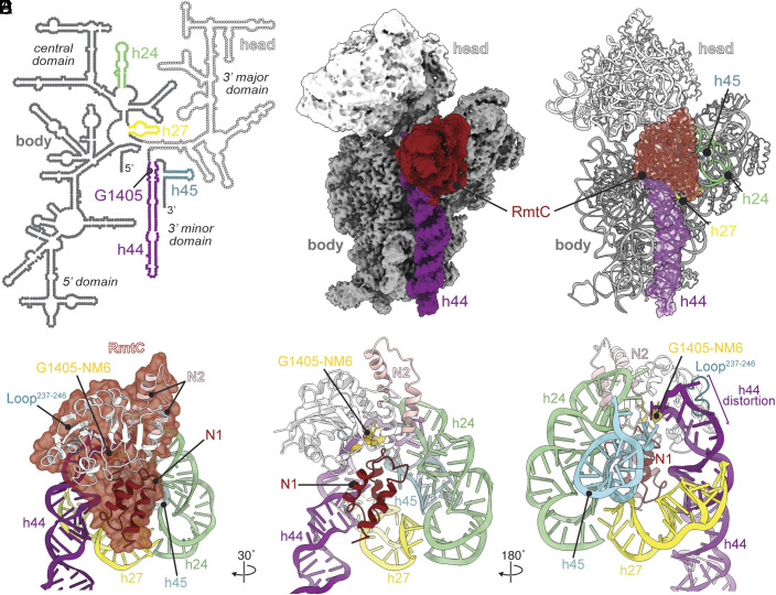
Fig. 1. RmtC engages with a conserved 16S rRNA tertiary surface. (A) Secondary structure of the E. coli 16S rRNA, highlighting the modified nucleotide G1405 within h44 (purple), and other rRNA helices, h24 (green), h27 (yellow), and h45 (cyan), that comprise the conserved 16S rRNA surface recognized by RmtC. (B) Postprocessed cryo-EM map of RmtC (red) bound to the 30S subunit with h44 (purple), body (dark gray) and head (white) domains indicated. (C) Model of the 30S–RmtC complex, with RmtC and h44 shown as cartoon with a semitransparent surface representation. (D) RmtC (red semitransparent surface) docks on the 30S subunit via interactions with helices h24, h27, h44, and h45 made by both N-terminal subdomains (N1, dark red and N2, pink), and the C-terminal domain (white), including the Loop237–246 region (teal). (E and F), Two additional views of RmtC on the 16S rRNA surface.
Fig. 1. RmtC engages with a conserved 16S rRNA tertiary surface. (A) Secondary structure of the E. coli 16S rRNA, highlighting the modified nucleotide G1405 within h44 (purple), and other rRNA helices, h24 (green), h27 (yellow), and h45 (cyan), that comprise the conserved 16S rRNA surface recognized by RmtC. (B) Postprocessed cryo-EM map of RmtC (red) bound to the 30S subunit with h44 (purple), body (dark gray) and head (white) domains indicated. (C) Model of the 30S–RmtC complex, with RmtC and h44 shown as cartoon with a semitransparent surface representation. (D) RmtC (red semitransparent surface) docks on the 30S subunit via interactions with helices h24, h27, h44, and h45 made by both N-terminal subdomains (N1, dark red and N2, pink), and the C-terminal domain (white), including the Loop237–246 region (teal). (E and F), Two additional views of RmtC on the 16S rRNA surface.CC BY-NC-ND · PubMed Central
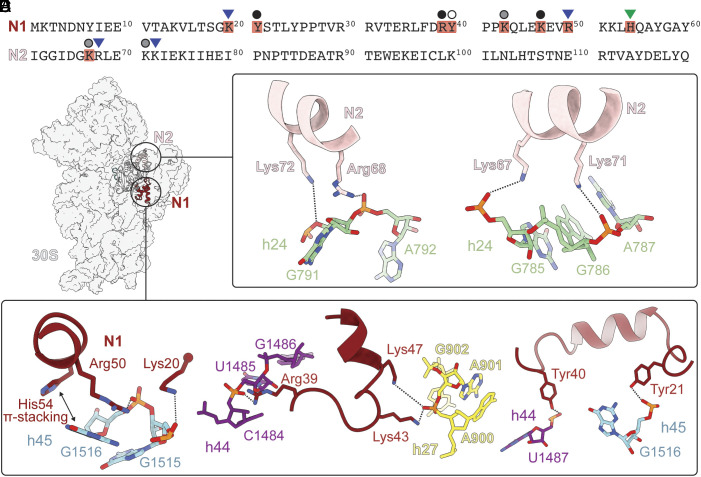
Fig. 2. RmtC N-terminal domain residues direct recognition and binding to the 16S rRNA. (A) Sequences of the RmtC N1 and N2 subdomains, highlighting conserved residues among m7G1405 methyltransferases (SI Appendix, Table S1) observed to interact with 16S rRNA (red shading) and indicating those previously (24) found to be important for both binding and RmtC activity (blue triangle) or activity only (green triangle). New interactions identified and tested in MIC assays in the present work (Table 2) are indicated with circles with shading indicating the impact of substitutions on RmtC activity: major loss (black), modest loss (gray), or no effect (white). (B) N1 subdomain residues Lys20 and Arg50 (red) form electrostatic interactions with the phosphate backbone of h45 nucleotides G1515 and G1516 (cyan), respectively, while His54 forms a face-edge base interaction with G1516. (C) Arg39, Lys43, and Lys47 form electrostatic interactions with U1485 (h44; purple) and A901 (h27; yellow). (D) Tyr21 and Tyr40 (red) interact with nucleotides G1516 (h45; cyan) and G1487 (h44; purple), respectively. (E) N2 subdomain residues Lys72 and Arg68 (pink) form electrostatic interactions with the phosphate backbone of h25 nucleotides A792 and G791 (green). (F) Lys67 and Lys71 interact with the phosphate backbones of h24 nucleotides G785 and A787, respectively.
Fig. 2. RmtC N-terminal domain residues direct recognition and binding to the 16S rRNA. (A) Sequences of the RmtC N1 and N2 subdomains, highlighting conserved residues among m7G1405 methyltransferases (SI Appendix, Table S1) observed to interact with 16S rRNA (red shading) and indicating those previously (24) found to be important for both binding and RmtC activity (blue triangle) or activity only (green triangle). New interactions identified and tested in MIC assays in the present work (Table 2) are indicated with circles with shading indicating the impact of substitutions on RmtC activity: major loss (black), modest loss (gray), or no effect (white). (B) N1 subdomain residues Lys20 and Arg50 (red) form electrostatic interactions with the phosphate backbone of h45 nucleotides G1515 and G1516 (cyan), respectively, while His54 forms a face-edge base interaction with G1516. (C) Arg39, Lys43, and Lys47 form electrostatic interactions with U1485 (h44; purple) and A901 (h27; yellow). (D) Tyr21 and Tyr40 (red) interact with nucleotides G1516 (h45; cyan) and G1487 (h44; purple), respectively. (E) N2 subdomain residues Lys72 and Arg68 (pink) form electrostatic interactions with the phosphate backbone of h25 nucleotides A792 and G791 (green). (F) Lys67 and Lys71 interact with the phosphate backbones of h24 nucleotides G785 and A787, respectively.CC BY-NC-ND · PubMed Central
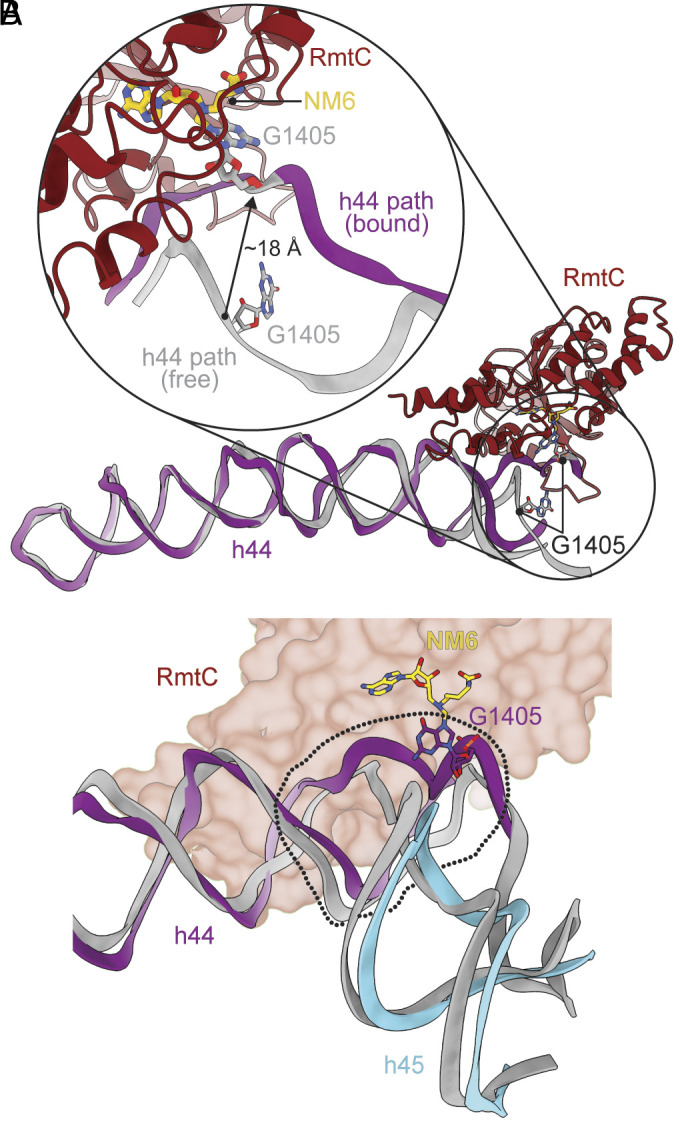
Fig. 3. RmtC binding induces distortion of h44 surrounding the G1405 target nucleotide. (A) RmtC (red) binds to h44 surrounding G1405, inducing a disruption of the h44 rRNA path that results in G1405 moving ~18 Å and with a rotation around its phosphodiester backbone to flip the nucleobase into the enzyme active site for modification. The region of h44 distal from RmtC does not significantly differ in structure from the free mature 30S subunit (PDB ID 7OE1). (B) Only the region of h44 proximal to G1405 (within the dotted outline) is distorted upon RmtC binding; the remainder of h44 (purple) and h45 (cyan) is essentially unchanged compared to the free mature 30S subunit.
Fig. 3. RmtC binding induces distortion of h44 surrounding the G1405 target nucleotide. (A) RmtC (red) binds to h44 surrounding G1405, inducing a disruption of the h44 rRNA path that results in G1405 moving ~18 Å and with a rotation around its phosphodiester backbone to flip the nucleobase into the enzyme active site for modification. The region of h44 distal from RmtC does not significantly differ in structure from the free mature 30S subunit (PDB ID 7OE1). (B) Only the region of h44 proximal to G1405 (within the dotted outline) is distorted upon RmtC binding; the remainder of h44 (purple) and h45 (cyan) is essentially unchanged compared to the free mature 30S subunit.CC BY-NC-ND · PubMed Central
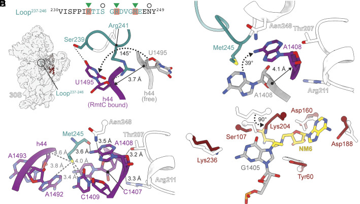
Fig. 4. RmtC Loop237–246 and adjacent CTD residues direct functionally essential alterations in h44 structure. (A) Sequence of RmtC Loop237–246, highlighting conserved residues among m7G1405 methyltransferases (SI Appendix, Table S1) observed to interact with 16S rRNA (red shading) and indicating those previously found (24) to be important for RmtC activity (green triangle). New interactions identified and tested in MIC assays in the present work (Table 2) are indicated with circles. (B) Overview of the 30S–RmtC complex with Loop237–246 (teal) circled. (C) Interaction of RmtC residues Ser239 and Arg241 promotes movement and flipping of nucleotide U1495, to accommodate the distorted position of G1405 required for modification. (D) The Met245 sidechain (teal) forms a network of van der Waals interactions with a h44 rRNA pocket comprising C1407, A1408, C1409, A1492, and A1493 (purple). The Met245 backbone and Thr208, Arg211, and Asn248 side chains also coordinate A1408 with multiple interactions. Thr207 and Asn248 (white) form interactions with h44 nucleotide A1408 (purple), while Arg211 (white) contacts the phosphate backbone. Note that Thr208, Arg211, and Asn248 are outside of Loop237–246 but are located in the CTD and close in the RmtC structure. (E) Changes in h44 structure around A1408 promoted by RmtC residues Met245, Thr207, Arg211, and Asn248. (F) Residues in the 30S–RmtC complex structure (red) are largely found in the same orientation as in the free RmtC–SAH complex (white; PDB ID 6PQB). Only Lys204 adopts a significantly altered position when RmtC binds to the 30S subunit, with the sidechain rotated ~90° and oriented toward to the G1405 (grey) base N7 modification site.
Fig. 4. RmtC Loop237–246 and adjacent CTD residues direct functionally essential alterations in h44 structure. (A) Sequence of RmtC Loop237–246, highlighting conserved residues among m7G1405 methyltransferases (SI Appendix, Table S1) observed to interact with 16S rRNA (red shading) and indicating those previously found (24) to be important for RmtC activity (green triangle). New interactions identified and tested in MIC assays in the present work (Table 2) are indicated with circles. (B) Overview of the 30S–RmtC complex with Loop237–246 (teal) circled. (C) Interaction of RmtC residues Ser239 and Arg241 promotes movement and flipping of nucleotide U1495, to accommodate the distorted position of G1405 required for modification. (D) The Met245 sidechain (teal) forms a network of van der Waals interactions with a h44 rRNA pocket comprising C1407, A1408, C1409, A1492, and A1493 (purple). The Met245 backbone and Thr208, Arg211, and Asn248 side chains also coordinate A1408 with multiple interactions. Thr207 and Asn248 (white) form interactions with h44 nucleotide A1408 (purple), while Arg211 (white) contacts the phosphate backbone. Note that Thr208, Arg211, and Asn248 are outside of Loop237–246 but are located in the CTD and close in the RmtC structure. (E) Changes in h44 structure around A1408 promoted by RmtC residues Met245, Thr207, Arg211, and Asn248. (F) Residues in the 30S–RmtC complex structure (red) are largely found in the same orientation as in the free RmtC–SAH complex (white; PDB ID 6PQB). Only Lys204 adopts a significantly altered position when RmtC binds to the 30S subunit, with the sidechain rotated ~90° and oriented toward to the G1405 (grey) base N7 modification site.CC BY-NC-ND · PubMed Central
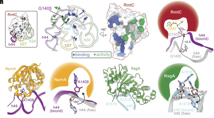
Fig. 5. Mechanism of substrate recognition and modification by RmtC and other structurally characterized 16S rRNA methyltransferases. (A) Views of the 16S rRNA and RmtC interaction interface surfaces highlighting locations of contacts important for both enzyme–substrate binding affinity and thus enzyme activity (blue), or enzyme activity only (green). (B) Major distortion of h44 is required to flip G1405 into the RmtC active site. (C) Overview of NpmA (orange) interaction with h44 (Left; PDB ID 4OX9) and zoomed view of the limited local distortion at A1408 necessary to flip the target nucleotide into the NpmA active site (Right). (D) Overview of KsgA (green) interaction with h45 (Left; PDB ID 7O5H) and zoomed view of the distortion at A1518/ A1519 necessary to flip the target nucleotide (A1519) into the KsgA active site.
Fig. 5. Mechanism of substrate recognition and modification by RmtC and other structurally characterized 16S rRNA methyltransferases. (A) Views of the 16S rRNA and RmtC interaction interface surfaces highlighting locations of contacts important for both enzyme–substrate binding affinity and thus enzyme activity (blue), or enzyme activity only (green). (B) Major distortion of h44 is required to flip G1405 into the RmtC active site. (C) Overview of NpmA (orange) interaction with h44 (Left; PDB ID 4OX9) and zoomed view of the limited local distortion at A1408 necessary to flip the target nucleotide into the NpmA active site (Right). (D) Overview of KsgA (green) interaction with h45 (Left; PDB ID 7O5H) and zoomed view of the distortion at A1518/ A1519 necessary to flip the target nucleotide (A1519) into the KsgA active site.CC BY-NC-ND · PubMed Central

Verification

Claims the paper makes

The claims and statistics of this paper haven’t been checked yet.

Run the check — free, no account needed. It reads the open-access text and checks the sources it cites.

A few free checks a day · papers already verified open instantly · results are public

Self-check

Percentages, sums and reported differences re-computed from the paper’s own numbers.

The self-check needs the paper’s full text.

Citations

Cites

Research works printed but not linked: 48

  1. E. Peterson , P. Kaur , Antibiotic Resistance Mechanisms in Bacteria: Relationships Between Resistance Determinants of Antibiotic Producers, Environmental Bacteria, and Clinical Pathogens. Frontiers in Microbiology 9 (2018). · DOI
  2. 10.1039/c5md00344j · DOI
  3. 10.1101/cshperspect.a027029 · DOI
  4. 10.1261/rna.2314311 · DOI
  5. 10.1128/AAC.00926-07 · DOI
  6. 10.1016/j.drup.2012.05.001 · DOI
  7. 10.1038/327389a0 · DOI · Paper page
  8. 1965, Misreading of RNA Codewords Induced by Aminoglycoside Antibiotics, Molecular Pharmacology, 1, 93
  9. 10.1016/0006-291X(78)91493-6 · DOI
  10. 10.1016/0006-291X(78)90178-X · DOI
  11. 10.1038/35030019 · DOI · Paper page
  12. 2002, Crystal structure of a complex between the aminoglycoside tobramycin and an oligonucleotide containing the ribosomal decoding a site, Cell Chemical Biology, 9, 747
  13. 10.1126/science.1060612 · DOI · Paper page
  14. 10.1016/j.idc.2020.06.002 · DOI
  15. 10.1016/j.idc.2016.02.011 · DOI
  16. 10.1093/nar/gkq1033 · DOI
  17. 10.1093/nar/gkq627 · DOI
  18. 10.1128/AAC.00056-15 · DOI
  19. 10.1093/nar/gkv1306 · DOI
  20. 10.1016/j.jmb.2009.03.034 · DOI
  21. 10.1093/nar/gkq122 · DOI
  22. 10.1073/pnas.1402789111 · DOI
  23. 10.1074/jbc.RA119.011181 · DOI
  24. 2017, Substrate Recognition and Modification by a Pathogen-Associated Aminoglycoside Resistance 16S rRNA Methyltransferase, Antimicrobial Agents and Chemotherapy, 61, e00077
  25. E. M. Maksimova et al., RbfA Is Involved in Two Important Stages of 30S Subunit Assembly: Formation of the Central Pseudoknot and Docking of Helix 44 to the Decoding Center. Int J Mol Sci 22 (2021). · DOI
  26. 10.1016/S0968-0004(03)00090-2 · DOI · Paper page
  27. S. Hong , X. Cheng , DNA base flipping: a general mechanism for writing, reading, and erasing DNA modifications. DNA Methyltransferases-Role and Function, 321-341 (2016). · DOI
  28. 2022, 50S subunit recognition and modification by the Mycobacterium tuberculosis ribosomal RNA methyltransferase TlyA, Proceedings of the National Academy of Sciences of the United States of America, 119, e2120352119, 10.1073/pnas.2120352119 · DOI
  29. 10.1093/nar/gkab430 · DOI
  30. 2022, Decoding the Mechanism of Specific RNA Targeting by Ribosomal Methyltransferases, ACS Chem Biol, 17, 829, 10.1021/acschembio.1c00732 · DOI
  31. 10.1093/nar/gkz505 · DOI
  32. 10.1261/rna.1618809 · DOI
  33. 10.1128/AAC.06066-11 · DOI
  34. 10.1261/rna.042572.113 · DOI
  35. 2017, Emergence of rmtC and rmtF 16S rRNA Methyltransferase in Clinical Isolates of Pseudomonas aeruginosa, Indian Journal of Medical Microbiology, 35, 282, 10.4103/ijmm.IJMM_16_231 · DOI
  36. 10.1016/j.jsb.2012.09.006 · DOI · Paper page
  37. 10.1038/nmeth.2472 · DOI
  38. 10.1016/j.jsb.2015.11.003 · DOI
  39. T. Nakane , D. Kimanius , E. Lindahl , S. H. Scheres , Characterisation of molecular motions in cryo-EM single-particle data by multi-body refinement in RELION. Elife 7 (2018). · DOI
  40. 10.1261/rna.075846.120 · DOI
  41. 10.1073/pnas.1413731111 · DOI
  42. 10.1016/j.ymeth.2011.07.005 · DOI
  43. 10.1107/S2059798317003382 · DOI
  44. 10.1107/S0907444910007493 · DOI · Paper page
  45. 2021, AlphaFold Protein Structure Database: massively expanding the structural coverage of protein-sequence space with high-accuracy models, Nucleic Acids Research, 50, D439
  46. 10.1002/jcc.20084 · DOI
  47. 10.1002/pro.3235 · DOI · Paper page
  48. 10.1093/molbev/msy096 · DOI

Metadata

Authors
Srinivas, P., Nosrati, M., Zelinskaya, N., Dey, D., Comstock, L. R., Dunham, C. M., & Conn, G. L.
Affiliations
  • Emory University (US)
  • Wake Forest University (US)
Corresponding author
  • Dunham, Christine M.
  • Conn, Graeme L.
Venue
Proceedings of the National Academy of Sciences
Publisher
openRxiv
Published
2023-03-13
Volume
120
Issue
25
Pages
e2304128120-e2304128120
ISSN
0027-8424 (ISSN-L)
OpenAlex
W4380302619
Cited by
20
References
1
Citations by year
2026: 5 · 2025: 7 · 2024: 6 · 2023: 2
FWCI
1.77
Type
journal article
Language
en
Open access
Licence
cc-by-nc-nd
Locations
  • Proceedings of the National Academy of Sciences (publishedVersion, cc-by-nc-nd)
  • bioRxiv (Cold Spring Harbor Laboratory) (acceptedVersion, cc-by-nd)
  • PubMed Central (publishedVersion, cc-by-nc-nd)
  • PubMed (publishedVersion)
  • OSTI OAI (U.S. Department of Energy Office of Scientific and Technical Information) (submittedVersion)
  • PubMed Central (submittedVersion, cc-by-nd)
Funders
  • U.S. Department of Energy (grid.436923.9, 436923.9)
  • Burroughs Wellcome Fund
  • Oregon Health and Science University (grid.436923.9, 436923.9)
  • Emory University
  • National Institutes of Health
  • Office of Science (grid.436923.9)
  • Emory College of Arts and Sciences, Emory University
  • National Institute of General Medical Sciences (U24GM129547, T32GM008602)
  • National Institute of Allergy and Infectious Diseases (R01AI088025, T32AI106699)
  • Biological and Environmental Research (grid.436923.9)
Topics
  • Life Sciences › Biochemistry, Genetics and Molecular Biology › Molecular Biology › RNA and protein synthesis mechanisms
  • Life Sciences › Biochemistry, Genetics and Molecular Biology › Molecular Biology › RNA modifications and cancer
  • Health Sciences › Medicine › Microbiology (medical) › Antibiotic Resistance in Bacteria
Keywords
rmtC, 16S rRNA methylase, aminoglycoside resistance, 30S ribosomal subunit, Escherichia coli, enzyme-substrate complex structure, N-terminal domain

About these checks

Registry, retraction, Cites and statistics results are computed, not judged, with one exception: a printed reference that no index lists is read once by a model before it counts as a citation error. Claims are extracted by a model and kept only when their quote appears verbatim in the text; each cited source is then checked, with a model’s help, for whether it says what the claim relies on. Those verdicts can be wrong — read the claim’s quote and the cited source before relying on one.

Statistics and arithmetic are recomputed, not judged.

Confidence bands are shown only for fields calibrated on our annotated test set. None is calibrated yet, so no band is shown.

What counts as a claim ›How we measure confidence ›How we check this ›

Are you an author, or do you see a problem with this page? Request a review