

The DOI matches its Crossref record; 1 of the 1 references it cites were found in a registry or an index.
PMIpred: A physics-informed web server for quantitative Protein-Membrane Interaction prediction
Open accessPreprint — published as 10.1093/bioinformatics/btae069 · Paper page
Abstract
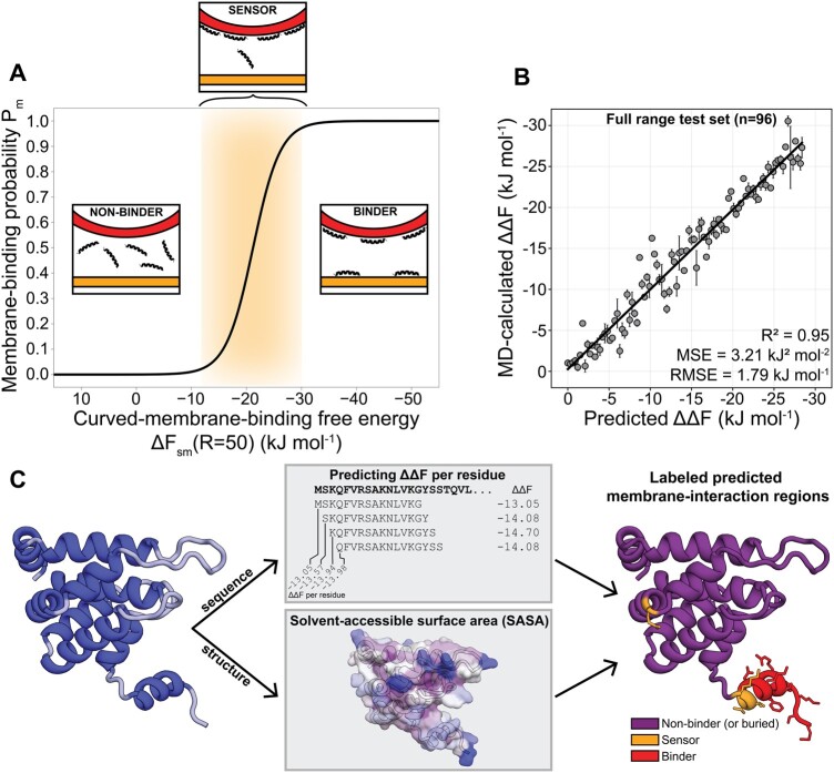
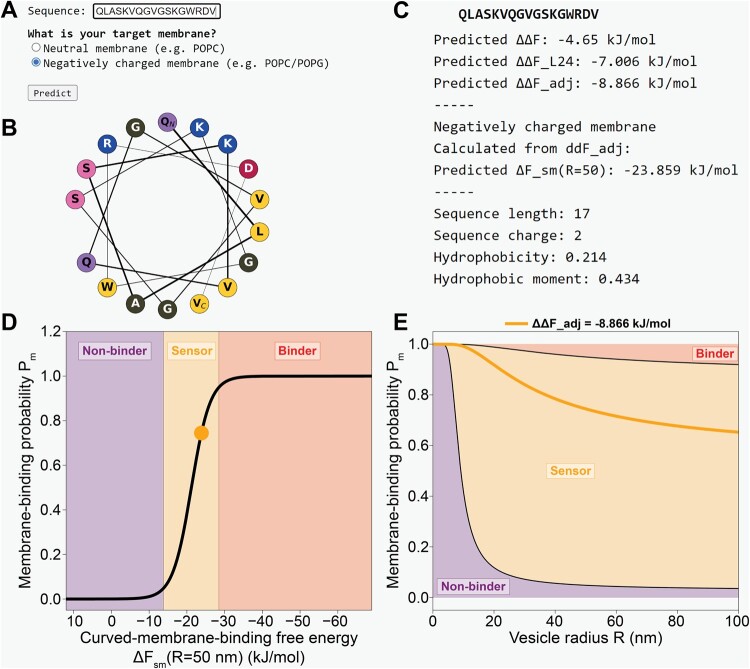
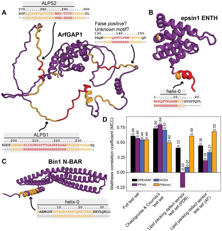
Motivation: Many membrane peripheral proteins have evolved to transiently interact with the surface of (curved) lipid bilayers. Currently, methods to quantitatively predict sensing and binding free energies for protein sequences or structures are lacking, and such tools could greatly benefit the discovery of membrane-interacting motifs, as well as their de novo design. Results: Here, we trained a transformer neural network model on molecular dynamics data for > 50,000 peptides that is able to accurately predict the (relative) membrane-binding free energy for any given amino acid sequence. Using this information, our physics-informed model is able to classify a peptide’s membrane-associative activity as either non-binding, curvature sensing, or membrane binding. Moreover, this method can be applied to detect membraneinteraction regions in a wide variety of proteins, with comparable predictive performance as state-of-the-art data-driven tools like DREAMM, PPM3, and MODA, but with a wider applicability regarding protein diversity, and the added feature to distinguish curvature sensing from general membrane binding. Availability: We made these tools available as a web server, coined Protein-Membrane Interaction predictor (PMIpred), which can be accessed at https://pmipred.fkt.physik.tu-dortmund.de .
Overview
The claims and statistics of this paper haven’t been checked yet.
Run the check — free, no account needed. It reads the open-access text and checks the sources it cites.
Integrity signals
Registry
Cites
Statistics
Figures
Verification
Claims the paper makes
The claims and statistics of this paper haven’t been checked yet.
Run the check — free, no account needed. It reads the open-access text and checks the sources it cites.
Self-check
Percentages, sums and reported differences re-computed from the paper’s own numbers.
The self-check needs the paper’s full text.
Citations
Cites
Highly accurate protein structure prediction with AlphaFold
Jumper, J., Evans, R., Pritzel, A., Green, T., Figurnov, M., Ronneberger, O., Tunyasuvunakool, K., Bates, R., Žídek, A., Potapenko, A., et al. (2021) · Nature · doi:10.1038/s41586-021-03819-2 · Cited by 43,764
Research works printed but not linked: 77
- 10.1016/j.bbamem.2016.02.018 · DOI
- 10.1016/j.chemphyslip.2015.07.015 · DOI
- 10.1038/nature02108 · DOI
- 10.1038/nsmb1194 · DOI
- 10.1074/jbc.273.16.9443 · DOI
- 10.1083/jcb.201011118 · DOI
- 10.1016/j.febslet.2009.10.022 · DOI
- 2014, Discovery of novel membrane binding structures and functions, Biochem. Cell Biol, 92, 555, 10.1139/bcb-2014-0074 · DOI
- 2022, Predicting protein-membrane interfaces of peripheral membrane proteins using ensemble machine learning, Brief. Bioinform, 23, bbab518, 10.1093/bib/bbab518 · DOI · Paper page
- 2022, DREAMM: a web-based server for drugging protein-membrane interfaces as a novel workflow for targeted drug design, Bioinformatics, 38, 5449, 10.1093/bioinformatics/btac680 · DOI
- 10.1002/pro.4219 · DOI
- 2023, Physics-based generative model of curvature sensing peptides; distinguishing sensors from binders, Sci. Adv, 9, eade8839, 10.1126/sciadv.ade8839 · DOI
- 2022, Efficient quantification of lipid packing defect sensing by amphipathic peptides: Comparing martini 2 and 3 with CHARMM36, J. Chem. Theory Comput, 18, 4503, 10.1021/acs.jctc.2c00222 · DOI
- 2005, An experiment-based algorithm for predicting the partitioning of unfolded peptides into phosphatidylcholine bilayer interfaces, Biochemistry, 44, 12614, 10.1021/bi051193b · DOI
- 2017, Polosukhin. Attention is all you need, Adv. Neural Inf. Process. Syst, 30, 6000
- 10.1016/0022-2836(73)90011-9 · DOI
- 10.1093/bioinformatics/btp163 · DOI · Paper page
- 10.1093/bioinformatics/btx285 · DOI · Paper page
- 10.1093/bioinformatics/btn392 · DOI
- 1983, Hydrophobic parameters-pi of amino-acid side-chains from the partitioning of n-acetyl-amino-acid amides, Eur. J. Med. Chem, 18, 369
- 10.1038/299371a0 · DOI
- 10.1021/bi062288w · DOI
- 10.1126/science.270.5244.1999 · DOI
- 10.1093/NAR/GKAB1061 · DOI · Paper page
- 10.1126/science.291.5506.1047 · DOI · Paper page
- 10.1038/nature01020 · DOI · Paper page
- 10.1021/ja907936c · DOI
- 10.1074/jbc.M302865200 · DOI
- 2006, Membrane topology of helix 0 of the epsin n-terminal homology domain, Mol. Cells, 21, 428, 10.1016/S1016-8478(23)12873-1 · DOI
- 10.1016/j.jmb.2012.08.010 · DOI
- 10.1529/biophysj.107.113118 · DOI
- 10.1529/biophysj.108.134155 · DOI
- 10.1126/science.1092586 · DOI · Paper page
- 10.1038/srep08026 · DOI
- 10.1016/j.tcb.2015.09.005 · DOI
- 10.1093/bioinformatics/btu829 · DOI
- J. Hallgren , K.D. Tsirigos , M.D. Pedersen , José J. Almagro A., P. Marcatili , H. Nielsen , A. Krogh , and O. Winther . DeepTMHMM predicts alpha and beta transmembrane proteins using deep neural networks. bioRxiv, 2022. · DOI
- 10.1093/nar/gkac928 · DOI
- T. Akiba , S. Sano , T. Yanase , T. Ohta , and M. Koyama . Optuna: A Next-generation Hyperparameter Optimization Framework, pages 2623–2631. 2019. · DOI
- 10.1016/j.bpj.2011.01.036 · DOI
- 2021, Martini 3: a general purpose force field for coarse-grained molecular dynamics, Nat. Methods, 18, 382, 10.1038/s41592-021-01098-3 · DOI · Paper page
- 10.1021/ct700324x · DOI
- 10.1021/cb1001558 · DOI
- 10.1073/pnas.1212025109 · DOI · Paper page
- 10.1021/bi0477034 · DOI
- 2015, Structural basis for the inhibition of voltage-dependent k+ channel by gating modifier toxin, Sci. Rep, 5, 14226, 10.1038/srep14226 · DOI
- 10.1038/srep34333 · DOI
- 10.1006/jmbi.2000.4283 · DOI
- 10.1074/jbc.M601089200 · DOI
- 10.1016/j.advenzreg.2007.11.009 · DOI
- 2011, Monitoring glycolipid transfer protein activity and membrane interaction with the surface plasmon resonance technique, Biochim. Biophys. Acta, 1808, 47, 10.1016/j.bbamem.2010.08.018 · DOI
- 10.1021/acs.biochem.5b00618 · DOI
- 10.1074/jbc.M112.367730 · DOI
- 10.1091/mbc.E06-01-0089 · DOI
- 2000, Use of the parallax-quench method to determine the position of the active-site loop of cholesterol oxidase in lipid bilayers, Biochemistry, 39, 13383, 10.1021/bi001407j · DOI
- 10.1021/ja4003525 · DOI
- 10.1046/j.1365-313X.2001.01102.x · DOI
- 10.1016/j.abb.2013.06.012 · DOI · Paper page
- 2017, The role of human monoacylglycerol lipase (hMAGL) binding pocket in breakup of unsaturated phospholipid membranes, Anal. Biochem, 536, 90, 10.1016/j.ab.2017.08.009 · DOI
- 10.1046/j.1432-1327.2000.01213.x · DOI
- 2018, Lipids shape the electron acceptor-binding site of the peripheral membrane protein dihydroorotate dehydrogenase, Cell Chem. Biol, 25, 309, 10.1016/j.chembiol.2017.12.012 · DOI
- 10.1016/S0092-8674(00)81663-3 · DOI
- 10.1038/onc.2013.507 · DOI
- 1999, A highly active, soluble mutant of the membrane-associated (s)-mandelate dehydrogenase from pseudomonas putida, Biochemistry, 38, 12367, 10.1021/bi990996u · DOI
- 2017, Membrane binding of the insertion sequence of proteus vulgaris l-amino acid deaminase stabilizes protein structure and increases catalytic activity, Sci. Rep, 7, 13719, 10.1038/s41598-017-14238-7 · DOI
- 10.1021/bi0357356 · DOI
- 10.1074/jbc.M511943200 · DOI
- 2022, Dissecting peripheral protein-membrane interfaces, PLOS Comput. Biol, 18, e1010346, 10.1371/journal.pcbi.1010346 · DOI · Paper page
- 2010, Structural effects of oncogenic PI3Kα mutations, Curr. Top. Microbiol. Immunol, 347, 43
- 10.1021/bi001076a · DOI
- 10.1038/sj.emboj.7600714 · DOI
- 10.1074/jbc.272.2.766 · DOI
- P. Papadopoulou , R. van der Pol , N. van Hilten , M.-A. Moradi , M. Ferraz , J. Aerts , H.J. Risselada , G.J.A. Sevink , and A. Kros . Lipase-mediated selective hydrolysis of lipid droplets in phase separated-liposomes. chemRxiv, 2023. · DOI
- 10.1073/pnas.1016472108 · DOI · Paper page
- 10.1073/pnas.1007974107 · DOI
- 10.1074/jbc.M111.271130 · DOI
- 2019, Envelope-deforming antiviral peptide derived from influenza virus M2 protein, Biochem. Biophys. Res. Commun, 517, 507, 10.1016/j.bbrc.2019.07.088 · DOI
Metadata
- Authors
- van Hilten, N., Verwei, N., Methorst, J., Nase, C., Bernatavicius, A., & Risselada, H. J.
- Affiliations
- Leiden University (NL)
- TU Dortmund University (DE)
- Corresponding author
- Risselada, Herre Jelger
- ORCID
- Venue
- Bioinformatics
- Publisher
- openRxiv
- Published
- 2023-04-10
- Volume
- 40
- Issue
- 2
- ISSN
- 1367-4803 (ISSN-L)
- PMC
- PMC11212490
- OpenAlex
- W4391575167
- Cited by
- 41
- References
- 1
- Citations by year
- 2026: 23 · 2025: 8 · 2024: 10
- FWCI
- 5.56
- Type
- journal article
- Language
- en
- Open access
- gold
- doi.org
- Licence
- cc-by
- Locations
- Bioinformatics (publishedVersion, cc-by)
- bioRxiv (Cold Spring Harbor Laboratory) (acceptedVersion)
- PubMed Central (publishedVersion, cc-by)
- PubMed (publishedVersion)
- Funders
- Deutsche Forschungsgemeinschaft (390677874, EXC2033390677874, EXC2033)
- Nederlandse Organisatie voor Wetenschappelijk Onderzoek (723.016.005)
- Gauss Centre for Supercomputing
- Topics
- Life Sciences › Biochemistry, Genetics and Molecular Biology › Molecular Biology › Machine Learning in Bioinformatics
- Life Sciences › Biochemistry, Genetics and Molecular Biology › Molecular Biology › Lipid Membrane Structure and Behavior
- Life Sciences › Biochemistry, Genetics and Molecular Biology › Molecular Biology › Protein Structure and Dynamics
- Keywords
- protein-membrane interactions, curvature sensing, peptide binding prediction, physics-informed machine learning, transformer models, molecular dynamics, web server
- MeSH terms
- Amino Acid Sequence, Membrane Proteins, Peptides, Physics, Neural Networks, Computer
About these checks
Registry, retraction, Cites and statistics results are computed, not judged, with one exception: a printed reference that no index lists is read once by a model before it counts as a citation error. Claims are extracted by a model and kept only when their quote appears verbatim in the text; each cited source is then checked, with a model’s help, for whether it says what the claim relies on. Those verdicts can be wrong — read the claim’s quote and the cited source before relying on one.
Statistics and arithmetic are recomputed, not judged.
Confidence bands are shown only for fields calibrated on our annotated test set. None is calibrated yet, so no band is shown.
What counts as a claim ›How we measure confidence ›How we check this ›
Are you an author, or do you see a problem with this page? Request a review
How to cite
Cite CoChat for the checks on this page, and the paper for its own findings.
- This page
- CoChat. (2026). Citation check: PMIpred: A physics-informed web server for quantitative Protein-Membrane Interaction prediction. https://app.cochat.ai/papers/10.1101/2023.04.10.536211
- The paper
- van Hilten, N., Verwei, N., Methorst, J., Nase, C., Bernatavicius, A., & Risselada, H. J. (2023). PMIpred: A physics-informed web server for quantitative Protein-Membrane Interaction prediction. Bioinformatics. https://doi.org/10.1101/2023.04.10.536211
