

The DOI matches its Crossref record; 1 of the 1 references it cites were found in a registry or an index.
Diversity of hydrodynamic radii of intrinsically disordered proteins
Open accessPreprint — published as 10.1007/s00249-023-01683-8 · Paper page
Abstract
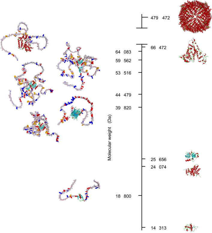
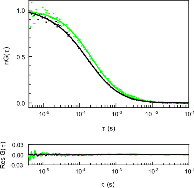
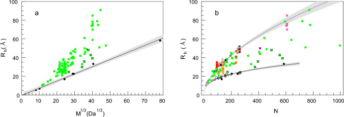
Intrinsically disordered proteins (IDPs) form an important class of biomolecules regulating biological processes in higher organisms. The lack of a fixed spatial structure facilitates them to perform their regulatory functions. Due to the possibility of large conformational changes of IDPs, the cellular milieu can also control productivity of biochemical reactions. From the biophysical point of view, IDPs are biopolymers with a broad configuration state space. The conformation of such a biopolymer depends on non-covalent interactions of its amino acid side chain groups at given temperature and chemical conditions. Thus, the hydrodynamic radius (R h ) of an IDP of a given polymer length (N) is a sequence- and environment-dependent variable. We have reviewed the literature values of hydrodynamic radii of IDPs determined experimentally by SEC, AUC, PFG NMR, DLS, and FCS, and complement them with our FCS results obtained for a series of protein fragments involved in regulation of human gene expression. The data collected herein show that the values of hydrodynamic radii of intrinsically disordered proteins can span the full space between the folded globular and denatured proteins in the R h (N) diagram.
Overview
The claims and statistics of this paper haven’t been checked yet.
Run the check — free, no account needed. It reads the open-access text and checks the sources it cites.
Integrity signals
Registry
Cites
Statistics
Figures
Verification
Claims the paper makes
The claims and statistics of this paper haven’t been checked yet.
Run the check — free, no account needed. It reads the open-access text and checks the sources it cites.
Self-check
Percentages, sums and reported differences re-computed from the paper’s own numbers.
The self-check needs the paper’s full text.
Citations
Cites
Highly accurate protein structure prediction with AlphaFold
Jumper, J., Evans, R., Pritzel, A., Green, T., Figurnov, M., Ronneberger, O., Tunyasuvunakool, K., Bates, R., Žídek, A., Potapenko, A., et al. (2021) · Nature · doi:10.1038/s41586-021-03819-2 · Cited by 43,764
Research works printed but not linked: 118
- 10.1021/acs.chemrev.1c00774 · DOI · Paper page
- 2002, Intrinsic structural disorder and sequence features of the cell cycle inhibitor p57Kip2. Proteins: Structure, Function, and Bioinformatics, 46, 1, 10.1002/prot.10018 · DOI
- 10.1016/j.cell.2018.12.035 · DOI · Paper page
- 10.1016/j.sbi.2011.03.011 · DOI
- 10.1038/nature13999 · DOI
- Baker JMR (2009) Structural Characterization and Interactons of the CFTR Regulatory Region (PhD Thesis)
- 10.1016/j.cell.2016.06.010 · DOI
- 10.1021/acs.jpcb.9b02575 · DOI
- 10.1101/gad.237289.113 · DOI
- 10.1038/nature25762 · DOI · Paper page
- 10.1021/bi0019545 · DOI
- 2015, Polymer physics of intracellular phase transitions, Nature Phys, 11, 899, 10.1038/nphys3532 · DOI
- 10.1007/978-1-4614-5107-5_9 · DOI
- 10.1021/bi9923555 · DOI
- 10.1021/acs.biochem.9b00217 · DOI
- 10.1016/j.str.2011.01.011 · DOI
- 10.1074/jbc.M404375200 · DOI
- 2019, Diffusion coefficients of elastic macromolecules, J Fluid Mech, 878, R3
- 2018, Structural Dynamics of the GW182 Silencing Domain Including its RNA Recognition motif (RRM) Revealed by Hydrogen-Deuterium Exchange Mass Spectrometry, J Am Soc Mass Spectrom, 29, 158, 10.1007/s13361-017-1830-9 · DOI
- 10.1073/pnas.2214064120 · DOI
- 10.1021/bi801040b · DOI
- 10.3389/fgene.2018.00158 · DOI
- 2018, Looking at the Disordered Proteins through the Computational Microscope, ACS Cent Sci, 4, 534, 10.1021/acscentsci.7b00626 · DOI
- 10.1073/pnas.0437902100 · DOI
- 10.1074/jbc.M203499200 · DOI
- 1992, Purification and characterization of the carboxyl-terminal transactivation domain of Vmw65 from herpes simplex virus type 1, Journal of Biological Chemistry, 267, 1411, 10.1016/S0021-9258(18)45957-1 · DOI
- 10.1038/nrm1589 · DOI · Paper page
- 10.1016/j.str.2018.10.016 · DOI
- 2019, The Biomineralization Proteome: Protein Complexity for a Complex Bioceramic Assembly Process, PROTEOMICS, 19, 1900036, 10.1002/pmic.201900036 · DOI
- 2002, A phosphorylated cytoplasmic autoantigen, GW182, associates with a unique population of human mRNAs within novel cytoplasmic speckles, Mol Biol Cell, 13, 1338–1351
- 10.1038/nsmb.2149 · DOI · Paper page
- 10.1038/nsmb.2572 · DOI
- 2021, A cryo-electron microscopy support film formed by 2D crystals of hydrophobin HFBI, Nat Commun, 12, 7257, 10.1038/s41467-021-27596-8 · DOI
- 10.1021/bi972494r · DOI
- 10.1002/pro.5560070720 · DOI
- 10.1063/1.1747243 · DOI
- 10.1110/ps.073062707 · DOI
- 10.1074/jbc.M701932200 · DOI
- 10.1101/gad.912401 · DOI · Paper page
- 10.1104/pp.106.092965 · DOI
- 10.1021/jacs.0c02088 · DOI
- 2000, The Anticodon-binding Domain of Tyrosyl-tRNA Synthetase: State of Folding and Origin of the Crystallographic Disorder, Biochemistry, 39, 1739, 10.1021/bi992382v · DOI
- 10.1074/jbc.M805024200 · DOI
- 10.1371/journal.pone.0011684 · DOI
- 10.1038/358209a0 · DOI
- 1984, DARPP-32, a dopamine- and adenosine 3’:5’-monophosphate-regulated phosphoprotein enriched in dopamine-innervated brain regions, II. Purification and characterization of the phosphoprotein from bovine caudate nucleus. J Neurosci, 4, 99
- 10.1529/biophysj.107.123596 · DOI
- 10.1073/pnas.1207719109 · DOI
- 10.1242/jcs.03429 · DOI
- 10.1021/bm800135m · DOI
- 10.1006/viro.2001.1296 · DOI
- 10.1021/pr070237v · DOI
- 2008, Intramolecular Cohesion of Coils Mediated by Phenylalanine–Glycine Motifs in the Natively Unfolded Domain of a Nucleoporin, PLOS Computational Biology, 4, e1000145, 10.1371/journal.pcbi.1000145 · DOI
- 10.1073/pnas.1814051115 · DOI
- 10.1002/prot.24449 · DOI
- 10.1103/PhysRevLett.39.95 · DOI
- 10.1074/jbc.M300518200 · DOI
- 10.1016/j.sbi.2019.07.012 · DOI
- 2008, Modeling the accessible conformations of the intrinsically unstructured transactivation domain of p53, Proteins: Structure, Function, and Bioinformatics, 71, 587, 10.1002/prot.21721 · DOI
- 1987, Smooth muscle caldesmon is an extended flexible monomeric protein in solution that can readily undergo reversible intra- and intermolecular sulfhydryl cross-linking. A mechanism for caldesmon’s F-actin bundling activity, J Biol Chem, 262, 7429, 10.1016/S0021-9258(18)48255-5 · DOI
- 10.1073/pnas.0704059104 · DOI
- 10.1016/S1097-2765(01)80003-4 · DOI
- 10.1016/j.bpj.2010.02.006 · DOI
- 10.1016/j.molcel.2014.03.036 · DOI
- 10.1016/j.jmb.2003.08.062 · DOI
- 10.1139/o85-102 · DOI
- 2022, ColabFold: making protein folding accessible to all, Nat Methods, 19, 679, 10.1038/s41592-022-01488-1 · DOI · Paper page
- 10.1016/j.jmb.2018.05.012 · DOI · Paper page
- 10.1021/jp308984e · DOI
- 2020, Revealing the Hidden Sensitivity of Intrinsically Disordered Proteins to their Chemical Environment, J Phys Chem Lett, 11, 10131, 10.1021/acs.jpclett.0c02822 · DOI
- Müller-Späth S , Soranno A , Hirschfeld V , et al (2010) Charge interactions can dominate the dimensions of intrinsically disordered proteins. Proceedings of the National Academy of Sciences of the United States of America 107:. https://doi.org/10.1073/pnas.1001743107 · DOI
- 10.15252/embj.2021109952 · DOI
- 10.1074/jbc.M110.199885 · DOI
- 10.1021/bi801933c · DOI
- 2011, Global architecture of human poly(A)-specific ribonuclease by atomic force microscopy in liquid and dynamic light scattering, Biophys Chem, 158, 141, 10.1016/j.bpc.2011.06.010 · DOI
- 10.1016/S0022-2836(02)00328-5 · DOI
- 10.1016/j.ceb.2020.12.007 · DOI
- 10.1016/j.bpj.2017.06.042 · DOI
- 10.1016/j.ceb.2020.12.012 · DOI
- 10.1074/jbc.M800747200 · DOI
- 10.1038/371762a0 · DOI
- 10.1529/biophysj.107.126995 · DOI
- 2014, Alanine and proline content modulate global sensitivity to discrete perturbations in disordered proteins, Proteins: Structure, Function, and Bioinformatics, 82, 3373, 10.1002/prot.24692 · DOI
- 2003, Natively unfolded C-terminal domain of caldesmon remains substantially unstructured after the effective binding to calmodulin, Proteins: Structure, Function, and Bioinformatics, 53, 855, 10.1002/prot.10481 · DOI · Paper page
- 2023, Assessment of models for calculating the hydrodynamic radius of intrinsically disordered proteins, Biophysical Journal, 122, 310, 10.1016/j.bpj.2022.12.013 · DOI
- 10.1007/978-1-4939-7386-6_17 · DOI
- 10.1529/biophysj.107.108811 · DOI
- 2000, Interactions and Aggregation of Apoferritin Molecules in Solution: Effects of Added Electrolytes, Biophysical Journal, 78, 2060, 10.1016/S0006-3495(00)76753-1 · DOI
- 2017, Structural properties of the intrinsically disordered, multiple calcium ion-binding otolith matrix macromolecule-64 (OMM-64), Biochimica et Biophysica Acta (BBA) - Proteins and Proteomics, 1865, 1358, 10.1016/j.bbapap.2017.08.019 · DOI
- 10.1021/acs.jctc.5b00736 · DOI
- 2012, Translational diffusion coefficients of macromolecules, Eur Phys J E, 35, 130, 10.1140/epje/i2012-12130-x · DOI
- 2011, FRET and FCS--friends or foes?, Chemphyschem, 12, 532, 10.1002/cphc.201000776 · DOI
- 10.1016/j.molcel.2004.11.019 · DOI
- 10.1110/ps.051368005 · DOI
- 10.1110/ps.43301 · DOI
- 2020, Orchestration of signaling by structural disorder in class 1 cytokine receptors, Cell Commun Signal, 18, 132, 10.1186/s12964-020-00626-6 · DOI
- 10.1016/j.cell.2018.02.051 · DOI
- 10.1016/j.jmb.2008.09.019 · DOI
- 10.1021/bi8008856 · DOI
- 2003, Biophysical Constraints for Protein Structure Prediction, J Proteome Res, 2, 37–42
- 10.1172/JCI78963 · DOI
- 10.1016/j.sbi.2016.10.015 · DOI · Paper page
- 10.1007/s10930-009-9201-4 · DOI
- 10.1021/bi990752+ · DOI
- 10.1074/jbc.M109541200 · DOI
- 10.1021/pr0155127 · DOI
- 10.3109/10409238.2013.771132 · DOI
- 1985, Bovine chymotrypsinogen A: X-ray crystal structure analysis and refinement of a new crystal form at 1.8 Å resolution, Journal of Molecular Biology, 185, 595–624
- 10.1016/j.jmb.2004.02.002 · DOI
- 2019, The intrinsically disordered C-terminal F domain of the ecdysteroid receptor from Aedes aegypti exhibits metal ion-binding ability, The Journal of Steroid Biochemistry and Molecular Biology, 186, 42, 10.1016/j.jsbmb.2018.09.008 · DOI
- 10.1021/bi991765q · DOI
- 10.1016/j.bpj.2017.12.033 · DOI
- 10.1016/j.str.2008.11.012 · DOI
- 10.1038/sj.emboj.7600869 · DOI
- 2007, Effects of Zinc Binding on the Structure and Dynamics of the Intrinsically Disordered Protein Prothymosin α: Evidence for Metalation as an Entropic Switch, Biochemistry, 46, 13120, 10.1021/bi7014822 · DOI
- 10.1016/j.bbrc.2006.02.153 · DOI
- 2003, The intracellular domain of the Drosophila cholinesterase-like neural adhesion protein, gliotactin, is natively unfolded, Proteins: Structure, Function, and Bioinformatics, 53, 758, 10.1002/prot.10471 · DOI
- 2008, Solution Conformation, Backbone Dynamics and Lipid Interactions of the Intrinsically Unstructured Malaria Surface Protein MSP2, Journal of Molecular Biology, 379, 105–121
Metadata
- Authors
- Białobrzewski, M. K., Klepka, B. P., Michaś, A., Cieplak-Rotowska, M. K., Staszałek, Z., & Niedźwiecka, A.
- Affiliations
- Institute of Physics (PL)
- ORCID
- Venue
- European Biophysics Journal
- Publisher
- openRxiv
- Published
- 2023-06-12
- Volume
- 52
- Issue
- 6-7
- Pages
- 607-618
- ISSN
- 0175-7571 (ISSN-L)
- PMC
- PMC10618399
- OpenAlex
- W4387599617
- Cited by
- 12
- References
- 1
- Citations by year
- 2026: 2 · 2025: 7 · 2024: 2 · 2023: 1
- FWCI
- 1.06
- Type
- journal article
- Language
- en
- Open access
- hybrid
- doi.org
- Licence
- cc-by
- Locations
- European Biophysics Journal (publishedVersion, cc-by)
- bioRxiv (Cold Spring Harbor Laboratory) (acceptedVersion)
- PubMed Central (publishedVersion, cc-by)
- PubMed (publishedVersion)
- Funders
- Narodowe Centrum Nauki (UMO-2016/22/E/NZ1/00656)
- European Regional Development Fund (POIG.02.02.00-00- 025/09)
- Topics
- Life Sciences › Biochemistry, Genetics and Molecular Biology › Molecular Biology › Protein Structure and Dynamics
- Life Sciences › Biochemistry, Genetics and Molecular Biology › Molecular Biology › Protein purification and stability
- Life Sciences › Biochemistry, Genetics and Molecular Biology › Molecular Biology › Protein Interaction Studies and Fluorescence Analysis
- Keywords
- intrinsically disordered proteins, hydrodynamic radius, protein conformation, sequence-dependent conformation, temperature dependence, non-covalent interactions, protein fragments, gene expression regulation
- MeSH terms
- Humans, Magnetic Resonance Spectroscopy, Protein Conformation, Radius, Temperature, Hydrodynamics, Intrinsically Disordered Proteins
About these checks
Registry, retraction, Cites and statistics results are computed, not judged, with one exception: a printed reference that no index lists is read once by a model before it counts as a citation error. Claims are extracted by a model and kept only when their quote appears verbatim in the text; each cited source is then checked, with a model’s help, for whether it says what the claim relies on. Those verdicts can be wrong — read the claim’s quote and the cited source before relying on one.
Statistics and arithmetic are recomputed, not judged.
Confidence bands are shown only for fields calibrated on our annotated test set. None is calibrated yet, so no band is shown.
What counts as a claim ›How we measure confidence ›How we check this ›
Are you an author, or do you see a problem with this page? Request a review
How to cite
Cite CoChat for the checks on this page, and the paper for its own findings.
- This page
- CoChat. (2026). Citation check: Diversity of hydrodynamic radii of intrinsically disordered proteins. https://app.cochat.ai/papers/10.1101/2023.06.12.543592
- The paper
- Białobrzewski, M. K., Klepka, B. P., Michaś, A., Cieplak-Rotowska, M. K., Staszałek, Z., & Niedźwiecka, A. (2023). Diversity of hydrodynamic radii of intrinsically disordered proteins. European Biophysics Journal. https://doi.org/10.1101/2023.06.12.543592
