

The DOI matches its Crossref record; 2 of the 2 references it cites were found in a registry or an index.
DONOR VARIABILITY IN HUMAN MESENCHYMAL STEM CELL OSTEOGENIC RESPONSE AS A FUNCTION OF PASSAGE CONDITIONS AND DONOR SEX
Open accessAbstract
Contemporary tissue engineering efforts often seek to use mesenchymal stem cells (MSCs) due to their potential to differentiate to various tissue-specific cells and generate a pro-regenerative secretome. While MSC differentiation and therapeutic potential can differ as a function of matrix environment, it may also be widely influenced as a function of donor-to-donor variability. Further, effects of passage number and donor sex may further convolute the identification of clinically effective MSC-mediated regeneration technologies. We report efforts to adapt a well-defined mineralized collagen scaffold platform to study the influence of MSC proliferation and osteogenic potential as a function of passage number and donor sex. Mineralized collagen scaffolds broadly support MSC osteogenic differentiation and regenerative potency in the absence of traditional osteogenic supplements for a wide range of MSCs (rabbit, rat, porcine, human). We obtained a library of bone marrow and adipose tissue derived stem cells to examine donor-variability of regenerative potency in mineralized collagen scaffolds. MSCs displayed reduced proliferative capacity as a function of passage duration. Further, MSCs showed significant sex-based differences. Notably, MSCs from male donors displayed significantly higher metabolic activity and proliferation while MSCs from female donor displayed significantly higher osteogenic response via increased alkaline phosphate activity, osteoprotegerin release, and mineral formation in vitro. Our study highlights the essentiality of considering MSC donor sex and culture expansion in future studies of biomaterial regenerative potential.
Overview
The claims and statistics of this paper haven’t been checked yet.
Run the check — free, no account needed. It reads the open-access text and checks the sources it cites.
Integrity signals
Registry
Cites
Statistics
Figures
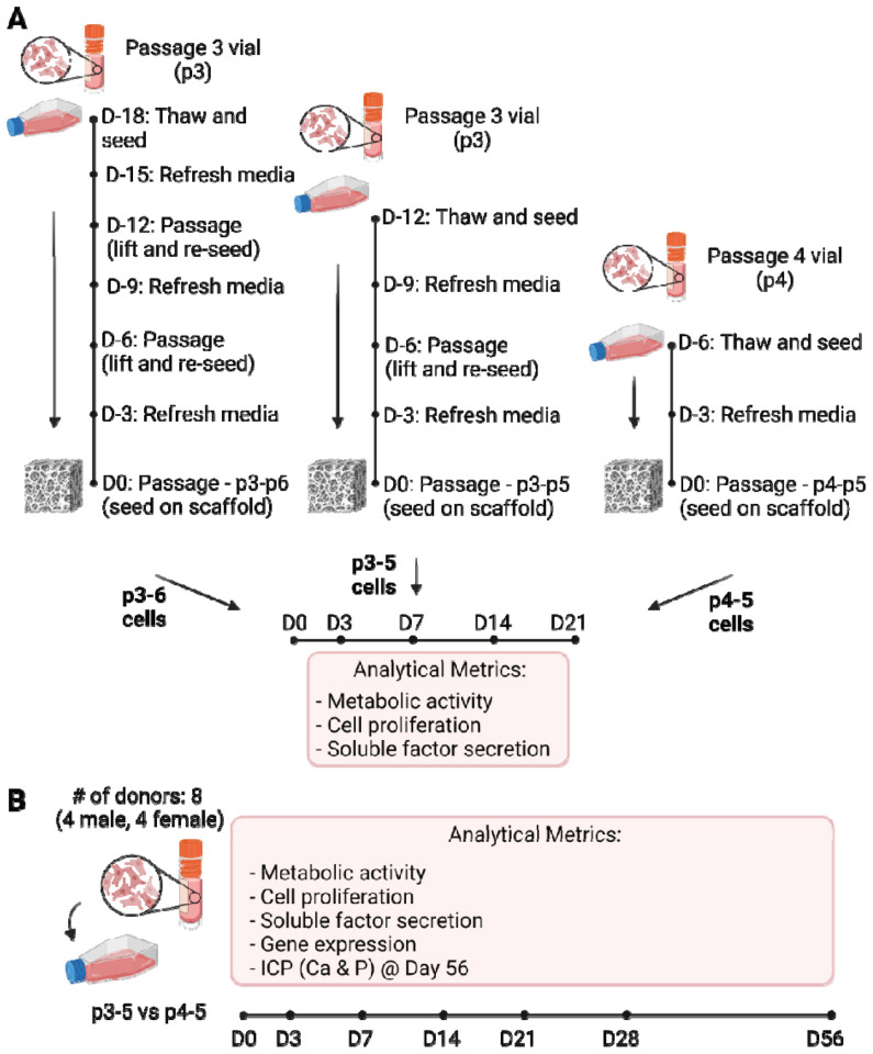
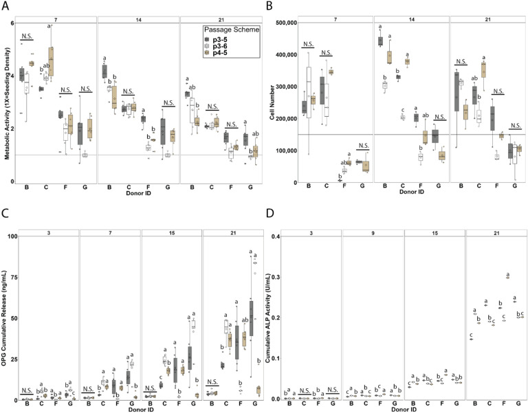
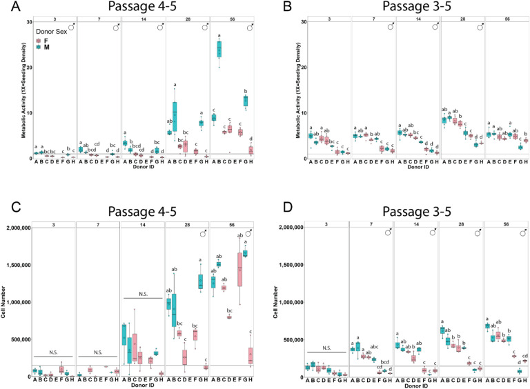
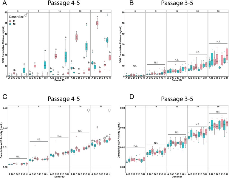
Verification
Claims the paper makes
The claims and statistics of this paper haven’t been checked yet.
Run the check — free, no account needed. It reads the open-access text and checks the sources it cites.
Claims other papers make about this one
Each is checked against this paper’s own text: does it say what the citing paper says it does?
The source backs part of the claim, not all of it — or backs it for a different population, condition or comparison.
In the citing paper
“In addition, the number of MSC passage [246], [247], [248] and the donor variability [246], [247], [248] cannot be ignored since MSC activity and, thus, the properties of MSC-secretome can be affected by donor age and sex [246], [248], [249]”
From Mesenchymal stem cell secretome for regenerative medicine: Where do we stand? (2025) · cites this paper as [249]
Experimental — accuracy not yet measuredThe source backs part of the claim, not all of it — or backs it for a different population, condition or comparison.
In the citing paper
“with older donors of BM-MSCs having lower proliferation rates [246], [249] and osteogenic [246], [248], adipogenic and neurogenic [248] capacities.”
From Mesenchymal stem cell secretome for regenerative medicine: Where do we stand? (2025) · cites this paper as [249]
Experimental — accuracy not yet measured
Self-check
Percentages, sums and reported differences re-computed from the paper’s own numbers.
The self-check needs the paper’s full text.
Citations
Cites
Zaim, M., Karaman, S., Cetin, G., & Isik, S. (2012) · Annals of Hematology · doi:10.1007/s00277-012-1438-x · Cited by 294
Mesenchymal stem cells: Amazing remedies for bone and cartilage defects
Kangari, P., Talaei-Khozani, T., Razeghian-Jahromi, I., & Razmkhah, M. (2020) · Stem Cell Research & Therapy · doi:10.1186/s13287-020-02001-1 · Cited by 286
Research works printed but not linked: 69
- 2021, Global, regional, and national burden of bone fractures in 204 countries and territories, 1990-2019: a systematic analysis from the Global Burden of Disease Study 2019, The Lancet Healthy Longevity, 2, e580, 10.1016/S2666-7568(21)00172-0 · DOI · Paper page
- Samarawickrama, K.G ., A Review on Bone Grafting, Bone Substitutes and Bone Tissue Engineering, in Proceedings of the 2nd International Conference on Medical and Health Informatics. 2018, Association for Computing Machinery: Tsukuba, Japan. p. 244–251. · DOI
- Finkemeier, C.G. , Bone-Grafting and Bone-Graft Substitutes. JBJS, 2002. 84(3). · DOI
- Baldwin, P. , et al., Autograft, Allograft, and Bone Graft Substitutes: Clinical Evidence and Indications for Use in the Setting of Orthopaedic Trauma Surgery. Journal of Orthopaedic Trauma, 2019. 33(4). · DOI
- 2019, Review of bone graft and bone substitutes with an emphasis on fracture surgeries, Biomaterials Research, 23, 9, 10.1186/s40824-019-0157-y · DOI
- 10.3928/01477447-20140825-54 · DOI
- 2017, Variant Purification of an Allogeneic Bone Block, Acta Stomatol Croat, 51, 141, 10.15644/asc51/2/7 · DOI
- 2023, Case report: Tuberculosis recall on bone graft patient, N Am Spine Soc J, 15, 100241
- 2013, Mesenchymal Stem Cells in Bone Regeneration, Adv Wound Care (New Rochelle, 2, 306, 10.1089/wound.2012.0420 · DOI
- Stamnitz, S. and A. Klimczak , Mesenchymal Stem Cells, Bioactive Factors, and Scaffolds in Bone Repair: From Research Perspectives to Clinical Practice. Cells, 2021. 10(8). · DOI
- 2021, Advancing application of mesenchymal stem cell-based bone tissue regeneration, Bioact Mater, 6, 666
- Ryan, H. , et al., Ancestral Background Is Underreported in Regenerative Engineering. Regenerative Engineering and Translational Medicine, 2021. · DOI
- Moore, E. , et al., Ancestry of cells must be considered in bioengineering. Nature Reviews Materials, 2021. · DOI
- 10.1385/JCD:4:2:147 · DOI
- 2017, Sex-Differences in Skeletal Growth and Aging, Current Osteoporosis Reports, 15, 70, 10.1007/s11914-017-0349-0 · DOI
- Fitzpatrick, L.A ., Sex differences in skeletal development, in Advances in Molecular and Cell Biology. 2004, Elsevier. p. 229–245. · DOI
- Ortona, E. , et al., The Role of Sex Differences in Bone Health and Healing. Biology (Basel), 2023. 12(7). · DOI · Paper page
- 2022, Sex-Based Difference in Bone Healing: A Review of Recent Pre-clinical Literature, Curr Rev Musculoskelet Med, 15, 651, 10.1007/s12178-022-09803-1 · DOI
- 2009, Sex-specific compromised bone healing in female rats might be associated with a decrease in mesenchymal stem cell quantity, Bone, 45, 1065, 10.1016/j.bone.2009.08.005 · DOI
- 2021, Differences in Fracture Healing Between Female and Male C57BL/6J Mice, Front Physiol, 12, 712494, 10.3389/fphys.2021.712494 · DOI
- 2021, Let’s Talk About Sex-Biological Sex Is Underreported in Biomaterial Studies, Adv Healthc Mater, 10, e2001034, 10.1002/adhm.202001034 · DOI
- 10.1007/s11914-017-0413-9 · DOI
- 2021, Influence of comorbidities on functional outcomes in patients with surgically treated fragility hip fractures: a retrospective cohort study, BMC Geriatrics, 21, 283, 10.1186/s12877-021-02227-5 · DOI
- 10.3109/17453674.2012.747054 · DOI
- 10.1038/nrrheum.2012.1 · DOI
- Jennifer, J.B. , et al., Concise Review: Bone Marrow-Derived Mesenchymal Stem Cells Change Phenotype Following In Vitro Culture: Implications for Basic Research and the Clinic. Stem Cells, 2014.
- 2013, Phenotype, donor age and gender affect function of human bone marrow-derived mesenchymal stromal cells, BMC Medicine, 11, 146, 10.1186/1741-7015-11-146 · DOI · Paper page
- 10.1016/j.mad.2007.12.002 · DOI · Paper page
- 2015, Correlation between in vitro expansion-related cell stiffening and differentiation potential of human mesenchymal stem cells, Differentiation, 90, 1, 10.1016/j.diff.2015.08.002 · DOI
- 2021, In Vitro Culture Expansion Shifts the Immune Phenotype of Human Adipose-Derived Mesenchymal Stem Cells, Front Immunol, 12, 621744, 10.3389/fimmu.2021.621744 · DOI
- 2019, The inclusion of zinc into mineralized collagen scaffolds for craniofacial bone repair applications, Acta biomaterialia, 93, 86, 10.1016/j.actbio.2019.05.031 · DOI
- 2019, Shape-fitting collagen-PLA composite promotes osteogenic differentiation of porcine adipose stem cells, Journal of the Mechanical Behavior of Biomedical Materials, 95, 21, 10.1016/j.jmbbm.2019.03.017 · DOI
- 2016, Evaluation of multi-scale mineralized collagen-polycaprolactone composites for bone tissue engineering, Journal of the mechanical behavior of biomedical materials, 61, 318, 10.1016/j.jmbbm.2016.03.032 · DOI
- 2020, Mineralized collagen scaffolds fabricated with amniotic membrane matrix increase osteogenesis under inflammatory conditions, Regenerative Biomaterials, 7, 247, 10.1093/rb/rbaa005 · DOI
- 2021, Glycosaminoglycan content of a mineralized collagen scaffold promotes mesenchymal stem cell secretion of factors to modulate angiogenesis and monocyte differentiation, Materialia, 18, 101149, 10.1016/j.mtla.2021.101149 · DOI
- 2020, Anisotropic mineralized collagen scaffolds accelerate osteogenic response in a glycosaminoglycan-dependent fashion, RSC Advances, 10, 15629, 10.1039/D0RA01336F · DOI
- 2021, Inclusion of a 3D-printed Hyperelastic Bone mesh improves mechanical and osteogenic performance of a mineralized collagen scaffold, Acta Biomaterialia, 121, 224, 10.1016/j.actbio.2020.11.028 · DOI
- 2022, Amnion and chorion matrix maintain hMSC osteogenic response and enhance immunomodulatory and angiogenic potential in a mineralized collagen scaffold, Front Bioeng Biotechnol, 10, 1034701, 10.3389/fbioe.2022.1034701 · DOI
- 2015, Mineralized collagen scaffolds induce hMSC osteogenesis and matrix remodeling, Biomaterials science, 3, 533, 10.1039/C4BM00397G · DOI
- 10.1016/j.biomaterials.2015.01.059 · DOI
- 2016, Nanoparticulate Mineralized Collagen Scaffolds and BMP-9 Induce a Long-Term Bone Cartilage Construct in Human Mesenchymal Stem Cells, Advanced Healthcare Materials, 5, 1821, 10.1002/adhm.201600187 · DOI
- 2017, Nonmineralized and Mineralized Collagen Scaffolds Induce Differential Osteogenic Signaling Pathways in Human Mesenchymal Stem Cells, Advanced Healthcare Materials, 6, 1700641, 10.1002/adhm.201700641 · DOI
- 2019, Nanoparticulate mineralized collagen glycosaminoglycan materials directly and indirectly inhibit osteoclastogenesis and osteoclast activation, Journal of tissue engineering and regenerative medicine, 13, 823, 10.1002/term.2834 · DOI
- 2022, Osteoprotegerin-eluting nanoparticulate mineralized collagen scaffolds improve skull regeneration, Biomater Adv, 145, 213262
- 10.1126/sciadv.aaw4991 · DOI
- 2016, Nanoparticulate mineralized collagen scaffolds induce in vivo bone regeneration independent of progenitor cell loading or exogenous growth factor stimulation, Biomaterials, 89, 67, 10.1016/j.biomaterials.2016.02.020 · DOI
- 2017, A Mineralized Collagen-Polycaprolactone Composite Promotes Healing of a Porcine Mandibular Defect, Tissue Engineering Part A, 24, 943
- 2021, Repair of critical-size porcine craniofacial bone defects using a collagen–polycaprolactone composite biomaterial, Biofabrication, 14, 014102
- 2023, Evaluation of bacterial attachment on mineralized collagen scaffolds and addition of manuka honey to increase mesenchymal stem cell osteogenesis, Biomaterials, 294, 122015, 10.1016/j.biomaterials.2023.122015 · DOI
- 2023, Inflammatory licensed hMSCs exhibit enhanced immunomodulatory capacity in a biomaterial mediated manner, ACS Biomaterials Science & Engineering, 9, 4916, 10.1021/acsbiomaterials.3c00290 · DOI
- 2010, Design of a multiphase osteochondral scaffold. II. Fabrication of a mineralized collagen-glycosaminoglycan scaffold, J Biomed Mater Res A, 92, 1066
- 2016, Nanoparticulate mineralized collagen scaffolds induce in vivo bone regeneration independent of progenitor cell loading or exogenous growth factor stimulation, Biomaterials, 89, 67, 10.1016/j.biomaterials.2016.02.020 · DOI
- Dewey, M.J. , et al., Glycosaminoglycan content of a mineralized collagen scaffold promotes mesenchymal stem cell secretion of factors to modulate angiogenesis and monocyte differentiation. Materialia (Oxf), 2021. 18. · DOI
- 2020, Orbital seeding of mesenchymal stromal cells increases osteogenic differentiation and bone-like tissue formation, Journal of Orthopaedic Research®, 38, 1228, 10.1002/jor.24583 · DOI
- 2008, Method to analyze three-dimensional cell distribution and infiltration in degradable scaffolds, Tissue Engineering Part C: Methods, 14, 319, 10.1089/ten.tec.2008.0221 · DOI
- 2015, Collagen scaffolds incorporating coincident gradations of instructive structural and biochemical cues for osteotendinous junction engineering, Advanced healthcare materials, 4, 831, 10.1002/adhm.201400809 · DOI
- 2017, Concise Review: Multifaceted Characterization of Human Mesenchymal Stem Cells for Use in Regenerative Medicine, Stem Cells Translational Medicine, 6, 2173, 10.1002/sctm.17-0129 · DOI
- 2008, Cell passage and composition of culture medium effects proliferation and differentiation of human osteoblast-like cells from facial bone, J Physiol Pharmacol, 59, 47
- 2023, Influence of donor age and comorbidities on transduced human adipose-derived stem cell in vitro osteogenic potential, Gene Therapy, 30, 369, 10.1038/s41434-022-00367-w · DOI
- 10.1186/s41232-019-0111-3 · DOI
- 2020, Alkaline phosphatase: Structure, expression and its function in bone mineralization, Gene, 754, 144855, 10.1016/j.gene.2020.144855 · DOI
- 2020, RANKL/RANK/OPG Pathway: A Mechanism Involved in Exercise-Induced Bone Remodeling, BioMed Research International, 2020, 1
- 2021, Stiffness of Nanoparticulate Mineralized Collagen Scaffolds Triggers Osteogenesis via Mechanotransduction and Canonical Wnt Signaling, Macromolecular Bioscience, 21, 2000370, 10.1002/mabi.202000370 · DOI
- 2021, Graham Vesey & Sharon McCracken Immune modulation via adipose derived Mesenchymal Stem cells is driven by donor sex in vitro, Scientific Reports, 11, 12454, 10.1038/s41598-021-91870-4 · DOI
- 2019, Immunomodulation by mesenchymal stem cells is sex dependent, Cytotherapy, 21, S84, 10.1016/j.jcyt.2019.03.502 · DOI
- 2023, Inflammatory licensed hMSCs exhibit enhanced immunomodulatory capacity in a biomaterial mediated manner, ACS Biomaterials Science & Engineering, 9, 4916, 10.1021/acsbiomaterials.3c00290 · DOI
- 2019, Bone health in estrogen-free contraception, Osteoporos Int, 30, 2391, 10.1007/s00198-019-05103-6 · DOI
- 10.1210/er.2007-0001 · DOI · Paper page
- 10.1073/pnas.83.8.2496 · DOI · Paper page
Cited by
Sorted by citations
Mesenchymal stem cell secretome for regenerative medicine: Where do we stand?
Trigo, C. M., Rodrigues, J. S., Camões, S. P., Solá, S., & Miranda, J. P. (2025) · Journal of Advanced Research · doi:10.1016/j.jare.2024.05.004 · Cited by 252
“…variability [246–248] cannot be ignored since MSC activity and, thus, the properties of MSC-secretome can be affected by donor age and sex [246,248,249], with older donors of BM-MSCs having lower proliferation rates [246,249] and osteogenic [246,248], adipogenic and neurogenic [248] capacities.”
Metadata
- Authors
- Kolliopoulos, V., Tiffany, A., Polanek, M., & Harley, B. A. C.
- Affiliations
- University of Illinois Urbana-Champaign (US)
- Corresponding author
- Harley, Brendan A.C.
- Venue
- bioRxiv (Cold Spring Harbor Laboratory)
- Publisher
- openRxiv
- Published
- 2023-11-15
- PMC
- PMC10680622
- OpenAlex
- W4388714160
- Cited by
- 17
- References
- 2
- Citations by year
- 2026: 6 · 2025: 6 · 2024: 5
- Type
- posted content
- Language
- en
- Open access
- green
- doi.org
- Licence
- cc-by-nd
- Locations
- bioRxiv (Cold Spring Harbor Laboratory) (acceptedVersion, cc-by-nd)
- PubMed (publishedVersion)
- PubMed Central (submittedVersion, cc-by-nd)
- Funders
- National Science Foundation (1144245, 1746047, DGE‐1 144 245)
- National Institutes of Health (1144245)
- University of Illinois at Urbana-Champaign (DGE-1144245)
- National Institute of General Medical Sciences (T32GM070421)
- National Institute of Arthritis and Musculoskeletal and Skin Diseases (R01AR077858)
- National Institute of Dental and Craniofacial Research (R21DE026582, R01DE030491)
- Carl R. Woese Institute for Genomic Biology
- Topics
- Life Sciences › Biochemistry, Genetics and Molecular Biology › Cell Biology › Mesenchymal stem cell research
- Physical Sciences › Engineering › Biomedical Engineering › Bone Tissue Engineering Materials
- Health Sciences › Dentistry › Periodontics › Periodontal Regeneration and Treatments
- Keywords
- mesenchymal stem cells, donor variability, osteogenic differentiation, donor sex, passage number, bone marrow-derived stem cells, adipose-derived stem cells, cell proliferation, alkaline phosphatase activity, osteoprotegerin, mineral formation, tissue engineering
About these checks
Registry, retraction, Cites and statistics results are computed, not judged, with one exception: a printed reference that no index lists is read once by a model before it counts as a citation error. Claims are extracted by a model and kept only when their quote appears verbatim in the text; each cited source is then checked, with a model’s help, for whether it says what the claim relies on. Those verdicts can be wrong — read the claim’s quote and the cited source before relying on one.
Statistics and arithmetic are recomputed, not judged.
Confidence bands are shown only for fields calibrated on our annotated test set. None is calibrated yet, so no band is shown.
What counts as a claim ›How we measure confidence ›How we check this ›
Are you an author, or do you see a problem with this page? Request a review
