PreprintCited by 11Cholesterol and Lipid MetabolismGenomics and Phylogenetic StudiesProtein Structure and Dynamics

The DOI matches its Crossref record; 1 of the 1 references it cites were found in a registry or an index.

Phylogenetic and protein structure analyses provide insight into the evolution and diversification of the CD36 domain ‘apex’ among scavenger receptor class B proteins across Eukarya

Open access
Boohar, Reed T., Vandepas, Lauren E., Traylor-Knowles, Nikki, Browne, William E.·2023·Genome Biology and Evolution

Preprint — published as 10.1093/gbe/evad218

Abstract

The cluster of differentiation 36 (CD36) domain defines the characteristic ectodomain associated with scavenger receptor class B (SR-B) proteins. In bilaterians, SR-Bs play critical roles in diverse biological processes including innate immunity functions such as pathogen recognition and apoptotic cell clearance, as well as metabolic sensing associated with fatty acid uptake and cholesterol transport. While previous studies suggest this protein family is ancient, SR-B diversity across Eukarya has not been robustly characterized. We analyzed SR-B homologs identified from the genomes and transcriptomes of 165 diverse eukaryotic species. The presence of highly conserved amino acid motifs across major eukaryotic supergroups supports the presence of a SR-B homolog in the last eukaryotic common ancestor (LECA). Our comparative analyses of SR-B protein structure identifies the retention of a canonical asymmetric beta barrel tertiary structure within the CD36 ectodomain across Eukarya. We also identify multiple instances of independent lineage-specific sequence expansions in the apex region of the CD36 ectodomain — a region functionally associated with ligand-sensing. We hypothesize that a combination of both sequence expansion and structural variation in the CD36 apex region may reflect the evolution of SR-B ligand sensing specificity between diverse eukaryotic clades. Significance Statement: SR-Bs are well-described in bilaterians, however the diversity and evolution of this ancient receptor protein family is poorly understood across Eukarya. Our analyses reveal a conserved beta barrel tertiary structure across eukaryotic SR-Bs that correlates with the presence of an intramolecular tunnel. In contrast, the putative ligand-sensing region associated with the apex of the protein can be highly divergent in both sequence and structure within and between taxa. Our data confirm the antiquity of the SR-B receptor protein family in Eukaryota and improve characterization of SR-B diversity outside of Metazoa. We hypothesize that eukaryotic SR-B diversity may be used as a model to explore receptor protein evolution driven by lineage-specific ligand specificity.

Overview

The claims and statistics of this paper haven’t been checked yet.

Run the check — free, no account needed. It reads the open-access text and checks the sources it cites.

A few free checks a day · papers already verified open instantly · results are public

Integrity signals

Registry

Crossref · 10.1101/2023.11.13.566873

Cites

1 of 1 references verified0 citation errors · 0 not checkable

Statistics

Figures

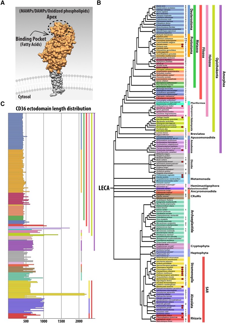
Fig. 1. Distribution of SR-Bs across Eukarya. (A) Space-filling model of SCARB3. SR-Bs are integral membrane proteins with two transmembrane domains, cytoplasmic N- and C-terminal tails (gray shading) and a CD36 ectodomain (orange shading) characterized by a ligand-sensing apex and binding pocket and containing an intramolecular tunnel. (B) Phylogenetic distribution of SR-Bs. Asterisk indicates taxa for which transcriptomic data were used. An oval next to a row indicates obligate parasitic life history. Number indicates how many SR-B homologs were detected in each representative taxon. Color-coded taxonomic groupings are on the right. Phylogeny based on Burki et al. (2020), Carr et al. (2017), Derelle et al. (2016), Ettahi et al. (2021), Li, Hou, et al. (2021), Li, Shen, et al. (2021), and Strassert et al. (2021). Raw CD36 Pfam domain hits following isoform removal for each taxon can be found in supplementary table S2, Supplementary Material online. (C) Length distribution for each of the 279 CD36 ectodomains. Includes aa comprising the predicted noncytoplasmic region containing CD36 ectodomain sequence between transmembrane helices. Colors follow the taxonomic scheme in panel (B).
Fig. 1. Distribution of SR-Bs across Eukarya. (A) Space-filling model of SCARB3. SR-Bs are integral membrane proteins with two transmembrane domains, cytoplasmic N- and C-terminal tails (gray shading) and a CD36 ectodomain (orange shading) characterized by a ligand-sensing apex and binding pocket and containing an intramolecular tunnel. (B) Phylogenetic distribution of SR-Bs. Asterisk indicates taxa for which transcriptomic data were used. An oval next to a row indicates obligate parasitic life history. Number indicates how many SR-B homologs were detected in each representative taxon. Color-coded taxonomic groupings are on the right. Phylogeny based on Burki et al. (2020), Carr et al. (2017), Derelle et al. (2016), Ettahi et al. (2021), Li, Hou, et al. (2021), Li, Shen, et al. (2021), and Strassert et al. (2021). Raw CD36 Pfam domain hits following isoform removal for each taxon can be found in supplementary table S2, Supplementary Material online. (C) Length distribution for each of the 279 CD36 ectodomains. Includes aa comprising the predicted noncytoplasmic region containing CD36 ectodomain sequence between transmembrane helices. Colors follow the taxonomic scheme in panel (B).CC BY · PubMed Central
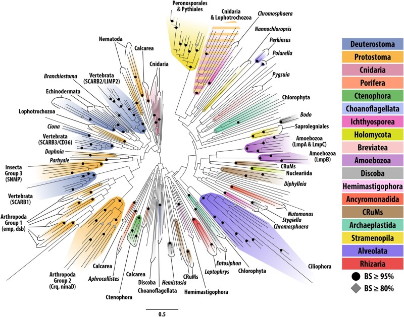
Fig. 2. ML (RAxML) gene tree for eukaryotic CD36 domains. Circles indicate nodes with BS support ≥ 95%. Diamonds indicate nodes with BS support ≥ 80%. Clades with ≥ 80% BS support are colored according to fig. 1B. Unannotated tree with node support values is supplementary fig. 2, Supplementary Material online.
Fig. 2. ML (RAxML) gene tree for eukaryotic CD36 domains. Circles indicate nodes with BS support ≥ 95%. Diamonds indicate nodes with BS support ≥ 80%. Clades with ≥ 80% BS support are colored according to fig. 1B. Unannotated tree with node support values is supplementary fig. 2, Supplementary Material online.CC BY · PubMed Central
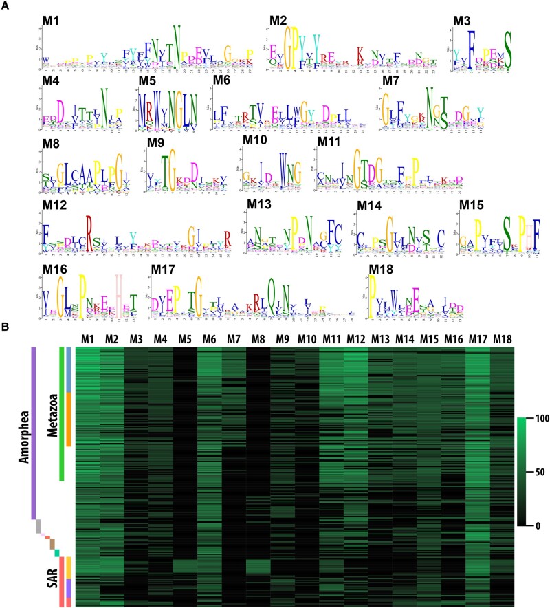
Fig. 3. CD36 ectodomain aa motifs. (A) Sequence logos for 18 aa motifs recovered from our set of 279 eukaryotic CD36 domains by STREME v5.5.3. Letter height represents the relative aa frequency. (B) A heatmap depicting the sequence score for each motif to each eukaryotic CD36 ectodomain is shown. Raw sequence scores calculated by STREME were transformed to a 0–100 scale represented on the right. The full matrix of motif sequence scores is found in supplementary table S5, Supplementary Material online. Colors correspond with the taxonomic scheme in fig. 1B.
Fig. 3. CD36 ectodomain aa motifs. (A) Sequence logos for 18 aa motifs recovered from our set of 279 eukaryotic CD36 domains by STREME v5.5.3. Letter height represents the relative aa frequency. (B) A heatmap depicting the sequence score for each motif to each eukaryotic CD36 ectodomain is shown. Raw sequence scores calculated by STREME were transformed to a 0–100 scale represented on the right. The full matrix of motif sequence scores is found in supplementary table S5, Supplementary Material online. Colors correspond with the taxonomic scheme in fig. 1B.CC BY · PubMed Central
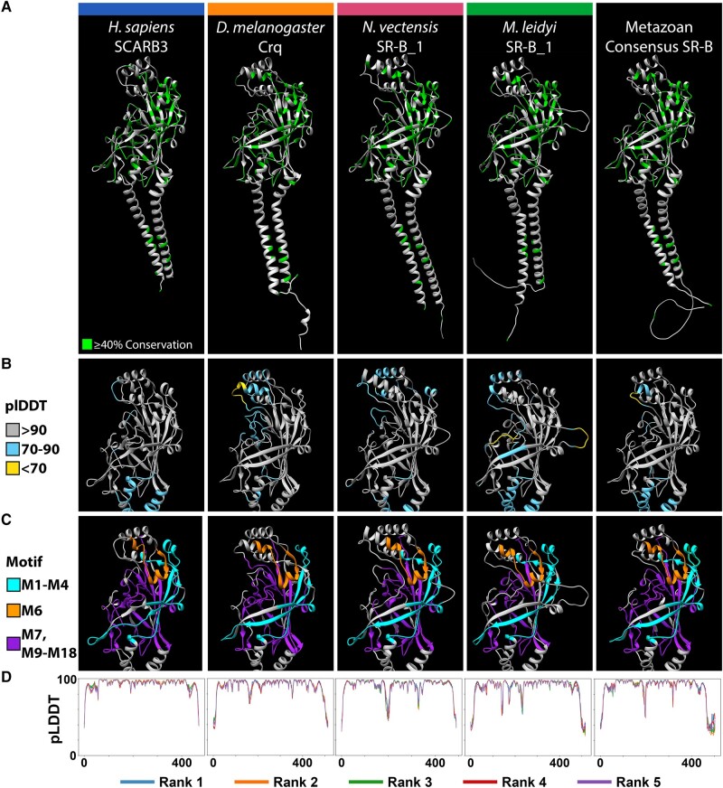
Fig. 4. Metazoan SR-Bs share a highly conserved tertiary structure. The highest ranked AlphaFold2 structure predictions for four exemplar metazoan SR-B proteins. From left to right, H. sapiens SCARB3, Drosophila melanogaster Crq, Nematostella vectensis SR-B_1, and M. leidyi SR-B_1, alongside the consensus structure prediction derived from an all-metazoan SR-B sequence alignment. Protein structure visualized with Chimera. (A) Green residues in the ribbon models reflect ≥40% sequence conservation across eukaryotic SR-Bs. (B) pLDDT scores computed by AlphaFold2 assess per-residue confidence across the CD36 ectodomain region of each model. Residue color reflects high-confidence (gray), moderate-confidence (light blue), and low-confidence (yellow) pLDDT (Varadi et al. 2021). (C) The CD36 ectodomain region of each model is annotated with the aa motifs discovered via STREME. Motifs M1–M4 (cyan) lie more N-terminal to the three-helix bundle at the apex, and motifs M7 and M9–M18 (purple) lie more C-terminal to the three-helix bundle at the apex. Motif M6 (orange) connects α5 to α7 but is not part of the bundle itself. (D) Graphs depict AlphaFold2 pLDDT scores across residue positions for each SR-B. AlphaFold2 via ColabFold generates five protein structure models per input protein sequence and ranks each by global accuracy using the pTM metric (pTM score). Line color indicates the model rank, with rank 1 being the best.
Fig. 4. Metazoan SR-Bs share a highly conserved tertiary structure. The highest ranked AlphaFold2 structure predictions for four exemplar metazoan SR-B proteins. From left to right, H. sapiens SCARB3, Drosophila melanogaster Crq, Nematostella vectensis SR-B_1, and M. leidyi SR-B_1, alongside the consensus structure prediction derived from an all-metazoan SR-B sequence alignment. Protein structure visualized with Chimera. (A) Green residues in the ribbon models reflect ≥40% sequence conservation across eukaryotic SR-Bs. (B) pLDDT scores computed by AlphaFold2 assess per-residue confidence across the CD36 ectodomain region of each model. Residue color reflects high-confidence (gray), moderate-confidence (light blue), and low-confidence (yellow) pLDDT (Varadi et al. 2021). (C) The CD36 ectodomain region of each model is annotated with the aa motifs discovered via STREME. Motifs M1–M4 (cyan) lie more N-terminal to the three-helix bundle at the apex, and motifs M7 and M9–M18 (purple) lie more C-terminal to the three-helix bundle at the apex. Motif M6 (orange) connects α5 to α7 but is not part of the bundle itself. (D) Graphs depict AlphaFold2 pLDDT scores across residue positions for each SR-B. AlphaFold2 via ColabFold generates five protein structure models per input protein sequence and ranks each by global accuracy using the pTM metric (pTM score). Line color indicates the model rank, with rank 1 being the best.CC BY · PubMed Central
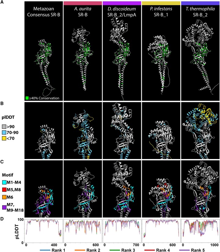
Fig. 5. Divergent eukaryotic SR-Bs retain a conserved CD36 domain beta barrel core with lineage-specific expansions in the apex region. The highest ranked AlphaFold2 structure predictions for four exemplar eukaryote SR-B proteins with divergent, lineage-specific alpha-helix structures at the apex of the CD36 ectodomain are shown, from left to right, the metazoan consensus structure, A. aurita SR-B, D. discoideum SR-B_2 (LmpA), Phytophthora infestans SR-B_1, and Tetrahymena thermophila SR-B_2. Protein structure visualization was in Chimera. (A) Green residues reflect ≥40% conservation across the alignment of all eukaryotic SR-Bs. (B) pLDDT scores computed by AlphaFold2 assess per-residue confidence across the CD36 ectodomain region of each model. Residue color reflects high-confidence (gray), moderate-confidence (light blue), and low-confidence (yellow) pLDDT (Varadi et al. 2021). (C) The CD36 ectodomain region of each model is annotated with the aa motifs discovered via STREME. Motifs M1–M4 (cyan) lie more N-terminal to the three-helix bundle at the apex, and motifs M7 and M9–M18 (purple) lie more C-terminal to the three-helix bundle at the apex. Motif M6 (orange) connects two apex α-helices but is not part of the bundle itself. M5 and M8 represent the two lowest significance STREME motifs and are short motifs found within the lineage-specific sequence expansions at the apex of the CD36 ectodomain of P. infestans. (D) Graphs depict AlphaFold2 pLDDT scores across residue positions for each SR-B. AlphaFold2 via ColabFold generates five protein structure models per input protein sequence and ranks each by global accuracy using the pTM metric (pTM score). Line color indicates the model rank, with rank 1 being the best.
Fig. 5. Divergent eukaryotic SR-Bs retain a conserved CD36 domain beta barrel core with lineage-specific expansions in the apex region. The highest ranked AlphaFold2 structure predictions for four exemplar eukaryote SR-B proteins with divergent, lineage-specific alpha-helix structures at the apex of the CD36 ectodomain are shown, from left to right, the metazoan consensus structure, A. aurita SR-B, D. discoideum SR-B_2 (LmpA), Phytophthora infestans SR-B_1, and Tetrahymena thermophila SR-B_2. Protein structure visualization was in Chimera. (A) Green residues reflect ≥40% conservation across the alignment of all eukaryotic SR-Bs. (B) pLDDT scores computed by AlphaFold2 assess per-residue confidence across the CD36 ectodomain region of each model. Residue color reflects high-confidence (gray), moderate-confidence (light blue), and low-confidence (yellow) pLDDT (Varadi et al. 2021). (C) The CD36 ectodomain region of each model is annotated with the aa motifs discovered via STREME. Motifs M1–M4 (cyan) lie more N-terminal to the three-helix bundle at the apex, and motifs M7 and M9–M18 (purple) lie more C-terminal to the three-helix bundle at the apex. Motif M6 (orange) connects two apex α-helices but is not part of the bundle itself. M5 and M8 represent the two lowest significance STREME motifs and are short motifs found within the lineage-specific sequence expansions at the apex of the CD36 ectodomain of P. infestans. (D) Graphs depict AlphaFold2 pLDDT scores across residue positions for each SR-B. AlphaFold2 via ColabFold generates five protein structure models per input protein sequence and ranks each by global accuracy using the pTM metric (pTM score). Line color indicates the model rank, with rank 1 being the best.CC BY · PubMed Central
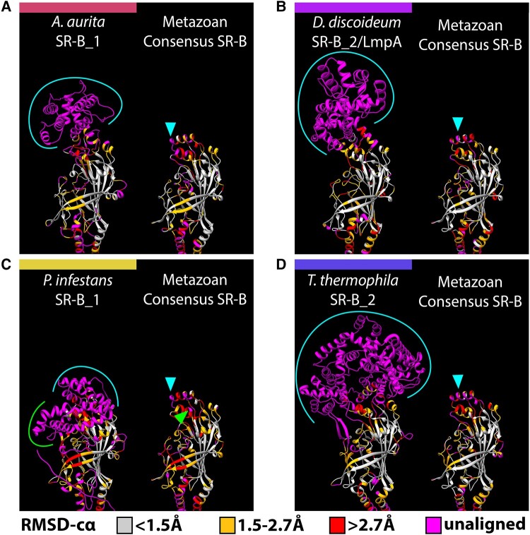
Fig. 6. Sequence expansions coincident with the SR-B apex are divergent and have independent origins. AlphaFold2 structure predictions for four eukaryotic SR-B exemplars with lineage-specific apex structures, (A) A. aurita SR-B 1, (B) D. discoideum SR-B_2 (LmpA), (C) P. infestans SR-B_1, and (D) T. thermophila SR-B_2, are each presented alongside the metazoan consensus SR-B structure, which serves as a structural proxy for the “three-helix bundle” SR-B apex. Structural superpositions were first performed for each exemplar SR-B with the metazoan consensus SR-B sequence, and alignments were generated based on the most spatially adjacent residues for each superposition. Residues in each model are color-coded by the RMSD-cα (root mean square deviation between the alpha carbons in each residue pair) to visualize structurally conserved versus divergent regions. RMSD-cα values are binned according to the categories of “high” (<1.5 Å), “medium” (1.5–2.7 Å), and “low” (>2.7 Å) resolution used to assess the quality of crystallographic protein models (Wlodawer et al. 2007). Residues that fail to align are “unaligned” and colored magenta. Brackets highlight stretches of unaligned, lineage-specific residues that correspond with arrowheads in the metazoan consensus SR-B and represent the location of sequence expansion. Positionally, lineage-specific apex sequence expansions are consistently found to correspond with the three-helix bundle at the apex of the canonical SR-B structure.
Fig. 6. Sequence expansions coincident with the SR-B apex are divergent and have independent origins. AlphaFold2 structure predictions for four eukaryotic SR-B exemplars with lineage-specific apex structures, (A) A. aurita SR-B 1, (B) D. discoideum SR-B_2 (LmpA), (C) P. infestans SR-B_1, and (D) T. thermophila SR-B_2, are each presented alongside the metazoan consensus SR-B structure, which serves as a structural proxy for the “three-helix bundle” SR-B apex. Structural superpositions were first performed for each exemplar SR-B with the metazoan consensus SR-B sequence, and alignments were generated based on the most spatially adjacent residues for each superposition. Residues in each model are color-coded by the RMSD-cα (root mean square deviation between the alpha carbons in each residue pair) to visualize structurally conserved versus divergent regions. RMSD-cα values are binned according to the categories of “high” (<1.5 Å), “medium” (1.5–2.7 Å), and “low” (>2.7 Å) resolution used to assess the quality of crystallographic protein models (Wlodawer et al. 2007). Residues that fail to align are “unaligned” and colored magenta. Brackets highlight stretches of unaligned, lineage-specific residues that correspond with arrowheads in the metazoan consensus SR-B and represent the location of sequence expansion. Positionally, lineage-specific apex sequence expansions are consistently found to correspond with the three-helix bundle at the apex of the canonical SR-B structure.CC BY · PubMed Central

Verification

Claims the paper makes

The claims and statistics of this paper haven’t been checked yet.

Run the check — free, no account needed. It reads the open-access text and checks the sources it cites.

A few free checks a day · papers already verified open instantly · results are public

Self-check

Percentages, sums and reported differences re-computed from the paper’s own numbers.

The self-check needs the paper’s full text.

Citations

Cites

Research works printed but not linked: 76

  1. 10.1038/ni1253 · DOI
  2. 10.1126/science.abj8754 · DOI · Paper page
  3. 10.1093/bioinformatics/btab203 · DOI
  4. 10.1016/j.dci.2015.02.001 · DOI
  5. 10.1016/j.tree.2019.08.008 · DOI · Paper page
  6. 10.1093/bioinformatics/btp348 · DOI
  7. 10.1016/j.ympev.2016.10.011 · DOI
  8. 10.1084/jem.20211314 · DOI
  9. 10.1371/journal.pcbi.1002708 · DOI
  10. 10.1038/s41467-017-02044-8 · DOI
  11. 10.1093/molbev/msz189 · DOI
  12. 10.1093/molbev/msw168 · DOI
  13. 10.1016/j.molbiopara.2005.12.010 · DOI
  14. 10.1016/j.neuroscience.2020.11.003 · DOI
  15. 10.1093/bioinformatics/14.9.755 · DOI · Paper page
  16. 10.4049/jimmunol.0901374 · DOI
  17. 10.1093/gbe/evaa241 · DOI
  18. 10.1016/S1074-7613(00)80410-0 · DOI
  19. 10.1093/bioinformatics/bts565 · DOI · Paper page
  20. 10.1194/jlr.R082933 · DOI
  21. 10.1038/ncomms11866 · DOI
  22. 10.1038/s41592-022-01685-y · DOI · Paper page
  23. 10.1038/s41467-019-11425-0 · DOI · Paper page
  24. 10.1038/s41467-021-21511-x · DOI · Paper page
  25. 10.1038/ncomms12837 · DOI
  26. 10.1002/prot.22458 · DOI
  27. 10.1074/jbc.M114.627026 · DOI
  28. 10.1074/jbc.M709195200 · DOI
  29. 10.1083/jcb.145.1.167 · DOI
  30. 10.1093/molbev/mst010 · DOI · Paper page
  31. 10.1093/nar/gkh468 · DOI
  32. 10.1126/science.1102218 · DOI
  33. 10.1006/jmbi.2000.4315 · DOI · Paper page
  34. 10.1074/jbc.M113.473298 · DOI
  35. 10.1093/gbe/evab101 · DOI
  36. 10.1093/molbev/msab170 · DOI · Paper page
  37. 10.1093/bioinformatics/btt473 · DOI · Paper page
  38. 10.1096/fasebj.10.1.8566545 · DOI
  39. 10.1084/jem.20082109 · DOI
  40. 10.1038/s41592-022-01488-1 · DOI · Paper page
  41. 10.1093/nar/gkaa913 · DOI · Paper page
  42. 10.1186/1471-2164-15-316 · DOI
  43. 10.1093/database/baaa029 · DOI
  44. 10.1242/jcs.01083 · DOI
  45. 10.1038/nature12684 · DOI
  46. 10.7717/peerj.2692 · DOI
  47. 10.1016/j.ibmb.2007.11.003 · DOI
  48. 10.1091/mbc.E10-03-0251 · DOI · Paper page
  49. 10.1021/bi2005625 · DOI
  50. 10.1073/pnas.1300021110 · DOI
  51. 10.1002/jcc.20084 · DOI
  52. 10.4049/jimmunol.1700373 · DOI
  53. 10.1371/journal.pone.0009490 · DOI · Paper page
  54. 10.1093/nar/gki442 · DOI
  55. 10.1046/j.1432-1327.1998.2570488.x · DOI
  56. 10.7554/eLife.34226 · DOI
  57. 10.1016/j.gde.2019.09.002 · DOI
  58. 10.24072/pcjournal.173 · DOI
  59. 10.1093/bioinformatics/btg180 · DOI
  60. 10.1242/jcs.218040 · DOI
  61. 10.1074/jbc.R114.619304 · DOI
  62. 10.1038/ni.2639 · DOI · Paper page
  63. 10.1126/scisignal.272re3 · DOI · Paper page
  64. 10.1093/bioinformatics/btu033 · DOI · Paper page
  65. 10.1038/ni.1836 · DOI · Paper page
  66. 10.1038/s41467-021-22044-z · DOI · Paper page
  67. 10.1074/jbc.M702887200 · DOI
  68. 10.1186/s12964-021-00812-0 · DOI
  69. 10.1006/bbrc.1997.7755 · DOI
  70. 10.1093/nar/gky473 · DOI · Paper page
  71. 10.1093/nar/gkab1061 · DOI · Paper page
  72. 10.1534/genetics.107.085936 · DOI
  73. 10.1111/j.1742-4658.2007.06178.x · DOI
  74. 10.1006/jmbi.2000.3975 · DOI
  75. 10.1021/bi301203x · DOI
  76. 10.1002/prot.20264 · DOI · Paper page

Metadata

Authors
Boohar, R. T., Vandepas, L. E., Traylor-Knowles, N., & Browne, W. E.
Affiliations
University of Miami (US)
Corresponding author
Browne, William E.
Venue
Genome Biology and Evolution
Publisher
openRxiv
Published
2023-11-16
Volume
15
Issue
12
ISSN
1759-6653 (ISSN-L)
OpenAlex
W4389208207
Cited by
11
References
1
Citations by year
2026: 2 · 2024: 9
FWCI
5.07
Type
journal article
Language
en
Open access
Licence
cc-by
Locations
  • Genome Biology and Evolution (publishedVersion, cc-by)
  • bioRxiv (Cold Spring Harbor Laboratory) (acceptedVersion, cc-by-nc-nd)
  • PubMed Central (publishedVersion, cc-by)
  • PubMed (publishedVersion)
Funders
National Science Foundation (2013692)
Topics
  • Life Sciences › Biochemistry, Genetics and Molecular Biology › Biochemistry › Cholesterol and Lipid Metabolism
  • Life Sciences › Biochemistry, Genetics and Molecular Biology › Molecular Biology › Genomics and Phylogenetic Studies
  • Life Sciences › Biochemistry, Genetics and Molecular Biology › Molecular Biology › Protein Structure and Dynamics
Keywords
scavenger receptor class B, protein evolution, last eukaryotic common ancestor, eukaryotic supergroups, innate immunity, fatty acid uptake, cholesterol transport, efferocytosis, pathogen recognition
MeSH terms
Ligands, Phylogeny, CD36 Antigens, Scavenger Receptors, Class B, Eukaryota

About these checks

Registry, retraction, Cites and statistics results are computed, not judged, with one exception: a printed reference that no index lists is read once by a model before it counts as a citation error. Claims are extracted by a model and kept only when their quote appears verbatim in the text; each cited source is then checked, with a model’s help, for whether it says what the claim relies on. Those verdicts can be wrong — read the claim’s quote and the cited source before relying on one.

Statistics and arithmetic are recomputed, not judged.

Confidence bands are shown only for fields calibrated on our annotated test set. None is calibrated yet, so no band is shown.

What counts as a claim ›How we measure confidence ›How we check this ›

Are you an author, or do you see a problem with this page? Request a review