

The DOI matches its Crossref record; 1 of the 1 references it cites were found in a registry or an index.
SPDesign: protein sequence designer based on structural sequence profile using ultrafast shape recognition
Open accessPreprint — published as 10.1093/bib/bbae146
Abstract
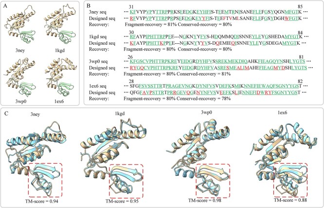
Designing protein with specified structure and function involves a key component named sequence design, which can provide valuable insights into understanding the life systems as well for the diagnosis and therapy of diseases. Although deep learning methods have made great progress in protein sequence design, most of these studies focus on network structure optimization, while ignoring protein-specific physicochemical features. Inspired by the successful application of structure templates and pre-trained models in the field of protein structure prediction, we explored whether the representation of structural sequence profile can be used for protein sequence design. In this work, we proposed SPDesign, a method for protein sequence design based on structural sequence profile using ultrafast shape recognition. Given an input back-bone structure, SPDesign utilizes ultrafast shape recognition vectors to accelerate the search for similar protein structures (aka, structural analogs) in our in-house PAcluster80 structure database, and then extracts the sequence profile from the analogs through structure alignment. Combined with structural pre-trained knowledge and geometric features, they are further feed into an enhanced graph neural network to predict the sequence. Experimental results show that SPDesign significantly outperforms the state-of-the-art methods, such as ProteinMPNN, Pifold and LM-Design, leading to 21.89%, 15.54% and 11.4% accuracy gains in sequence recovery rate on CATH 4.2 benchmark, respectively. Encouraging results also have been achieved on the TS50 and TS500 benchmarks, with performance reaching 68.64% and 71.63%. Furthermore, detailed analysis conducted by the PDBench tool suggest that SPDesign performs well in subdivided structures such as buried residues and solenoid. More interestingly, we found that SPDesign can well reconstruct the sequences of some proteins that have similar structures but different sequences. Finally, the structural modeling verification experiment bears out that the sequences designed by our method can fold into the native structures more accurately.
Overview
The claims and statistics of this paper haven’t been checked yet.
Run the check — free, no account needed. It reads the open-access text and checks the sources it cites.
Integrity signals
Registry
Cites
Statistics
Figures
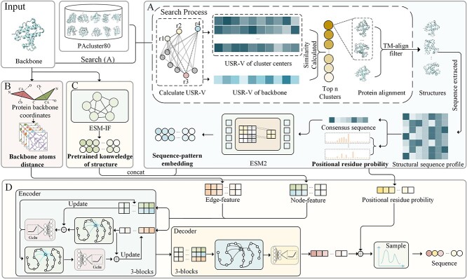
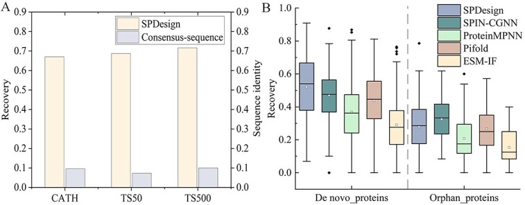
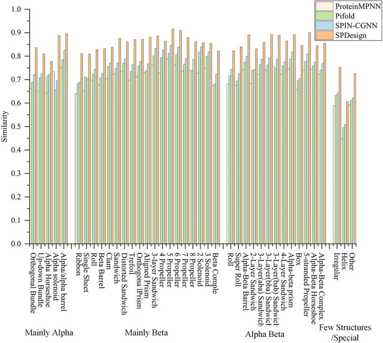
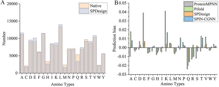
Verification
Claims the paper makes
The claims and statistics of this paper haven’t been checked yet.
Run the check — free, no account needed. It reads the open-access text and checks the sources it cites.
Self-check
Percentages, sums and reported differences re-computed from the paper’s own numbers.
The self-check needs the paper’s full text.
Citations
Cites
Highly accurate protein structure prediction with AlphaFold
Jumper, J., Evans, R., Pritzel, A., Green, T., Figurnov, M., Ronneberger, O., Tunyasuvunakool, K., Bates, R., Žídek, A., Potapenko, A., et al. (2021) · Nature · doi:10.1038/s41586-021-03819-2 · Cited by 43,764
Research works printed but not linked: 46
- 2019, Unified rational protein engineering with sequence-based deep representation learning, Nat. Methods, 16, 1315, 10.1038/s41592-019-0598-1 · DOI · Paper page
- 10.1093/nar/gkz1064 · DOI
- 2019, Accurate prediction of protein structures and interactions using a threetrack neural network, Science, 373, 871
- 2019, Ultrafast shape recognition to search compound databases for similar molecular shapes, J Comput Chem, 28, 1711
- 10.1006/jmbi.2000.4427 · DOI
- Burley, S. K. et al. (2017) Protein Data Bank (PDB): the single global macromolecular structure archive. Protein crystallography: methods and protocols, 627–641. · DOI · Paper page
- 2022, ProteinBERT: a universal deep-learning model of protein sequence and function, Bioinformatics, 38, 2102, 10.1093/bioinformatics/btac020 · DOI · Paper page
- 10.1093/bioinformatics/btad027 · DOI
- 10.1101/628917 · DOI
- 2022, Single-sequence protein structure prediction using a language model and deep learning, Nat. Biotechnol, 40, 1617, 10.1038/s41587-022-01432-w · DOI · Paper page
- 2021, High-level expression of palmitoylated MPP1 recombinant protein in mammalian cells, Membranes, 11, 715, 10.3390/membranes11090715 · DOI
- 10.1126/science.add2187 · DOI · Paper page
- 10.3390/ijms222111741 · DOI
- 10.1093/bib/bbac102 · DOI · Paper page
- 10.48550/arXiv.2202.01079 · DOI
- 10.48550/arXiv.2305.15151 · DOI
- 10.48550/arXiv.2209.12643 · DOI
- 2017, Neural message passing for quantum chemistry, ICML 2017, 3, 2053
- 2022, DeepUMQA: ultrafast shape recognition-based protein model quality assessment using deep learning, Bioinformatics, 38, 1895, 10.1093/bioinformatics/btac056 · DOI · Paper page
- Hsu, C. et al. (2022) Learning inverse folding from millions of predicted structures. ICML 2022, 8946–8970. · DOI
- 2023, Accurate and efficient protein sequence design through learning concise local environment of residues, Bioinformatics, 39, btad122, 10.1093/bioinformatics/btad122 · DOI · Paper page
- 10.48550/arXiv.2009.01411 · DOI
- 2020, De novo protein design for novel folds using guided conditional wasserstein generative adversarial networks, J Chem Inf Model, 60, 5667, 10.1021/acs.jcim.0c00593 · DOI
- 10.1074/jbc.M110792200 · DOI
- 10.1002/prot.24620 · DOI
- 10.1101/2022.07.20.500902 · DOI · Paper page
- 2018, Minimalist de novo design of protein catalysts, ACS Catal, 9, 9265
- 2018, Spin2: Predicting sequence profiles from protein structures using deep neural networks, Proteins, 86, 629, 10.1002/prot.25489 · DOI
- 2021, Structure-based protein design with deep learning, Curr Opin Chem Biol, 65, 136, 10.1016/j.cbpa.2021.08.004 · DOI · Paper page
- 10.1016/j.sbi.2021.01.007 · DOI · Paper page
- 10.1021/acs.jcim.0c00043 · DOI
- 2019, Evaluating Protein Transfer Learning with TAPE, NeurIPS 2019, 32, 9689
- 2020, CATH: increased structural coverage of functional space, Nucleic Acids Res, 49, D266
- 10.1016/j.sbi.2021.11.008 · DOI
- 10.48550/arXiv.2204.10673 · DOI
- 2021, AlphaFold Protein Structure Database: massively expanding the structural coverage of protein-sequence space with high-accuracy models, Nucleic Acids Res, 50, D439
- Vaswani, A. et al. (2017) Attention is all you need. NeurIPS 2017, 30.
- 10.1038/s41598-018-23276-8 · DOI
- 2021, Representing long-range context for graph neural networks with global attention, NeurIPS 2021, 34, 13266
- 2023, SPIN-CGNN: Improved fixed backbone protein design with contact map-based graph construction and contact graph neural network, PLOS COMPUT BIOL, 19, e1011330, 10.1371/journal.pcbi.1011330 · DOI · Paper page
- 10.1093/nar/gki524 · DOI · Paper page
- 2020, Prodconn: Protein design using a convolutional neural network, Proteins, 88, 819, 10.1002/prot.25868 · DOI
- 2023, Protein structure and folding pathway prediction based on remote homologs recognition using pathreader, Commun. Biol, 6, 243, 10.1038/s42003-023-04605-8 · DOI
- 2021, MMpred: a distance-assisted multimodal conformation sampling for de novo protein structure prediction, Bioinformatics, 37, 4350, 10.1093/bioinformatics/btab484 · DOI
- 10.48550/arXiv.2302.01649 · DOI
- 10.1038/emboj.2011.428 · DOI
Metadata
- Authors
- Wang, H., Liu, D., Zhao, K.-L., Wang, Y.-J., & Zhang, G.-J.
- Venue
- Briefings in Bioinformatics
- Publisher
- openRxiv
- Published
- 2023-12-14
- Volume
- 25
- Issue
- 3
- ISSN
- 1467-5463 (ISSN-L)
- PMC
- PMC11006797
- OpenAlex
- W4394741301
- Cited by
- 6
- References
- 1
- Citations by year
- 2026: 2 · 2025: 2 · 2024: 2
- FWCI
- 0.81
- Type
- journal article
- Language
- en
- Open access
- gold
- doi.org
- Licence
- cc-by-nc
- Locations
- Briefings in Bioinformatics (publishedVersion, cc-by-nc)
- bioRxiv (Cold Spring Harbor Laboratory) (acceptedVersion)
- PubMed Central (publishedVersion, cc-by-nc)
- PubMed (publishedVersion)
- Funders
- National Natural Science Foundation of China (LZ20F030002, 62173304)
- National Science and Technology Major Project (2022ZD0115103)
- Natural Science Foundation of Zhejiang Province (LZ20F030002)
- Topics
- Life Sciences › Biochemistry, Genetics and Molecular Biology › Molecular Biology › Protein Structure and Dynamics
- Life Sciences › Biochemistry, Genetics and Molecular Biology › Molecular Biology › Machine Learning in Bioinformatics
- Life Sciences › Pharmacology, Toxicology and Pharmaceutics › Drug Discovery › Computational Drug Discovery Methods
- Keywords
- protein sequence design, graph neural networks, structural alignment, sequence recovery, de novo protein design, orphan proteins, protein structure prediction, geometric features
- MeSH terms
- Amino Acid Sequence, Proteins, Sequence Alignment, Neural Networks, Computer, Sequence Analysis, Protein
About these checks
Registry, retraction, Cites and statistics results are computed, not judged, with one exception: a printed reference that no index lists is read once by a model before it counts as a citation error. Claims are extracted by a model and kept only when their quote appears verbatim in the text; each cited source is then checked, with a model’s help, for whether it says what the claim relies on. Those verdicts can be wrong — read the claim’s quote and the cited source before relying on one.
Statistics and arithmetic are recomputed, not judged.
Confidence bands are shown only for fields calibrated on our annotated test set. None is calibrated yet, so no band is shown.
What counts as a claim ›How we measure confidence ›How we check this ›
Are you an author, or do you see a problem with this page? Request a review
