Student or educator? Sign up with your school email and get 6 months of Pro free.
PreprintCited by 9Metalloenzymes and iron-sulfur proteinsMetal Extraction and BioleachingMicrobial metabolism and enzyme function

The DOI matches its Crossref record; 1 of the 1 references it cites were found in a registry or an index.

A cascade of sulfur transferases delivers sulfur to the sulfur-oxidizing heterodisulfide reductase-like complex

Open access

Preprint — published as 10.1002/pro.5014 · Paper page

Abstract

A heterodisulfide reductase-like complex (sHdr) and novel lipoate-binding proteins (LbpAs) are central players of a wide-spread pathway of dissimilatory sulfur oxidation. Bioinformatic analysis demonstrate that the cytoplasmic sHdr-LbpA systems are always accompanied by sets of sulfur transferases (DsrE proteins, TusA, rhodaneses). The exact composition of these sets may vary depending on the organism and sHdr system type. To enable generalizations, we studied model sulfur oxidizers from distant bacterial phyla, i.e. Aquificota and Pseudomonadota. DsrE3C of the chemoorganotrophic Alphaproteobacterium Hyphomicrobium denitrificans and DsrE3B from the Gammaproteobacteria Thioalkalivibrio sp. K90mix, an obligate chemolithotroph, and Thiorhodospira sibirica , an obligate photolithotroph, are homotrimers that donate sulfur to TusA. Additionally, the hyphomicrobial rhodanese-like protein Rhd442 exchanges sulfur with both TusA and DsrE3C. The latter is essential for sulfur oxidation in Hm. denitrificans . TusA from Aquifex aeolicus (AqTusA) interacts physiologically with AqDsrE, AqLbpA and AqsHdr proteins. This is particularly significant as it establishes a direct link between sulfur transferases and the sHdr-LbpA complex that oxidizes sulfane sulfur to sulfite. In vivo, it is unlikely that there is a strict unidirectional transfer between the sulfur-binding enzymes studied. Rather, the sulfur transferases form a network, each with a pool of bound sulfur. Sulfur flux can then be shifted in one direction or the other depending on metabolic requirements. A single pair of sulfur-binding proteins with a preferred transfer direction, such as a DsrE3-type protein towards TusA, may be sufficient to push sulfur into the sink where it is further metabolized or needed. SIGNIFICANCE STATEMENT: A network of bacterial sulfur transferases is uncovered and characterized that ultimately delivers sulfur to a complex cytoplasmic sulfur-oxidizing metalloenzyme, sHdr, that resembles heterodisulfide reductase from methanogenic archaea and interacts with lipoate-binding proteins. Similar sets of sulfur transferases occur in phylogenetically distant bacteria, underscoring the fundamental importance of the work.

Overview

The claims and statistics of this paper haven’t been checked yet.

Run the check — free, no account needed. It reads the open-access text and checks the sources it cites.

A few free checks a day · papers already verified open instantly · results are public

Integrity signals

Registry

Crossref · 10.1101/2023.12.18.572138

Cites

1 of 1 references verified0 citation errors · 0 not checkable

Statistics

Figures

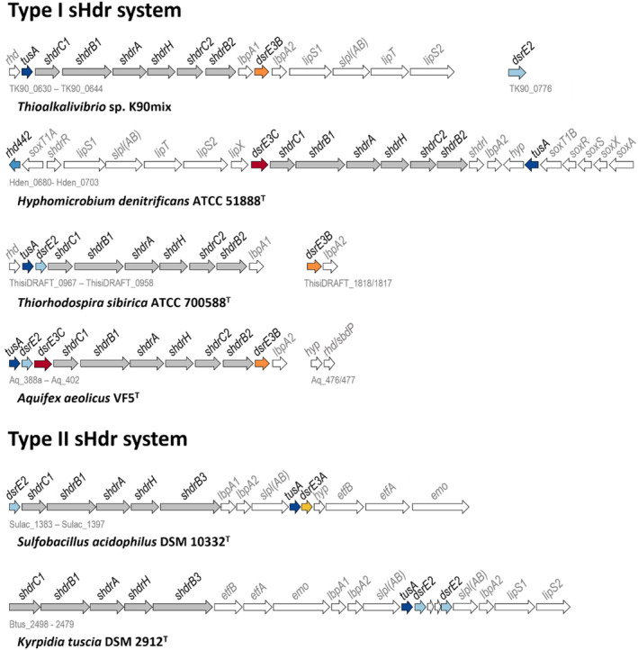
FIGURE 1 Representative shdr gene clusters in sulfur oxidizers. The KEGG/NCBI locus tag identifiers for the first and last genes are shown below each cluster. For Ts. sibirica, locus tags are given according to JGI IMG. Genes for TusA, DsrE2, DsrE3A, DsrE3B, and DsrE3C are indicated in dark blue, light blue, yellow, orange, and dark red, respectively. Genes for probable components of the sulfur‐oxidizing heterodisulfide reductase‐like (sHdr) complex are shaded in gray. EMO, ETF:(methyl)menaquinone oxidoreductase; Etf, electron transfer protein. EtfAB and EMO have been proposed to direct electrons stemming from sulfane sulfur oxidation to menaquinone (Kümpel et al., 2024). LbpA, lipoate‐binding protein; sLpl(AB), lipoate: protein ligase; LipS1/S2, lipoyl synthase; LipT, FAD‐binding NAD(P)H‐dependent oxidoreductase possibly delivering electrons for the LipS1/S2‐catalyzed sulfur insertion step (Kümpel et al., 2024; Tanabe et al., 2023); Rhd, rhodanese; SbdP, sulfur‐binding‐donating protein.
FIGURE 1 Representative shdr gene clusters in sulfur oxidizers. The KEGG/NCBI locus tag identifiers for the first and last genes are shown below each cluster. For Ts. sibirica, locus tags are given according to JGI IMG. Genes for TusA, DsrE2, DsrE3A, DsrE3B, and DsrE3C are indicated in dark blue, light blue, yellow, orange, and dark red, respectively. Genes for probable components of the sulfur‐oxidizing heterodisulfide reductase‐like (sHdr) complex are shaded in gray. EMO, ETF:(methyl)menaquinone oxidoreductase; Etf, electron transfer protein. EtfAB and EMO have been proposed to direct electrons stemming from sulfane sulfur oxidation to menaquinone (Kümpel et al., 2024). LbpA, lipoate‐binding protein; sLpl(AB), lipoate: protein ligase; LipS1/S2, lipoyl synthase; LipT, FAD‐binding NAD(P)H‐dependent oxidoreductase possibly delivering electrons for the LipS1/S2‐catalyzed sulfur insertion step (Kümpel et al., 2024; Tanabe et al., 2023); Rhd, rhodanese; SbdP, sulfur‐binding‐donating protein.CC BY-NC · PubMed Central
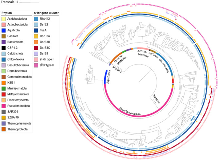
FIGURE 2 Taxonomic distribution of type I or type II sHdr systems in sulfur‐oxidizing prokaryotes. The distribution of TusA, Rhd442, and DsrE‐type sulfur transferases is also visualized. The data underlying the figure is provided in Table S1.
FIGURE 2 Taxonomic distribution of type I or type II sHdr systems in sulfur‐oxidizing prokaryotes. The distribution of TusA, Rhd442, and DsrE‐type sulfur transferases is also visualized. The data underlying the figure is provided in Table S1.CC BY-NC · PubMed Central
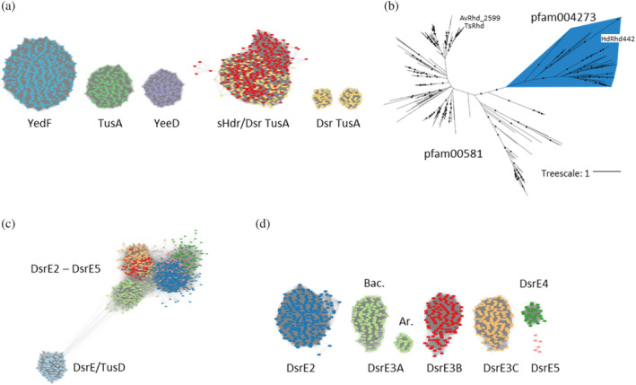
FIGURE 3 Sequence similarity network (SSN) and phylogenetic analyses for sulfur transferases relevant for dissimilatory sulfur oxidation. (a) SSN for the TusA family (pfam01206) members clustering at 50% identity with TusA, YedF, and YeeD from E. coli and TusA‐like proteins that are genetically linked with sHdr and/or Dsr sulfur‐oxidation systems. Connections below a threshold score of 100 were removed. (b) Maximum likelihood tree for ~100‐aa single domain rhodaneses that are encoded in synteny with DsrE2 and TusA or that are related to Rhd442 from Hm. denitrificans XT. Bootstrap values exceeding 95% are indicated by dots. (c) SSN for all DsrE‐type sequences from the representative genomes of GTDB (release R207). (d) SSN for all DsrE homologs associated with rDsr and/or sHdr systems, excluding the divergent TusD/DsrE proteins. Connections below a threshold score of 135 were removed. Ar, Archaea; Bac., Bacteria.
FIGURE 3 Sequence similarity network (SSN) and phylogenetic analyses for sulfur transferases relevant for dissimilatory sulfur oxidation. (a) SSN for the TusA family (pfam01206) members clustering at 50% identity with TusA, YedF, and YeeD from E. coli and TusA‐like proteins that are genetically linked with sHdr and/or Dsr sulfur‐oxidation systems. Connections below a threshold score of 100 were removed. (b) Maximum likelihood tree for ~100‐aa single domain rhodaneses that are encoded in synteny with DsrE2 and TusA or that are related to Rhd442 from Hm. denitrificans XT. Bootstrap values exceeding 95% are indicated by dots. (c) SSN for all DsrE‐type sequences from the representative genomes of GTDB (release R207). (d) SSN for all DsrE homologs associated with rDsr and/or sHdr systems, excluding the divergent TusD/DsrE proteins. Connections below a threshold score of 135 were removed. Ar, Archaea; Bac., Bacteria.CC BY-NC · PubMed Central
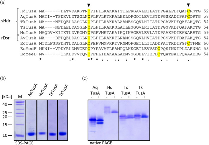
FIGURE 4 (a) Alignment of a portion of TusA and related proteins from sulfur oxidizers and E. coli. Ec, E. coli (TusA, b3470, YedF, b1930, YeeD, and b2012); Ts, Ts. sibirica (ThisiDRAFT_0966, according to JGI IMG); Tk, Thioalkalivibrio sp. K90mix (TK90_0631); Av, Ac. vinosum (Alvin_2600); Hd, Hm. denitrificans (Hden_0698); Aq, Aq. aeolicus (Aq_388a); Mc, Ms. cuprina (Mcup_0683). Triangles indicate the cysteines that were exchanged to serine in this work. Asterisk, fully conserved residues; colon, conservation between groups of strongly similar properties; dot, conservation between groups of weakly similar properties. (b) 20% SDS–PAGE of recombinant TusAs HdTusA, TkTusA, TsTusA, and AqTusA reduced with DTT. (c) 20% native PAGE of 3.5 μg recombinant TusAs HdTusA, TkTusA, and TsTusA and AqTusA as purified (−) and reduced with 5 mM DTT (+).
FIGURE 4 (a) Alignment of a portion of TusA and related proteins from sulfur oxidizers and E. coli. Ec, E. coli (TusA, b3470, YedF, b1930, YeeD, and b2012); Ts, Ts. sibirica (ThisiDRAFT_0966, according to JGI IMG); Tk, Thioalkalivibrio sp. K90mix (TK90_0631); Av, Ac. vinosum (Alvin_2600); Hd, Hm. denitrificans (Hden_0698); Aq, Aq. aeolicus (Aq_388a); Mc, Ms. cuprina (Mcup_0683). Triangles indicate the cysteines that were exchanged to serine in this work. Asterisk, fully conserved residues; colon, conservation between groups of strongly similar properties; dot, conservation between groups of weakly similar properties. (b) 20% SDS–PAGE of recombinant TusAs HdTusA, TkTusA, TsTusA, and AqTusA reduced with DTT. (c) 20% native PAGE of 3.5 μg recombinant TusAs HdTusA, TkTusA, and TsTusA and AqTusA as purified (−) and reduced with 5 mM DTT (+).CC BY-NC · PubMed Central
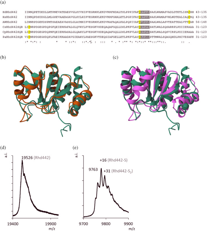
FIGURE 5 (a) Alignment of single domain Rhd442 from Hm. denitrificans (Hd), Hyphomicrobium sp018242215 (Hs), and AWTP1‐13‐sp008933705 (AW) with Rhd442 domains fused to sulfide: quinone oxidoreductase (SQR) from Cupriavidus sp. amp6 (Cs), Cupriavidus pinatubonensis (Cp), Pseudomonas veronii (Pa) (Xin et al., 2016). Partially and fully conserved cysteines are highlighted in yellow. Sequences conforming to the rhodanese active site sequence logos (Ran et al., 2022) are shaded in gray. Asterisk, fully conserved residues; colon, conservation between groups of strongly similar properties; dot, conservation between groups of weakly similar properties. (b) HdRhd442 Alphafold structure (green) overlayed with Rhd442 domain of Cv. pinatubonensis SQR (brown) and (c) non‐classical phosphatase from Ns. meningitidis (PDB 2F46, purple). (d) Mass spectrum of recombinant HdRhd442 as isolated. (e) HdRhd442 mass spectrum after incubation with 0.5 mM polysulfide. Note that a distinguishable signal for Rhd442 and its persulfidated species was only observable for the double ionized protein.
FIGURE 5 (a) Alignment of single domain Rhd442 from Hm. denitrificans (Hd), Hyphomicrobium sp018242215 (Hs), and AWTP1‐13‐sp008933705 (AW) with Rhd442 domains fused to sulfide: quinone oxidoreductase (SQR) from Cupriavidus sp. amp6 (Cs), Cupriavidus pinatubonensis (Cp), Pseudomonas veronii (Pa) (Xin et al., 2016). Partially and fully conserved cysteines are highlighted in yellow. Sequences conforming to the rhodanese active site sequence logos (Ran et al., 2022) are shaded in gray. Asterisk, fully conserved residues; colon, conservation between groups of strongly similar properties; dot, conservation between groups of weakly similar properties. (b) HdRhd442 Alphafold structure (green) overlayed with Rhd442 domain of Cv. pinatubonensis SQR (brown) and (c) non‐classical phosphatase from Ns. meningitidis (PDB 2F46, purple). (d) Mass spectrum of recombinant HdRhd442 as isolated. (e) HdRhd442 mass spectrum after incubation with 0.5 mM polysulfide. Note that a distinguishable signal for Rhd442 and its persulfidated species was only observable for the double ionized protein.CC BY-NC · PubMed Central
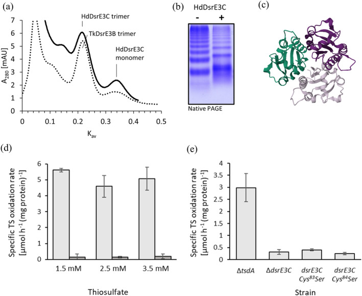
FIGURE 6 Function and properties of DsrE3B and DsrE3C proteins. (a) Gel permeation chromatography of HdDsrE3C and TkDsrE3B on Hiload 16/60 Superdex 75. (b) Native‐PAGE of 3.5 μg HdDsrE3C as purified (−) and reduced with 5 mM DTT (+). (c) Alphafold model of the HdDsrE3C trimer. (d) Specific thiosulfate (TS) oxidation rates for Hm. denitrificans ΔtsdA (light gray columns) and Hm. denitrificans ΔtsdA ΔdsrE3C (dark gray columns) at the indicated initial thiosulfate concentrations. The corresponding growth curves are provided in Figure S1. Precultures contained 2 mM thiosulfate. (e) Specific thiosulfate oxidation rates of Hm. denitrificans ΔtsdA compared to a strain lacking the complete dsrE3C gene and two strains carrying dsrE3C genes encoding the indicating cysteine to serine exchanges grown with 2.5 mM thiosulfate. Note that specific thiosulfate oxidation rates are not fully comparable to the experiments shown in (d) because the growth experiments shown here were performed in a plate reader.
FIGURE 6 Function and properties of DsrE3B and DsrE3C proteins. (a) Gel permeation chromatography of HdDsrE3C and TkDsrE3B on Hiload 16/60 Superdex 75. (b) Native‐PAGE of 3.5 μg HdDsrE3C as purified (−) and reduced with 5 mM DTT (+). (c) Alphafold model of the HdDsrE3C trimer. (d) Specific thiosulfate (TS) oxidation rates for Hm. denitrificans ΔtsdA (light gray columns) and Hm. denitrificans ΔtsdA ΔdsrE3C (dark gray columns) at the indicated initial thiosulfate concentrations. The corresponding growth curves are provided in Figure S1. Precultures contained 2 mM thiosulfate. (e) Specific thiosulfate oxidation rates of Hm. denitrificans ΔtsdA compared to a strain lacking the complete dsrE3C gene and two strains carrying dsrE3C genes encoding the indicating cysteine to serine exchanges grown with 2.5 mM thiosulfate. Note that specific thiosulfate oxidation rates are not fully comparable to the experiments shown in (d) because the growth experiments shown here were performed in a plate reader.CC BY-NC · PubMed Central
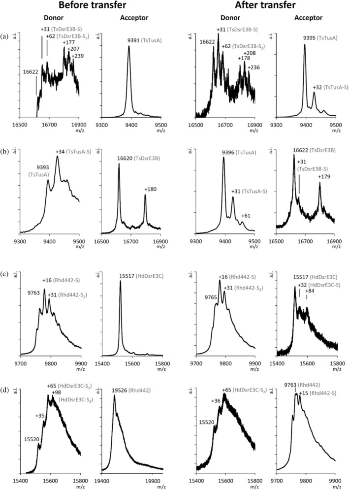
FIGURE 7 Sulfur transfer between TusA, DsrE3 proteins, and Rhd442. Sulfur transfer reaction between TusA and DsrE3B from Ts. sibirica and between Rhd442 and DsrE3C from Hm. denitrificans are shown as examples. The full set of experiments is available as Figures S2, S3, and S4. (a) Left panels: TsDsrE3B as persulfidated donor after treatment with polysulfide and unmodified reduced TsTusA as acceptor; right panels: DsrE3B (donor) and TusA (acceptor) after the transfer reaction. The TsDsrE3B species exhibiting additional 178 Da are due to glucosylation of the His‐Tagged protein (Geoghegan et al., 1999). (b) Left panels: TsTusA as persulfidated donor after treatment with polysulfide and unmodified reduced TsDsrE3B as acceptor; right panels: TsTusA and TsDsrE3B after the transfer reaction. (c) Left panels: HdRhd442 as persulfidated donor after treatment with polysulfide and unmodified reduced HdDsrE3C as acceptor. Right panels: HdRhd442 (donor) and HdDsrE3C (acceptor) after the transfer reaction. (d) Left panels: HdDsrE3C as persulfidated donor after treatment with polysulfide and unmodified reduced HdRhd442 as acceptor. Right panels: HdDsrE3C (donor) and HdRhd442 (acceptor) after the transferreaction. Note that the spectrum on the far right shows the doubly charged ion.
FIGURE 7 Sulfur transfer between TusA, DsrE3 proteins, and Rhd442. Sulfur transfer reaction between TusA and DsrE3B from Ts. sibirica and between Rhd442 and DsrE3C from Hm. denitrificans are shown as examples. The full set of experiments is available as Figures S2, S3, and S4. (a) Left panels: TsDsrE3B as persulfidated donor after treatment with polysulfide and unmodified reduced TsTusA as acceptor; right panels: DsrE3B (donor) and TusA (acceptor) after the transfer reaction. The TsDsrE3B species exhibiting additional 178 Da are due to glucosylation of the His‐Tagged protein (Geoghegan et al., 1999). (b) Left panels: TsTusA as persulfidated donor after treatment with polysulfide and unmodified reduced TsDsrE3B as acceptor; right panels: TsTusA and TsDsrE3B after the transfer reaction. (c) Left panels: HdRhd442 as persulfidated donor after treatment with polysulfide and unmodified reduced HdDsrE3C as acceptor. Right panels: HdRhd442 (donor) and HdDsrE3C (acceptor) after the transfer reaction. (d) Left panels: HdDsrE3C as persulfidated donor after treatment with polysulfide and unmodified reduced HdRhd442 as acceptor. Right panels: HdDsrE3C (donor) and HdRhd442 (acceptor) after the transferreaction. Note that the spectrum on the far right shows the doubly charged ion.CC BY-NC · PubMed Central
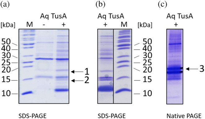
FIGURE 8 Re‐purification experiments of recombinant AqTusA after incubation with Aq. aeolicus soluble extract in different conditions. (a) Proteins eluted after 30 min incubation of AqTusA with Aq. aeolicus soluble extract and separated on a 18% SDS–PAGE. ‘‐’ corresponds to the proteins retained in the control experiment lacking AqTusA (extract only, negative control). ‘+’ corresponds to retained proteins after incubation of soluble cell extract with recombinant AqTusA. Equal volumes of elution fractions, corresponding to 2 and 10 μg of total protein, were loaded into the lanes. Proteins in bands 1 and 2 were analyzed by mass spectrometry (Table S5). (b) Proteins (6 μg) eluted after 10 min incubation of AqTusA with Aq. aeolicus soluble extract and separated on a 18% SDS–PAGE. (c) Proteins (6 μg) eluted after 10 min incubation of AqTusA with Aq. aeolicus soluble extract and separated on a 15% native PAGE. Proteins in band 3 were analyzed by mass spectrometry (Table S6). Molecular weight markers (kDa) for the SDS gels are shown in lanes ‘M'.
FIGURE 8 Re‐purification experiments of recombinant AqTusA after incubation with Aq. aeolicus soluble extract in different conditions. (a) Proteins eluted after 30 min incubation of AqTusA with Aq. aeolicus soluble extract and separated on a 18% SDS–PAGE. ‘‐’ corresponds to the proteins retained in the control experiment lacking AqTusA (extract only, negative control). ‘+’ corresponds to retained proteins after incubation of soluble cell extract with recombinant AqTusA. Equal volumes of elution fractions, corresponding to 2 and 10 μg of total protein, were loaded into the lanes. Proteins in bands 1 and 2 were analyzed by mass spectrometry (Table S5). (b) Proteins (6 μg) eluted after 10 min incubation of AqTusA with Aq. aeolicus soluble extract and separated on a 18% SDS–PAGE. (c) Proteins (6 μg) eluted after 10 min incubation of AqTusA with Aq. aeolicus soluble extract and separated on a 15% native PAGE. Proteins in band 3 were analyzed by mass spectrometry (Table S6). Molecular weight markers (kDa) for the SDS gels are shown in lanes ‘M'.CC BY-NC · PubMed Central
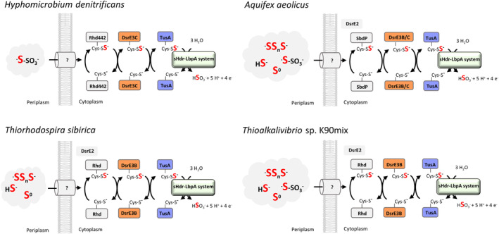
FIGURE 9 Sulfur relay systems in the four bacterial model organisms studied in this work. Hm. denitrificans oxidizes thiosulfate and dimethyl sulfide. Thiosulfate is an intermediate of DMS oxidation (Cao et al., 2018; Koch & Dahl, 2018). Aq. aeolicus (Deckert et al., 1998) and Thioalkalivibrio sp. K90mix (Muyzer et al., 2011) metabolize a wide range of inorganic sulfur substrates including thiosulfate, whereas Ts. sibirica is unable to oxidize thiosulfate (Bryantseva et al., 1999). It was originally predicted that DsrE2A from Aq. aeolicus (Aq_389) is bound to the membrane via two transmembrane segments. However, recent deep‐learning based programs like DeepTMHMM (Hallgren et al., 2022) challenge this view not only for the Aquifex protein but also for the corresponding enzymes found in Ts. sibirica and Thioalkalivibrio sp. K90mix.
FIGURE 9 Sulfur relay systems in the four bacterial model organisms studied in this work. Hm. denitrificans oxidizes thiosulfate and dimethyl sulfide. Thiosulfate is an intermediate of DMS oxidation (Cao et al., 2018; Koch & Dahl, 2018). Aq. aeolicus (Deckert et al., 1998) and Thioalkalivibrio sp. K90mix (Muyzer et al., 2011) metabolize a wide range of inorganic sulfur substrates including thiosulfate, whereas Ts. sibirica is unable to oxidize thiosulfate (Bryantseva et al., 1999). It was originally predicted that DsrE2A from Aq. aeolicus (Aq_389) is bound to the membrane via two transmembrane segments. However, recent deep‐learning based programs like DeepTMHMM (Hallgren et al., 2022) challenge this view not only for the Aquifex protein but also for the corresponding enzymes found in Ts. sibirica and Thioalkalivibrio sp. K90mix.CC BY-NC · PubMed Central

Verification

Claims the paper makes

The claims and statistics of this paper haven’t been checked yet.

Run the check — free, no account needed. It reads the open-access text and checks the sources it cites.

A few free checks a day · papers already verified open instantly · results are public

Self-check

Percentages, sums and reported differences re-computed from the paper’s own numbers.

The self-check needs the paper’s full text.

Citations

Cites

Show all 1 references

Research works printed but not linked: 68

  1. 10.1016/bs.ampbs.2019.07.004 · DOI
  2. 10.1002/iub.1371 · DOI
  3. 10.1038/nchembio779 · DOI · Paper page
  4. 10.1111/j.1574-6976.2006.00036.x · DOI
  5. 2020, On H2S prodrugs, Antioxid Redox Signal, 33, 999, 10.1089/ars.2020.8078 · DOI
  6. Dahl C. A biochemical view on the biological sulfur cycle. In: Lens P , Ed. (2020) Environmental technologies to treat sulfur pollution: principles and engineering. IWA Publishing, London, pp. 55–96. · DOI
  7. -. Sulfur metabolism in phototrophic bacteria. In: Hallenbeck PC , Ed. (2017) Modern topics in the phototrophic prokaryotes: Metabolism, bioenergetics and omics. Springer International Publishing, Cham, pp. 27–66. · DOI
  8. 10.1074/jbc.M113.536425 · DOI
  9. 10.1371/journal.pone.0040785 · DOI
  10. 10.1016/j.jmb.2008.10.016 · DOI
  11. 2018, Lipoate-binding proteins and specific lipoate-protein ligases in microbial sulfur oxidation reveal an atypical role for an old cofactor, eLife, 7, e37439, 10.7554/eLife.37439 · DOI
  12. 10.1038/s41396-018-0209-7 · DOI
  13. 2023, Identification of a novel lipoic acid biosynthesis pathway reveals the complex evolution of lipoate assembly in prokaryotes, PLos Biol, 21, e3002177, 10.1371/journal.pbio.3002177 · DOI
  14. 10.1186/1471-2164-15-1107 · DOI
  15. 10.1074/jbc.M114.591669 · DOI
  16. 10.1093/femsle/fnw156 · DOI
  17. Kümpel C , Grosser M , Tanabe TS , Dahl C (2023) Fe/S proteins in microbial sulfur oxidation. Biochimica et Biophysica Acta (BBA) - Molecular Cell Research submitted. · DOI
  18. 2021, Structural and spectroscopic characterization of a HdrA-like subunit from Hyphomicrobium denitrificans, FEBS J, 288, 1664, 10.1111/febs.15505 · DOI
  19. 2020, The rise of diversity in metabolic platforms across the Candidate Phyla Radiation, BMC Biology, 18, 69, 10.1186/s12915-020-00804-5 · DOI
  20. 10.1186/2049-2618-1-22 · DOI
  21. 2023, HMSS2: an advanced tool for the analysis of sulfur metabolism, including organosulfur compound transformation, in genome and metagenome assemblies, Mol Ecol Resour, 23, 1930, 10.1111/1755-0998.13848 · DOI
  22. 10.1099/00221287-142-4-775 · DOI
  23. 10.1128/aem.03098-16 · DOI
  24. 2019, Genomes of neutrophilic sulfur-oxidizing chemolithoautotrophs representing 9 proteobacterial species from 8 genera, Front Microbiol, 10, 316, 10.3389/fmicb.2019.00316 · DOI
  25. 10.1371/journal.pone.0004345 · DOI · Paper page
  26. 10.1074/jbc.M112.431569 · DOI
  27. 10.1016/j.molcel.2005.11.001 · DOI · Paper page
  28. 10.1271/bbb.64.799 · DOI
  29. Yildiz T , Leimkühler S (2021) TusA is a versatile protein that links translation efficiency to cell division in Escherichia coli . J Bacteriol 203. · DOI
  30. 10.1126/sciadv.aba7637 · DOI
  31. 2017, First insights into the pleiotropic role of vrf (yedF), a newly characterized gene of Salmonella Typhimurium, Sci Rep, 7, 15291, 10.1038/s41598-017-15369-7 · DOI
  32. 1973, Rhodanese, Advances in Enzymology and Related Areas of Molecular Biology, 39, 327
  33. 10.1093/embo-reports/kvf150 · DOI · Paper page
  34. 10.1074/jbc.M111.324863 · DOI
  35. 2022, HMS-S-S: a tool for the identification of sulphur metabolism-related genes and analysis of operon structures in genome and metagenome assemblies, Mol Ecol Resour, 22, 2758, 10.1111/1755-0998.13642 · DOI
  36. 10.1371/journal.pbio.1000354 · DOI · Paper page
  37. 10.1038/32831 · DOI
  38. 10.1111/1462-2920.13511 · DOI
  39. 2022, Rhodaneses minimize the accumulation of cellular sulfane sulfur to avoid disulfide stress during sulfide oxidation in bacteria, Redox Biology, 53, 102345, 10.1016/j.redox.2022.102345 · DOI
  40. 10.1002/prot.21314 · DOI
  41. 10.1128/JB.182.8.2277-2284.2000 · DOI
  42. 10.1006/abio.1998.2990 · DOI
  43. 2023, A metabolic puzzle: consumption of C1 compounds and thiosulfate in Hyphomicrobium denitrificans XT, Biochim Biophys Acta Bioenerget, 1864, 148932, 10.1016/j.bbabio.2022.148932 · DOI
  44. 2023, Carbon fixation in the chemolithoautotrophic bacterium Aquifex aeolicus involves two low-potential ferredoxins as partners of the PFOR and OGOR enzymes, Life, 13, 627, 10.3390/life13030627 · DOI
  45. 10.4056/sigs.2315092 · DOI
  46. 10.1099/00207713-49-2-697 · DOI
  47. Hallgren J , Tsirigos KD , Pedersen MD , Almagro Armenteros JJ , Marcatili P , Nielsen H , Krogh A , Winther O (2022) DeepTMHMM predicts alpha and beta transmembrane proteins using deep neural networks. bioRxiv. · DOI · Paper page
  48. 2023, Stepwise pathway for early evolutionary assembly of dissimilatory sulfite and sulfate reduction, ISME J, 17, 1680, 10.1038/s41396-023-01477-y · DOI · Paper page
  49. 2020, Sulfite oxidation by the quinone-reducing molybdenum sulfite dehydrogenase SoeABC from the bacterium Aquifex aeolicus, Biochim Biophys Acta Bioenerg, 1861, 148279, 10.1016/j.bbabio.2020.148279 · DOI
  50. 2023, In the Alphaproteobacterium Hyphomicrobium denitrificans SoxR serves as a sulfane sulfur-responsive repressor of sulfur oxidation, Antioxidants, 12, 1620, 10.3390/antiox12081620 · DOI
  51. 10.1128/JB.186.3.595-600.2004 · DOI · Paper page
  52. Ausubel FA , Brent R , Kingston RE , Moore DD , Seidman JG , Smith JA , Struhl K . 1997. Current protocols in molecular biology, John Wiley & Sons, New York.
  53. 10.1099/13500872-142-12-3363 · DOI
  54. 10.1021/pr8007946 · DOI
  55. 10.1016/0039-9140(72)80240-6 · DOI
  56. 10.1186/1471-2105-11-119 · DOI · Paper page
  57. 10.1093/nar/gkaa1105 · DOI · Paper page
  58. 10.1093/NAR/GKAA913 · DOI · Paper page
  59. 10.1093/molbev/mst010 · DOI · Paper page
  60. 10.1186/1471-2148-10-210 · DOI · Paper page
  61. 10.1093/molbev/msu300 · DOI · Paper page
  62. 10.1038/nmeth.4285 · DOI · Paper page
  63. 10.1093/sysbio/syq010 · DOI · Paper page
  64. 10.1093/sysbio/syr041 · DOI · Paper page
  65. 10.1093/molbev/msx281 · DOI · Paper page
  66. 10.1093/NAR/GKAB301 · DOI · Paper page
  67. 10.1038/nbt.3988 · DOI · Paper page
  68. 10.1093/bioinformatics/bty1057 · DOI · Paper page

Metadata

Authors
Tanabe, T. S., Bach, E., D’Ermo, G., Mohr, M. G., Hager, N., Pfeiffer, N., Guiral, M., & Dahl, C.
Affiliations
  • University of Vienna (AT)
  • University of Bonn (DE)
  • Centre National de la Recherche Scientifique (FR)
Corresponding author
Dahl, Christiane
Venue
Protein Science
Publisher
openRxiv
Published
2023-12-18
Volume
33
Issue
6
Pages
e5014-e5014
ISSN
0961-8368 (ISSN-L)
OpenAlex
W4396937845
Cited by
9
References
1
Citations by year
2026: 4 · 2025: 3 · 2024: 2
FWCI
0.60
Type
journal article
Language
en
Open access
Licence
cc-by-nc
Locations
  • Protein Science (publishedVersion, cc-by-nc)
  • bioRxiv (Cold Spring Harbor Laboratory) (acceptedVersion, cc-by-nc-nd)
  • PubMed Central (publishedVersion, cc-by-nc)
  • PubMed (publishedVersion)
  • HAL (submittedVersion, cc-by-nc)
  • bonndoc (University of Bonn) (submittedVersion)
  • bonndoc (University of Bonn)
Funders
  • Deutscher Akademischer Austauschdienst (57388731, 40444VM)
  • Deutsche Forschungsgemeinschaft (351/13‐1)
  • Studienstiftung des Deutschen Volkes
Topics
  • Life Sciences › Biochemistry, Genetics and Molecular Biology › Biochemistry › Metalloenzymes and iron-sulfur proteins
  • Physical Sciences › Materials Science › Metals and Alloys › Metal Extraction and Bioleaching
  • Life Sciences › Biochemistry, Genetics and Molecular Biology › Molecular Biology › Microbial metabolism and enzyme function
Keywords
Tusa, rhodanese, sulfur fluxes, sulfane sulfur, sulfite, Thioalkalivibrio, Aquifex aeolicus
MeSH terms
Bacterial Proteins, Oxidation-Reduction, Oxidoreductases, Sulfur, Sulfurtransferases

About these checks

Registry, retraction, Cites and statistics results are computed, not judged, with one exception: a printed reference that no index lists is read once by a model before it counts as a citation error. Claims are extracted by a model and kept only when their quote appears verbatim in the text; each cited source is then checked, with a model’s help, for whether it says what the claim relies on. Those verdicts can be wrong — read the claim’s quote and the cited source before relying on one.

Statistics and arithmetic are recomputed, not judged.

Confidence bands are shown only for fields calibrated on our annotated test set. None is calibrated yet, so no band is shown.

What counts as a claim ›How we measure confidence ›How we check this ›

Are you an author, or do you see a problem with this page? Request a review

How to cite

Cite CoChat for the checks on this page, and the paper for its own findings.

This page
CoChat. (2026). Citation check: A cascade of sulfur transferases delivers sulfur to the sulfur-oxidizing heterodisulfide reductase-like complex. https://app.cochat.ai/papers/10.1101/2023.12.18.572138
The paper
Tanabe, T. S., Bach, E., D’Ermo, G., Mohr, M. G., Hager, N., Pfeiffer, N., Guiral, M., & Dahl, C. (2023). A cascade of sulfur transferases delivers sulfur to the sulfur-oxidizing heterodisulfide reductase-like complex. Protein Science. https://doi.org/10.1101/2023.12.18.572138

Data sources

Bibliographic data: Semantic Scholar, OpenAlex, Crossref.

Contains information from the Semantic Scholar Academic Graph Datasets, made available under the ODC Attribution License (ODC-BY 1.0).

Semantic Scholar’s data is described in: Kinney, R., et al. (2023). The Semantic Scholar Open Data Platform. arXiv:2301.10140.