

The DOI matches its Crossref record; 1 of the 1 references it cites were found in a registry or an index.
An ancient lysozyme in placozoans participates in acidic extracellular digestion
Open accessPreprint — published as 10.1038/s42003-025-09409-6
Abstract
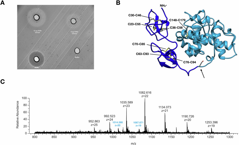
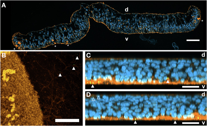
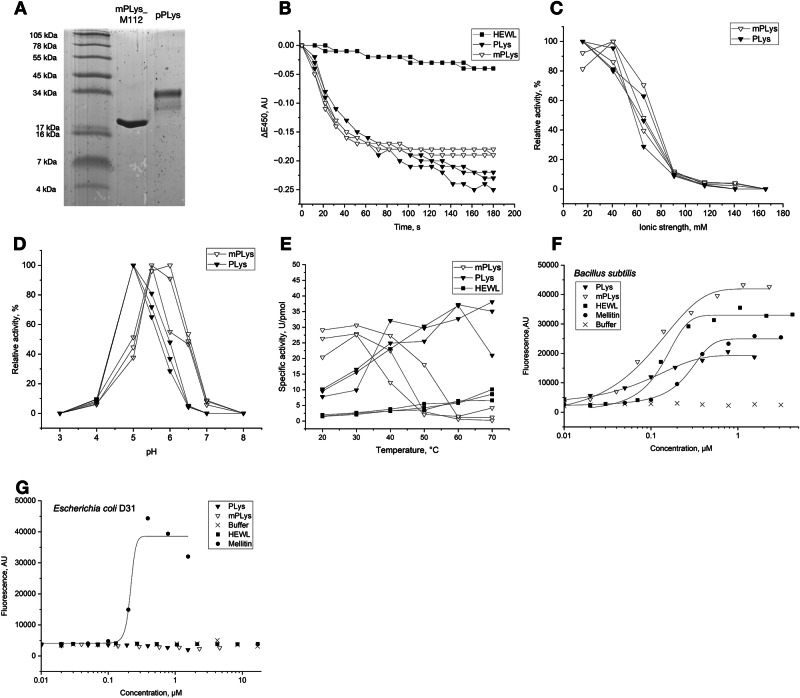
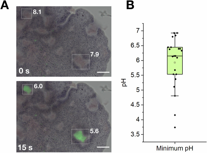
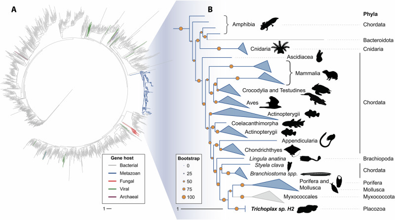
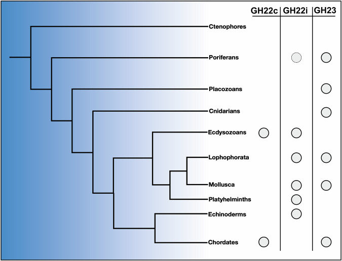
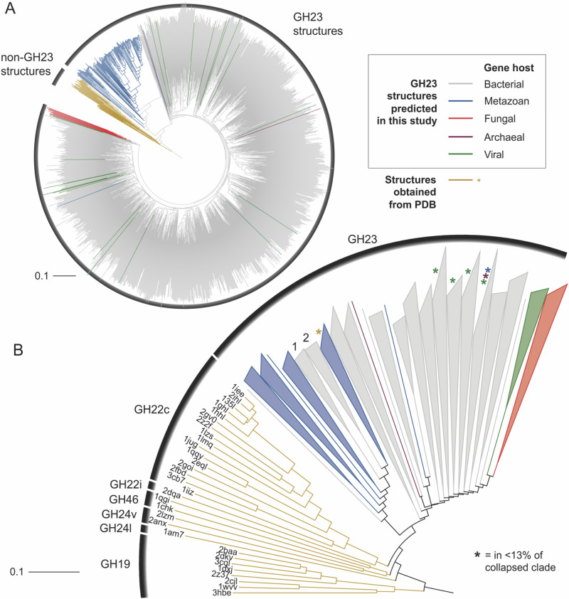
Lysozymes are an essential part of immunity and nutrition in metazoans, degrading bacterial cell walls via the hydrolysis of peptidoglycan. Although various lysozymes have been reported for higher animals, the origin of animal lysozymes remains elusive as they seem to be lacking in all early branching phyla. In this study, we investigated a putative goose-type lysozyme (PLys, glycoside hydrolase family 23, GH23) of the placozoan Trichoplax sp. H2. We show that PLys is highly active and produced in gland cells of the ventral epithelium. PLys contains a protective and non-conserved cysteine-rich domain N-terminal of the conserved GH23 lysozyme domain. A truncation of this N-terminal domain in the maturation process of PLys leads to a drastic increase in enzymatic activity at the cost of stability. As the lysozyme is most active under acidic conditions, we investigated the pH trajectories during extracellular digestion in situ . Using a pH-senstive fluorescence reporter, we show that Trichoplax sp. H2 acidifies its temporary feeding grooves pulsatively during digestive events close to the optimum pH for PLys activity. To elucidate the evolutionary origin of the metazoan GH23 lysozyme family, we applied a structure-based phylogenetics approach to show that the metazoan g-type GH23 lysozymes originated from a horizontal gene transfer event from bacteria to an early pre-bilaterian ancestor. GH23 lysozymes have then been retained and expanded in many phyla, including Porifera, Cnidaria, Placozoa and chordates, acting as first animal lysozyme and a key component in the antibacterial arsenal since early animal evolution.
Overview
The claims and statistics of this paper haven’t been checked yet.
Run the check — free, no account needed. It reads the open-access text and checks the sources it cites.
Integrity signals
Registry
Cites
Statistics
Figures
Verification
Claims the paper makes
The claims and statistics of this paper haven’t been checked yet.
Run the check — free, no account needed. It reads the open-access text and checks the sources it cites.
Self-check
Percentages, sums and reported differences re-computed from the paper’s own numbers.
The self-check needs the paper’s full text.
Citations
Cites
Highly accurate protein structure prediction with AlphaFold
Jumper, J., Evans, R., Pritzel, A., Green, T., Figurnov, M., Ronneberger, O., Tunyasuvunakool, K., Bates, R., Žídek, A., Potapenko, A., et al. (2021) · Nature · doi:10.1038/s41586-021-03819-2 · Cited by 43,764
Research works printed but not linked: 55
- 10.1073/pnas.1218525110 · DOI
- 10.1007/s12038-010-0015-5 · DOI
- 10.1007/s002390010136 · DOI
- Wohlkönig A , Huet J , Looze Y , Wintjens R. Structural relationships in the lysozyme superfamily: Significant evidence for glycoside hydrolase signature motifs. Uversky VN , editor. PLoS ONE. 2010 Nov 9;5(11):e15388. · DOI
- 10.1016/j.cell.2023.08.027 · DOI
- Laumer CE , Fernández R , Lemer S , Combosch D , Kocot KM , Riesgo A , et al. Revisiting metazoan phylogeny with genomic sampling of all phyla. Proc R Soc B Biol Sci. 2019 Jul 10;286(1906):20190831. · DOI · Paper page
- 10.1038/ncomms2424 · DOI
- DuBuc TQ , Ryan JF , Martindale MQ . “Dorsal–Ventral” genes are part of an ancient axial patterning system: Evidence from Trichoplax adhaerens (Placozoa). True J , editor. Mol Biol Evol. 2019 May 1;36(5):966–73. · DOI
- 10.1097/WNR.0000000000001436 · DOI
- 10.1038/s41564-019-0475-9 · DOI
- 10.1038/s41598-019-54037-w · DOI
- 10.1016/j.cub.2014.05.046 · DOI · Paper page
- 10.1038/s41598-021-02735-9 · DOI
- 15. Smith CL , Pivovarova N , Reese TS. Coordinated feeding behavior in Trichoplax, an animal without synapses. Steele RE , editor. PLOS ONE. 2015 Sep 2;10(9):e0136098. · DOI · Paper page
- Mayorova TD , Hammar K , Winters CA , Reese TS , Smith CL . The ventral epithelium of Trichoplax adhaerens deploys in distinct patterns cells that secrete digestive enzymes, mucus or diverse neuropeptides. Biol Open. 2019 Jan 1;bio.045674. · DOI
- 10.1186/s12864-018-5377-3 · DOI
- 10.1111/j.1742-4658.2008.06422.x · DOI
- 10.1110/ps.26801 · DOI
- 10.1186/s13100-019-0146-7 · DOI
- 10.1186/s12859-020-03599-y · DOI
- Schierwater B , Eitel M , Jakob W , Osigus HJ , Hadrys H , Dellaporta SL , et al. Concatenated Analysis Sheds Light on Early Metazoan Evolution and Fuels a Modern “Urmetazoon” Hypothesis. Penny D , editor. PLoS Biol. 2009 Jan 27;7(1):e1000020. · DOI · Paper page
- Mayorova TD , Koch TL , Kachar B , Jung JH , Reese TS , Smith CL. Placozoan secretory cell types implicated in feeding, innate immunity and regulation of behavior [Internet]. 2024 [cited 2025 Feb 1]. Available from: http://biorxiv.org/lookup/doi/10.1101/2024.09.18.613768 · DOI
- 10.1016/j.cub.2021.06.093 · DOI
- Weston A , Vladescu SC , Reddyhoff T , Griffiths A , Crouzier T , Fielden M , et al. The influence of ions on the lubricative abilities of mucin and the role of sialic acids. Colloids Surf B Biointerfaces. 2023 Jul;227:113327. · DOI
- 10.7554/eLife.36278 · DOI · Paper page
- Mukherjee K , Moroz LL . Evolution of g-type lysozymes in metazoa: insights into immunity and digestive adaptations. Front Cell Dev Biol. 2024 Nov 6;12:1487920. · DOI · Paper page
- 10.1186/s13568-022-01393-y · DOI
- Schultz DT , Haddock SHD , Bredeson JV , Green RE , Simakov O , Rokhsar DS . Ancient gene linkages support ctenophores as sister to other animals. Nature. 2023 Jun 1;618(7963):110– 7. · DOI · Paper page
- Berndt H , Fuchs S , Kraus-Stojanowic I , Pees B , Gelhaus C , Leippe M . Molecular and functional characterization of ILYS-5, a major invertebrate lysozyme of Caenorhabditis elegans . Dev Comp Immunol. 2024 Oct;159:105220. · DOI
- 10.1074/jbc.M504049200 · DOI
- 10.1002/prot.22458 · DOI
- 10.1016/j.str.2013.06.020 · DOI
- 10.1038/S41592-022-01488-1 · DOI · Paper page
- Moi D , Bernard C , Steinegger M , Nevers Y , Langleib M , Dessimoz C. Structural phylogenetics unravels the evolutionary diversification of communication systems in gram-positive bacteria and their viruses [Internet]. 2023 [cited 2024 May 31]. Available from: http://biorxiv.org/lookup/doi/10.1101/2023.09.19.558401 · DOI
- 10.1038/nsb0296-133 · DOI
- 10.1016/0003-2697(87)90587-2 · DOI
- 10.1084/jem.124.5.921 · DOI
- 10.7717/peerj.1860 · DOI
- 10.1038/s41587-019-0036-z · DOI
- 10.1016/0006-3002(52)90045-0 · DOI
- Ellis DA . A new universal buffer system. Nature. 1961 Sep;191(4793):1099–100. · DOI
- 10.1042/bj20030250 · DOI
- 10.1128/jb.121.2.455-464.1975 · DOI
- Huynh K , Partch CL . Analysis of protein stability and ligand interactions by thermal shift assay. Curr Protoc Protein Sci [Internet]. 2015 Feb [cited 2024 May 31];79(1). Available from: https://currentprotocols.onlinelibrary.wiley.com/doi/10.1002/0471140864.ps2809s79 · DOI
- 10.1093/nar/gkab1045 · DOI · Paper page
- 10.1093/bioinformatics/bts565 · DOI · Paper page
- 10.1038/s41467-022-34630-w · DOI
- 10.1093/bioinformatics/btu031 · DOI
- 10.1093/molbev/msu300 · DOI · Paper page
- 10.1038/nmeth.4285 · DOI · Paper page
- 10.1093/NAR/GKAB301 · DOI
- 10.1093/bioinformatics/bts578 · DOI
- 10.1093/sysbio/syy054 · DOI
- 10.1002/jcc.20084 · DOI
- 10.1038/nbt.2839 · DOI
Metadata
- Authors
- Berndt, H., Duarte, I., Repnik, U., Struwe, M., Abukhalaf, M., Scheidig, A., Tholey, A., Gruber-Vodicka, H., & Leippe, M.
- Affiliations
- Christian-Albrechts-Universität zu Kiel (DE)
- Corresponding author
- Leippe, Matthias
- Venue
- Communications Biology
- Publisher
- openRxiv
- Published
- 2024-10-06
- Volume
- 9
- Issue
- 1
- Pages
- 130-130
- ISSN
- 2399-3642 (ISSN-L)
- PMC
- PMC12855814
- OpenAlex
- W7119052418
- Cited by
- 2
- References
- 1
- Citations by year
- 2025: 2
- FWCI
- 0.00
- Type
- journal article
- Language
- en
- Open access
- gold
- doi.org
- Licence
- Locations
- Communications Biology (publishedVersion, cc-by)
- bioRxiv (Cold Spring Harbor Laboratory) (acceptedVersion)
- Europe PMC (PubMed Central) (publishedVersion, cc-by)
- PubMed (publishedVersion)
- PubMed Central (publishedVersion, cc-by)
- DOAJ (DOAJ: Directory of Open Access Journals) (submittedVersion, cc-by-sa)
- Funders
- Deutsche Forschungsgemeinschaft (777‐1 FUGG, 152/777-1, 5028/1-1, INST 152/772-1, INST 257/650-1 FUGG, INST 152/772-1 | 152/774-1 | 152/775-1 | 152/776-1 | 152/777-1 FUGG, 152/777 -1 FUGG, 152/775-1, 152/772-1, INST 152/774-1 FUGG, INST 152/772-1|152/774-1|152/775-1|152/776-1, 774-1, 261376515, 152/774-1, INST 152/776-1 FUGG, 776-1, 775-1, 152/776-1, INST 152/772-1 FUGG)
- Universität Hamburg (INST 152/772-1, 774-1)
- Topics
- Life Sciences › Immunology and Microbiology › Immunology › Invertebrate Immune Response Mechanisms
- Life Sciences › Agricultural and Biological Sciences › Animal Science and Zoology › Marine Invertebrate Physiology and Ecology
- Life Sciences › Biochemistry, Genetics and Molecular Biology › Molecular Biology › Developmental Biology and Gene Regulation
- Keywords
- placozoans, Trichoplax, extracellular digestion, pH-dependent fluorescence, horizontal gene transfer, antibacterial defense, peptidoglycan hydrolysis, cysteine-rich domain
- MeSH terms
- Amino Acid Sequence, Animals, Hydrogen-Ion Concentration, Muramidase, Phylogeny, Evolution, Molecular, Placozoa
About these checks
Registry, retraction, Cites and statistics results are computed, not judged, with one exception: a printed reference that no index lists is read once by a model before it counts as a citation error. Claims are extracted by a model and kept only when their quote appears verbatim in the text; each cited source is then checked, with a model’s help, for whether it says what the claim relies on. Those verdicts can be wrong — read the claim’s quote and the cited source before relying on one.
Statistics and arithmetic are recomputed, not judged.
Confidence bands are shown only for fields calibrated on our annotated test set. None is calibrated yet, so no band is shown.
What counts as a claim ›How we measure confidence ›How we check this ›
Are you an author, or do you see a problem with this page? Request a review
How to cite
Cite CoChat for the checks on this page, and the paper for its own findings.
- This page
- CoChat. (2026). Citation check: An ancient lysozyme in placozoans participates in acidic extracellular digestion. https://app.cochat.ai/papers/10.1101/2024.10.06.616844
- The paper
- Berndt, H., Duarte, I., Repnik, U., Struwe, M., Abukhalaf, M., Scheidig, A., Tholey, A., Gruber-Vodicka, H., & Leippe, M. (2024). An ancient lysozyme in placozoans participates in acidic extracellular digestion. Communications Biology. https://doi.org/10.1101/2024.10.06.616844
