

The DOI matches its Crossref record; 1 of the 1 references it cites were found in a registry or an index.
Unravelling the functional diversity of type III polyketide synthases in fungi
Open accessPreprint — published as 10.1002/anie.202514786 · Paper page
Abstract
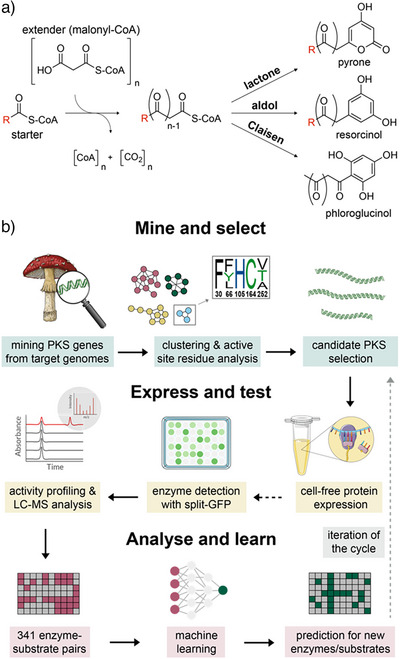
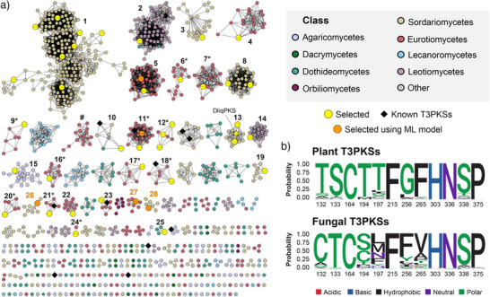
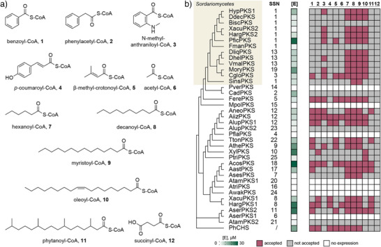
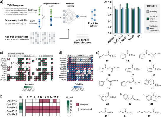
Type III polyketide synthases (T3PKSs) are enzymes that produce diverse compounds of ecological and clinical importance. While well-studied in plants and bacteria, only a handful of T3PKSs from fungi have been characterised to date. Here, we developed a comprehensive workflow for kingdom-wide characterisation of T3PKSs. Using publicly available genomes, we mined more than 1000 putative enzymes and analysed their active site architecture and genomic neighbourhood. From there, we selected 37 representative PKS candidates for cell-free expression and prototyping with a diverse set of Coenzyme A activated substrates, revealing unique patterns in substrate and cyclisation specificity, as well as the preferred number of malonyl-Coenzyme A extensions. Using the 341 enzyme-substrate pairs generated in this study, we trained a machine learning model to predict T3PKS substrate specificity and experimentally validated it with an extended panel of non-natural substrates. We anticipate that the model will be useful for in silico screening of T3PKSs, while the insight into the product scope of these enzymes offers interesting starting points for further exploration.
Overview
The claims and statistics of this paper haven’t been checked yet.
Run the check — free, no account needed. It reads the open-access text and checks the sources it cites.
Integrity signals
Registry
Cites
Statistics
Figures
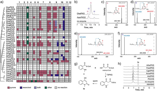
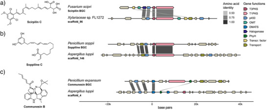
Verification
Claims the paper makes
The claims and statistics of this paper haven’t been checked yet.
Run the check — free, no account needed. It reads the open-access text and checks the sources it cites.
Self-check
Percentages, sums and reported differences re-computed from the paper’s own numbers.
The self-check needs the paper’s full text.
Citations
Cites
Highly accurate protein structure prediction with AlphaFold
Jumper, J., Evans, R., Pritzel, A., Green, T., Figurnov, M., Ronneberger, O., Tunyasuvunakool, K., Bates, R., Žídek, A., Potapenko, A., et al. (2021) · Nature · doi:10.1038/s41586-021-03819-2 · Cited by 43,764
Research works printed but not linked: 90
- 10.1038/s41570-018-0055-1 · DOI · Paper page
- 10.1002/anie.202006648 · DOI
- 10.1016/j.mattod.2015.04.002 · DOI
- 10.1002/1873-3468.14745 · DOI
- 10.1093/bioinformatics/bty065 · DOI
- Buller, R. et al. From nature to industry: Harnessing enzymes for biocatalysis. Science 382, eadh8615 (2023). · DOI
- 10.1016/j.cbpa.2010.02.018 · DOI · Paper page
- 10.1002/cbic.201600522 · DOI
- Bisht, R. , Bhattacharyya, A. , Shrivastava, A. & Saxena, P . An Overview of the Medicinally Important Plant Type III PKS Derived Polyketides. Frontiers in Plant Science 12, (2021). · DOI
- Katsuyama, Y. & Ohnishi, Y. Chapter Sixteen - Type III Polyketide Synthases in Microorganisms. in Methods in Enzymology (ed. Hopwood, D. A. ) vol. 515 359–377 (Academic Press, 2012). · DOI
- 10.1074/jbc.M701239200 · DOI
- 10.1016/j.bmcl.2011.08.058 · DOI
- 10.1016/j.bmcl.2010.06.119 · DOI
- 10.1016/j.bmcl.2013.08.036 · DOI
- 10.1039/c2mb25282a · DOI
- 10.1007/s00253-016-7444-5 · DOI
- 10.1016/j.enzmictec.2018.04.006 · DOI
- 10.1007/s12275-018-8278-x · DOI
- 10.1039/C9OB00807A · DOI
- Manoharan, G. et al. Identification and characterization of type III polyketide synthase genes from culturable endophytes of ethnomedicinal plants. Enzyme and Microbial Technology 131, (2019). · DOI
- Zhang, H. , Huang, Y. , Tang, Y. , Kong, D. & Zou, Y . Genome mining of multi-substituted alkylresorcinols from a hybrid highly reducing- and type III- polyketide pathway. Chinese Chemical Letters 35, 108968 (2024). · DOI
- Martinelli, L. et al. Identification and Characterization of a New Type III Polyketide Synthase from a Marine Yeast, Naganishia uzbekistanensis . Marine Drugs 18, 637 (2020). · DOI
- 10.3389/fmicb.2019.03018 · DOI · Paper page
- 10.1093/nar/gkt1183 · DOI · Paper page
- 10.1093/NAR/GKAC993 · DOI
- 10.1093/bioinformatics/btx469 · DOI
- Oberg, N. , Zallot, R. & Gerlt, J. A. EFI-EST , EFI-GNT, and EFI-CGFP: Enzyme Function Initiative (EFI) Web Resource for Genomic Enzymology Tools. Journal of Molecular Biology 435, 168018 (2023). · DOI
- 10.1016/S1074-5521(00)00041-7 · DOI
- 2022, Evolution-Informed Discovery of the Naphthalenone Biosynthetic Pathway in Fungi, mBio, 13, e00223
- 10.1038/11553 · DOI
- 10.1074/jbc.M112.429886 · DOI
- 10.1248/cpb.56.1505 · DOI
- 10.1021/acs.jafc.3c06785 · DOI
- 10.1021/acssynbio.1c00205 · DOI
- Garenne, D. , Thompson, S. , Brisson, A. , Khakimzhan, A. & Noireaux, V . The all-E. coli TXTL toolbox 3.0: new capabilities of a cell-free synthetic biology platform. Synthetic Biology 6, ysab017 (2021). · DOI
- 10.1038/s42003-020-01566-0 · DOI
- 10.1074/jbc.M114.569095 · DOI
- 10.1016/j.enzmictec.2010.02.011 · DOI
- 10.1016/j.bbrc.2016.04.023 · DOI
- 10.1007/s12155-010-9085-3 · DOI
- 10.1128/JB.01140-07 · DOI
- 10.1016/j.chembiol.2008.08.011 · DOI
- 10.1038/s42003-023-05690-5 · DOI
- 10.1074/jbc.M113.493155 · DOI
- 10.1021/ol0625233 · DOI
- 10.1016/j.bmc.2010.04.011 · DOI
- 10.1093/nar/gkac1049 · DOI
- 10.1021/acs.jnatprod.4c00619 · DOI · Paper page
- 10.1002/anie.201411297 · DOI
- 10.1073/pnas.97.16.8762 · DOI
- 10.1038/s41467-021-27479-y · DOI
- 10.1021/acs.jmedchem.9b01617 · DOI
- 10.1016/j.molstruc.2023.137098 · DOI
- Niu, Z.-X. et al. Application and synthesis of thiazole ring in clinically approved drugs. European Journal of Medicinal Chemistry 250, 115172 (2023). · DOI
- 10.1021/bi9009055 · DOI
- 10.1093/nar/gkr323 · DOI
- Robinson, S. L. , Smith, M. D. , Richman, J. E. , Aukema, K. G. & Wackett, L. P . Machine learning- based prediction of activity and substrate specificity for OleA enzymes in the thiolase superfamily. Synthetic Biology 5, ysaa004 (2020). · DOI
- 10.1021/acssynbio.3c00225 · DOI
- 10.1002/prot.26019 · DOI
- 10.1038/s41589-018-0154-9 · DOI
- 10.1038/s41467-023-38347-2 · DOI · Paper page
- 10.1002/cbdv.201700059 · DOI
- 10.1038/ja.2008.29 · DOI
- 10.1021/acs.jnatprod.9b00776 · DOI
- 2022, New Alkylpyridinium Anthraquinone, Isocoumarin, C-Glucosyl Resorcinol Derivative and Prenylated Pyranoxanthones from the Culture of a Marine Sponge-Associated Fungus, Aspergillus stellatus KUFA 2017. Marine Drugs, 20, 672
- 10.1515/znc-1978-11-1201 · DOI
- 2000, Production and Secretion of 5-n-Alkylresorcinols by Fusarium culmorum, Zeitschrift für Naturforschung C, 55, 846, 10.1515/znc-2000-9-1030 · DOI
- 2021, Plant metabolite 5-pentadecyl resorcinol is produced by the Amazonian fungus Penicillium sclerotiorum LM 5679, Braz J Biol, 82, e241863
- 1999, Effect of rye 5-n-alkylresorcinols on in vitro growth of phytopathogenic Fusarium and Rhizoctonia fungi, Bulletin of the Polish Academy of Sciences, 47, 231
- 10.1074/jbc.M113.487272 · DOI
- 10.1016/S0031-9422(98)00346-X · DOI
- 10.1128/am.19.5.718-720.1970 · DOI
- 10.1007/s10529-020-03042-5 · DOI
- Kaur, N. , Arora, D. S. , Kalia, N. & Kaur, M . Bioactive potential of endophytic fungus Chaetomium globosum and GC–MS analysis of its responsible components. Sci Rep 10, 18792 (2020). · DOI
- 10.1038/s41467-022-34150-7 · DOI
- 10.1002/anie.202215529 · DOI
- 10.1038/s41467-018-05221-5 · DOI
- 10.1080/14786419.2022.2139694 · DOI
- 10.1093/bioinformatics/bty121 · DOI
- 10.1038/nbt.3988 · DOI · Paper page
- 10.1101/gr.1239303 · DOI · Paper page
- 10.1371/journal.pone.0163962 · DOI · Paper page
- 10.1093/NAR/GKAD344 · DOI
- Cabantous, S. & Waldo, G. S . In vivo and in vitro protein solubility assays using split GFP. NATURE METHODS | 3, (2006). · DOI
- 10.1038/s41587-023-01690-2 · DOI · Paper page
- 10.1109/TPAMI.2021.3095381 · DOI · Paper page
- Pedregosa, F. et al. Scikit-learn: Machine Learning in Python. Journal of Machine Learning Research 12, (2012).
- 2015, Peptides: A Package for Data Mining of Antimicrobial Peptides, The R Journal, 7, 4
- Wickham, H. ggplot2: Elegant Graphics for Data Analysis. (2016). · DOI
- 10.1002/imt2.43 · DOI
Metadata
- Authors
- Sokolova, N., Denisov, S. S., Hackl, T., & Haslinger, K.
- Affiliations
- University of Groningen (NL)
- University of Vienna (AT)
- Corresponding author
- Haslinger, Kristina
- Venue
- Angewandte Chemie International Edition
- Publisher
- openRxiv
- Published
- 2024-11-25
- Volume
- 64
- Issue
- 44
- Pages
- e202514786-e202514786
- ISSN
- 1433-7851 (ISSN-L)
- PMC
- PMC12559476
- OpenAlex
- W4413995573
- Cited by
- 9
- References
- 1
- Citations by year
- 2026: 5 · 2025: 3 · 2024: 1
- FWCI
- 4.52
- Type
- journal article
- Language
- en
- Open access
- hybrid
- doi.org
- Licence
- Locations
- Angewandte Chemie International Edition (publishedVersion, cc-by)
- bioRxiv (Cold Spring Harbor Laboratory) (acceptedVersion, cc-by-nc)
- Europe PMC (PubMed Central) (publishedVersion, cc-by)
- PubMed (publishedVersion)
- University of Groningen research database (University of Groningen / Centre for Information Technology) (submittedVersion, cc-by)
- Funders
- European Commission (HORIZON2020, 893122)
- Rijksuniversiteit Groningen
- Universitair Medisch Centrum Groningen
- HORIZON EUROPE Marie Sklodowska-Curie Actions (101152560)
- Topics
- Life Sciences › Immunology and Microbiology › Applied Microbiology and Biotechnology › Microbial Natural Products and Biosynthesis
- Life Sciences › Biochemistry, Genetics and Molecular Biology › Biotechnology › Microbial Metabolism and Applications
- Life Sciences › Biochemistry, Genetics and Molecular Biology › Biotechnology › Enzyme Production and Characterization
- Keywords
- type III polyketide synthase, fungal secondary metabolites, substrate specificity prediction, machine learning, active site structure, non-natural substrates, promiscuous enzymes, cell-free expression
- MeSH terms
- Machine Learning, Fungi, Substrate Specificity, Catalytic Domain, Polyketide Synthases
About these checks
Registry, retraction, Cites and statistics results are computed, not judged, with one exception: a printed reference that no index lists is read once by a model before it counts as a citation error. Claims are extracted by a model and kept only when their quote appears verbatim in the text; each cited source is then checked, with a model’s help, for whether it says what the claim relies on. Those verdicts can be wrong — read the claim’s quote and the cited source before relying on one.
Statistics and arithmetic are recomputed, not judged.
Confidence bands are shown only for fields calibrated on our annotated test set. None is calibrated yet, so no band is shown.
What counts as a claim ›How we measure confidence ›How we check this ›
Are you an author, or do you see a problem with this page? Request a review
How to cite
Cite CoChat for the checks on this page, and the paper for its own findings.
- This page
- CoChat. (2026). Citation check: Unravelling the functional diversity of type III polyketide synthases in fungi. https://app.cochat.ai/papers/10.1101/2024.11.25.625244
- The paper
- Sokolova, N., Denisov, S. S., Hackl, T., & Haslinger, K. (2024). Unravelling the functional diversity of type III polyketide synthases in fungi. Angewandte Chemie International Edition. https://doi.org/10.1101/2024.11.25.625244
Data sources
Bibliographic data: Semantic Scholar, OpenAlex, Crossref.
Contains information from the Semantic Scholar Academic Graph Datasets, made available under the ODC Attribution License (ODC-BY 1.0).
Semantic Scholar’s data is described in: Kinney, R., et al. (2023). The Semantic Scholar Open Data Platform. arXiv:2301.10140.
