

The DOI matches its Crossref record; 1 of the 1 references it cites were found in a registry or an index.
Bacteria use processing body condensates to attenuate host translation during infection
Open accessPreprint — published as 10.1126/sciadv.aec4477
Abstract
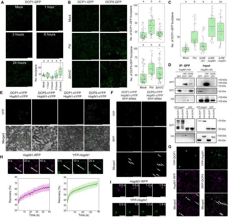
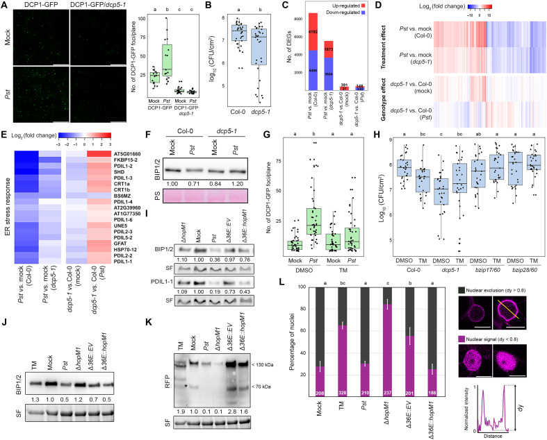
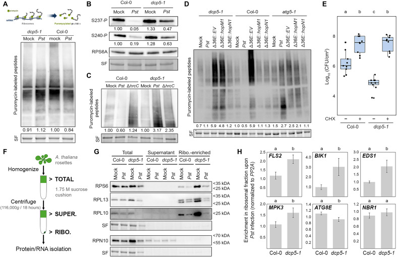
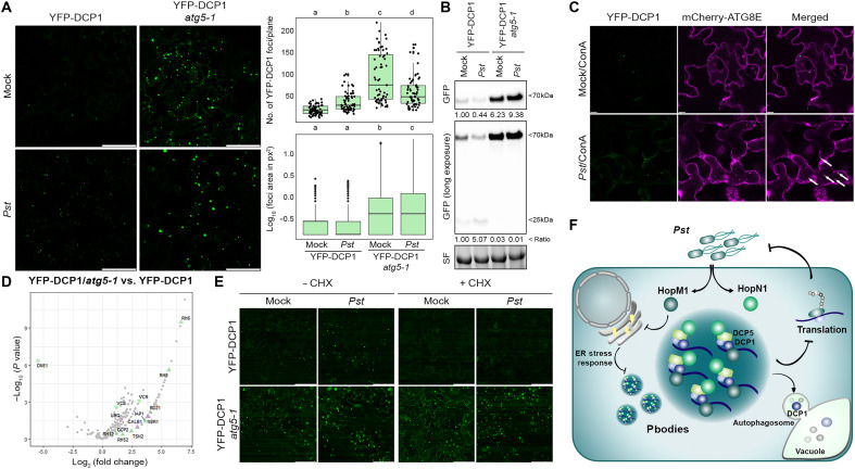
Pathogens employ sophisticated strategies to modulate host protein homeostasis by targeting proteolytic pathways, but their impact on protein synthesis remains elusive. We report that pathogenic bacteria Pseudomonas syringae ( Pst ) targets ribonucleoprotein condensates, known as processing bodies (P-bodies), to attenuate host translation through two effectors with liquid-like properties. We uncovered a previously unknown link that Pst -mediated repression of the ER stress response is required for P-body assembly. Furthermore, we identify a novel intersection between P-bodies and autophagy, demonstrating that autophagic clearance of P-bodies is crucial for maintaining the balance between translationally active and inactive mRNAs. Altogether, our discoveries provide novel insights on how host translation is attenuated by bacteria to dampen plant immunity and uncover unknown connections between ER stress responses and autophagy with P-body dynamics.
Overview
The claims and statistics of this paper haven’t been checked yet.
Run the check — free, no account needed. It reads the open-access text and checks the sources it cites.
Integrity signals
Registry
Cites
Statistics
Figures
Verification
Claims the paper makes
The claims and statistics of this paper haven’t been checked yet.
Run the check — free, no account needed. It reads the open-access text and checks the sources it cites.
Self-check
Percentages, sums and reported differences re-computed from the paper’s own numbers.
The self-check needs the paper’s full text.
Citations
Cites
Global quantification of mammalian gene expression control
Schwanhäusser, B., Busse, D., Li, N., Dittmar, G., Schuchhardt, J., Wolf, J., Chen, W., & Selbach, M. (2011) · Nature · doi:10.1038/nature10098 · Cited by 6,566
Research works printed but not linked: 55
- 10.1016/j.tplants.2023.01.013 · DOI
- 10.1146/annurev-phyto-021622-110443 · DOI
- 10.1146/annurev-arplant-060324-094912 · DOI
- 10.1146/annurev-arplant-060223-030224 · DOI
- 10.1016/j.molcel.2022.10.012 · DOI
- 10.1146/annurev-phyto-082718-100026 · DOI
- 10.1146/annurev-phyto-121423-041908 · DOI
- 10.1104/pp.111.182295 · DOI
- 10.1038/nature22371 · DOI
- 10.1016/j.cell.2022.06.037 · DOI
- 10.1021/acs.biochem.7b01162 · DOI
- 10.1104/pp.17.01468 · DOI
- 10.1016/j.semcdb.2022.11.008 · DOI
- C. L. Riggs , N. Kedersha , P. Ivanov , P. Anderson , Mammalian stress granules and P bodies at a glance. J Cell Sci 133 (2020). · DOI
- 10.1016/j.fgb.2011.10.003 · DOI
- 10.1016/j.celrep.2019.07.054 · DOI
- 10.1111/pce.15344 · DOI
- 10.1016/j.cell.2006.02.047 · DOI
- 10.1126/science.1082320 · DOI
- 2015, Pseudomonas syringae pv, tomato DC3000 Type III Secretion Effector Polymutants Reveal an Interplay between HopAD1 and AvrPtoB. Cell Host Microbe, 17, 752
- 10.1104/pp.16.00808 · DOI
- 10.1111/j.1462-5822.2012.01749.x · DOI
- 10.1038/s44318-024-00090-9 · DOI
- 10.1105/tpc.109.070078 · DOI
- 10.1038/s44319-024-00344-x · DOI
- 10.1104/pp.17.01414 · DOI
- J. E. Lee , P. I. Cathey , H. Wu , R. Parker , G. K. Voeltz , Endoplasmic reticulum contact sites regulate the dynamics of membraneless organelles. Science (1979) 367 (2020). · DOI
- 10.1016/j.chom.2022.02.006 · DOI
- 10.1105/tpc.106.050021 · DOI
- 10.1534/genetics.119.302376 · DOI
- 10.1094/MPMI-03-18-0062-R · DOI
- 10.1038/s41467-025-57231-9 · DOI
- 10.1186/s13007-016-0120-z · DOI
- 10.1146/annurev-arplant-043014-114648 · DOI
- 10.1038/s41586-021-03315-7 · DOI
- 10.1105/tpc.17.00815 · DOI
- 10.1242/dev.01096 · DOI
- 10.1104/pp.105.060673 · DOI · Paper page
- 10.1016/S0092-8674(03)00364-7 · DOI
- 10.7554/eLife.12248 · DOI
- 10.1016/j.it.2024.11.010 · DOI
- 10.1101/2024.11.20.624400 · DOI
- 10.1371/journal.ppat.1011080 · DOI
- Y. Gwon , B. A. Maxwell , R.-M. Kolaitis , P. Zhang , H. J. Kim , J. P. Taylor , Ubiquitination of G3BP1 mediates stress granule disassembly in a context-specific manner. Science (1979) 372 (2021). · DOI
- 10.1016/j.cell.2013.05.037 · DOI · Paper page
- 10.1093/nar/gkz205 · DOI
- 10.1186/s13059-014-0550-8 · DOI · Paper page
- 10.1101/2023.12.11.571118 · DOI
- 10.1016/j.molp.2024.10.003 · DOI
- 10.1038/nmeth.4228 · DOI
- 10.1186/s12864-019-5556-x · DOI
- J. X. Leong , M. Raffeiner , D. Spinti , G. Langin , M. Franz-Wachtel , A. R. Guzman , J. Kim , P. Pandey , A. E. Minina , B. Macek , A. Hafrén , T. O. Bozkurt , M. B. Mudgett , F. Börnke , D. Hofius , S. Üstün , A bacterial effector counteracts host autophagy by promoting degradation of an autophagy component. EMBO J 41 (2022). · DOI · Paper page
- 10.1093/BIOINFORMATICS/BTZ931 · DOI · Paper page
- 10.1093/NAR/GKAC1000 · DOI · Paper page
- 10.1101/gr.1239303 · DOI · Paper page
Metadata
- Authors
- González-Fuente, M., Schulz, N., Abdrakhmanov, A., Izzati, G., Zhu, S., Langin, G., Gouguet, P., Franz-Wachtel, M., Macek, B., Hafrén, A., Dagdas, Y., & Üstün, S.
- Affiliations
- Faculty of Biology and Biotechnology, Ruhr-University Bochum; Bochum, 44780, Germany
- Ruhr University Bochum (DE)
- University of Vienna (AT)
- University of Tübingen (DE)
- Proteome Center Tübingen, Interfaculty Institute for Cell Biology, University of Tübingen; Tübingen, 72076, Germany
- Swedish University of Agricultural Sciences (SE)
- Gregor Mendel Institute of Molecular Plant Biology (AT)
- ORCID
- Venue
- Science Advances
- Publisher
- openRxiv
- Published
- 2025-01-14
- Volume
- 12
- Issue
- 17
- Pages
- eaec4477-eaec4477
- ISSN
- 2375-2548 (ISSN-L)
- PMC
- PMC13108548
- OpenAlex
- W7155491829
- Cited by
- 4
- References
- 1
- Citations by year
- 2026: 1 · 2025: 3
- FWCI
- 3.70
- Type
- journal article
- Language
- en
- Open access
- gold
- doi.org
- Licence
- Locations
- Science Advances (publishedVersion, cc-by-nc)
- bioRxiv (Cold Spring Harbor Laboratory) (acceptedVersion, cc-by-nc-nd)
- PubMed Central (publishedVersion, cc-by-nc)
- PubMed (publishedVersion)
- Funders
- European Commission
- Deutsche Forschungsgemeinschaft (INST 37/965‐1 FUGG, UE188/2‐1, INST 213/1180-1 FUGG, DFG INST 37/819-1 FUGG, GO 3479/1-1)
- University of Toronto
- Université de Strasbourg
- Vetenskapsrådet (2022-03846)
- Boehringer Ingelheim Stiftung
- European Research Council (948996 DIVERSIPHAGY)
- Topics
- Life Sciences › Agricultural and Biological Sciences › Plant Science › Plant Pathogenic Bacteria Studies
- Life Sciences › Agricultural and Biological Sciences › Plant Science › Plant-Microbe Interactions and Immunity
- Life Sciences › Biochemistry, Genetics and Molecular Biology › Molecular Biology › RNA and protein synthesis mechanisms
- Keywords
- Pseudomonas syringae, P-bodies, ribonucleoprotein condensates, bacterial effector proteins, endoplasmic reticulum stress response, autophagy, plant immunity, plant-pathogen interaction, mRNA degradation
- MeSH terms
- Biomolecular Condensates, Autophagy, Plant Diseases, Ribonucleoproteins, RNA, Messenger, Protein Biosynthesis, Arabidopsis, Pseudomonas syringae, Host-Pathogen Interactions, Plant Immunity, Endoplasmic Reticulum Stress
About these checks
Registry, retraction, Cites and statistics results are computed, not judged, with one exception: a printed reference that no index lists is read once by a model before it counts as a citation error. Claims are extracted by a model and kept only when their quote appears verbatim in the text; each cited source is then checked, with a model’s help, for whether it says what the claim relies on. Those verdicts can be wrong — read the claim’s quote and the cited source before relying on one.
Statistics and arithmetic are recomputed, not judged.
Confidence bands are shown only for fields calibrated on our annotated test set. None is calibrated yet, so no band is shown.
What counts as a claim ›How we measure confidence ›How we check this ›
Are you an author, or do you see a problem with this page? Request a review
