PreprintCited by 5Proteoglycans and glycosaminoglycans researchGlycosylation and Glycoproteins ResearchPolysaccharides and Plant Cell Walls

The DOI matches its Crossref record; 1 of the 1 references it cites were found in a registry or an index.

Structural basis for human chondroitin sulfate chain polymerization

Open access
Dutta, Poushalee, Lorizolla Cordeiro, Rosa, Friedel-Arboleas, Mélanie, Bourgeais, Marie, Vallet, Sylvain D., Weber, Margot, Molinas, Margaux, Boeri-Erba, Elisabetta, Wild, Rebekka·2025·Nature Communications

Preprint — published as 10.1038/s41467-025-66787-5

Abstract

Chondroitin sulfates are complex polysaccharide chains that regulate various biological processes at the cell surface and within the extracellular matrix. Here, we identify four heterodimeric complexes responsible for chondroitin sulfate chain polymerization in humans: CHSY1-CHPF, CHSY1-CHPF2, CHSY3-CHPF and CHSY3-CHPF2. Using a novel in vitro glycosylation assay based on chemo-enzymatically synthesized fluorescent substrates, we demonstrate that all four complexes exhibit chain polymerization activity. The cryo-EM structure of the CHSY3-CHPF complex provides, for the first time, molecular insights into the chondroitin sulfate chain polymerization reaction. The architecture of the catalytic sites suggests that CHSY1 and CHSY3 are enzymatically active, while CHPF and CHPF2 primarily play a stabilizing role. Mutational analysis of purified enzyme complexes, combined with an in cellulo complementation assay, confirms that only CHSY1 and CHSY3 have bifunctional glycosyltransferase activities. Based on the spatial arrangement of the catalytic sites, we propose that chondroitin sulfates chain polymerization follows a non-processive, disruptive mechanism.

Overview

The claims and statistics of this paper haven’t been checked yet.

Run the check — free, no account needed. It reads the open-access text and checks the sources it cites.

A few free checks a day · papers already verified open instantly · results are public

Integrity signals

Registry

Crossref · 10.1101/2025.03.21.644485

Cites

1 of 1 references verified0 citation errors · 0 not checkable

Statistics

Figures

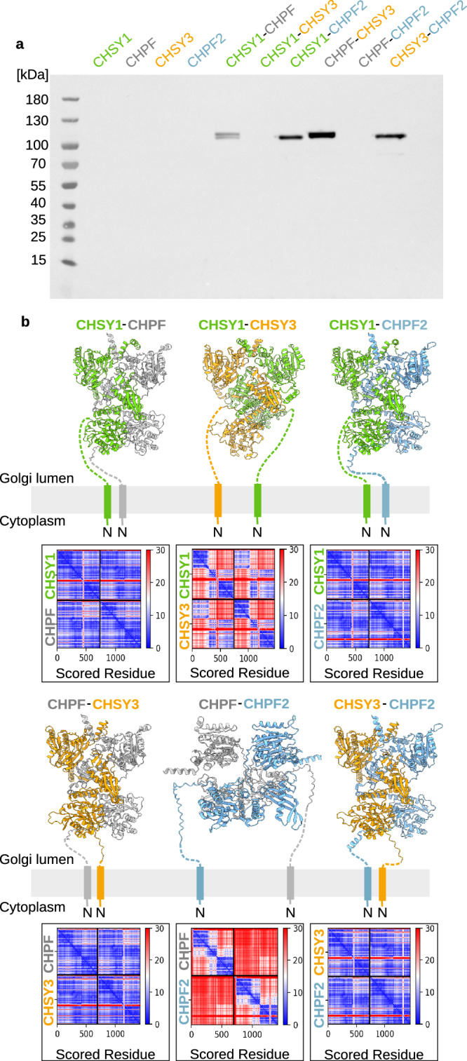
Fig. 1 CS synthases can form heterodimeric complexes. a Transient single- and co-expression of secreted CS synthase-encoding constructs in human embryonic kidney Freestyle 293-F cells. Proteins carrying an N-terminal secretion signal, SUMO (Small Ubiquitin-like Modifier) tag, and 6x His tag were detected in the cell culture medium by western blotting using an anti-His-HRP antibody. Expected molecular weights are 103 kDa for secreted CHSY1 (aa68-aa802), 98 kDa for CHPF (aa81-aa775), 102 kDa for CHSY3 (aa157-aa882), and 101 kDa for CHPF2 (aa57-aa772). Experiment was performed in duplicate (see Supplementary Fig 1b). Source data are provided as a Source Data file. b Structural models for the six potential heterodimeric CS polymerase complexes were predicted using AlphaFold 233,34. Models are shown in cartoon representation with CHSY1 in green, CHPF in gray, CHSY3 in orange, and CHPF2 in light blue. The N-terminal anchoring helices and flexible stem regions, which were omitted during model prediction, were drawn by hand. The corresponding predicted aligned error matrices are shown below. AlphaFold 2-predicted models are provided as a Source Data file.
Fig. 1 CS synthases can form heterodimeric complexes. a Transient single- and co-expression of secreted CS synthase-encoding constructs in human embryonic kidney Freestyle 293-F cells. Proteins carrying an N-terminal secretion signal, SUMO (Small Ubiquitin-like Modifier) tag, and 6x His tag were detected in the cell culture medium by western blotting using an anti-His-HRP antibody. Expected molecular weights are 103 kDa for secreted CHSY1 (aa68-aa802), 98 kDa for CHPF (aa81-aa775), 102 kDa for CHSY3 (aa157-aa882), and 101 kDa for CHPF2 (aa57-aa772). Experiment was performed in duplicate (see Supplementary Fig 1b). Source data are provided as a Source Data file. b Structural models for the six potential heterodimeric CS polymerase complexes were predicted using AlphaFold 233,34. Models are shown in cartoon representation with CHSY1 in green, CHPF in gray, CHSY3 in orange, and CHPF2 in light blue. The N-terminal anchoring helices and flexible stem regions, which were omitted during model prediction, were drawn by hand. The corresponding predicted aligned error matrices are shown below. AlphaFold 2-predicted models are provided as a Source Data file.CC BY · PubMed Central
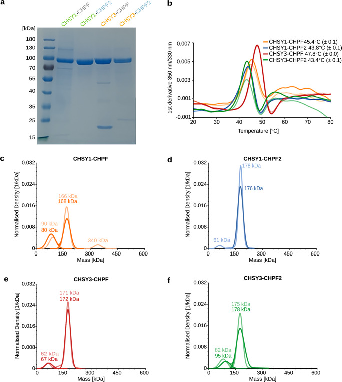
Fig. 2 Purified CS polymerase complexes are well-folded. a Coomassie-stained SDS-PAGE analysis of purified CS polymerase complexes. The expected molecular weight of 3C protease-cleaved proteins is 88 kDa for CHSY1 (aa68-aa802), 86 kDa for CHSY3 (aa157-aa882), 79 kDa for CHPF (aa81-aa775), and 81 kDa for CHPF2 (aa57-aa772). Comparable results were obtained from at least two independent purifications for each complex. b The melting temperature (Tm) of CS polymerase complexes was determined using nano-differential scanning fluorimetry (nanoDSF). The graph corresponds to the first derivative plot of the fluorescence 350 nm/330 nm ratio. Average melting temperatures from technical duplicate measurements are indicated, with corresponding standard deviation in brackets. c–f Mass photometry analysis of purified CS polymerase complexes. The expected molecular masses are 167 kDa for CHSY1–CHPF, 169 kDa for CHSY1–CHPF2, 165 kDa for CHSY3–CHPF, and 167 kDa for CHSY3–CHPF2. Molecular masses from duplicate experiments are shown in the graph; additional measured values are provided in Supplementary Table 2. Source data are provided as a Source Data file.
Fig. 2 Purified CS polymerase complexes are well-folded. a Coomassie-stained SDS-PAGE analysis of purified CS polymerase complexes. The expected molecular weight of 3C protease-cleaved proteins is 88 kDa for CHSY1 (aa68-aa802), 86 kDa for CHSY3 (aa157-aa882), 79 kDa for CHPF (aa81-aa775), and 81 kDa for CHPF2 (aa57-aa772). Comparable results were obtained from at least two independent purifications for each complex. b The melting temperature (Tm) of CS polymerase complexes was determined using nano-differential scanning fluorimetry (nanoDSF). The graph corresponds to the first derivative plot of the fluorescence 350 nm/330 nm ratio. Average melting temperatures from technical duplicate measurements are indicated, with corresponding standard deviation in brackets. c–f Mass photometry analysis of purified CS polymerase complexes. The expected molecular masses are 167 kDa for CHSY1–CHPF, 169 kDa for CHSY1–CHPF2, 165 kDa for CHSY3–CHPF, and 167 kDa for CHSY3–CHPF2. Molecular masses from duplicate experiments are shown in the graph; additional measured values are provided in Supplementary Table 2. Source data are provided as a Source Data file.CC BY · PubMed Central
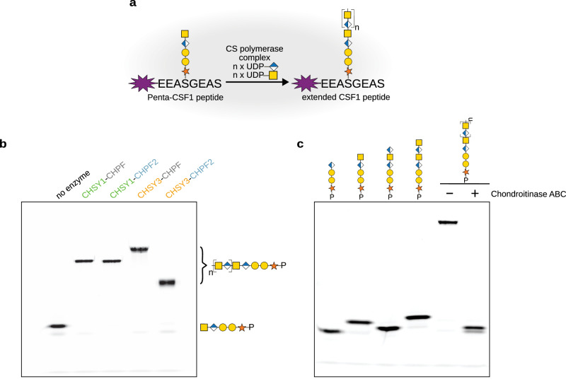
Fig. 3 In vitro chain elongation assay using purified CS polymerase complexes. a Schematic illustration of an in vitro glycosylation assay used to monitor chain elongation by the CS polymerase complexes. The fluorescently labelled Penta-CSF1 peptide, derived from the CSPG colony-stimulating factor 1, served as the acceptor substrate, while UDP-GlcA and UDP-GalNAc were used as donor substrates. Monosaccharide symbols follow the SNFG (Symbol Nomenclature for Glycans) system67. b Catalytic activity of purified CS polymerase complexes was studied using the in vitro glycosylation assay described in (a). Reaction products were separated by polyacrylamide gel electrophoresis, and fluorescent peptides were visualized using a fluorescence imager. This result is representative of three independent experiments. c Analysis of CHSY3-CHPF reaction product upon chondroitinase ABC treatment from a single experiment. Tetra-, penta-, hexa-, and heptasaccharide peptides were loaded alongside as length markers. Source data are provided as a Source Data file.
Fig. 3 In vitro chain elongation assay using purified CS polymerase complexes. a Schematic illustration of an in vitro glycosylation assay used to monitor chain elongation by the CS polymerase complexes. The fluorescently labelled Penta-CSF1 peptide, derived from the CSPG colony-stimulating factor 1, served as the acceptor substrate, while UDP-GlcA and UDP-GalNAc were used as donor substrates. Monosaccharide symbols follow the SNFG (Symbol Nomenclature for Glycans) system67. b Catalytic activity of purified CS polymerase complexes was studied using the in vitro glycosylation assay described in (a). Reaction products were separated by polyacrylamide gel electrophoresis, and fluorescent peptides were visualized using a fluorescence imager. This result is representative of three independent experiments. c Analysis of CHSY3-CHPF reaction product upon chondroitinase ABC treatment from a single experiment. Tetra-, penta-, hexa-, and heptasaccharide peptides were loaded alongside as length markers. Source data are provided as a Source Data file.CC BY · PubMed Central
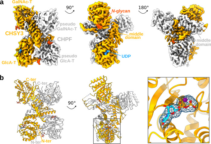
Fig. 4 Structure of the human CS polymerase complex CHSY3-CHPF. a Cryo-EM map of the heterodimeric CHSY3-CHPF complex. Density covering CHSY3 is shown in orange and CHPF in gray. A UDP molecule is highlighted in blue, and N-glycans are depicted in dark orange. Both proteins contain an N-terminal GT domain with putative D-glucuronic acid transferase (GlcA-T) activity and a C-terminal GT domain with putative N-acetyl-D-galactosamine transferase (GalNAc-T) activity. The complex is shown from three rotated views. b Structural model of the complex, shown in cartoon representation with the same color scheme as in (a). UDP and N-glycans are shown in stick representation. A close-up view shows EM density around UDP and Mn2+ ion at a contour level of 0.125.
Fig. 4 Structure of the human CS polymerase complex CHSY3-CHPF. a Cryo-EM map of the heterodimeric CHSY3-CHPF complex. Density covering CHSY3 is shown in orange and CHPF in gray. A UDP molecule is highlighted in blue, and N-glycans are depicted in dark orange. Both proteins contain an N-terminal GT domain with putative D-glucuronic acid transferase (GlcA-T) activity and a C-terminal GT domain with putative N-acetyl-D-galactosamine transferase (GalNAc-T) activity. The complex is shown from three rotated views. b Structural model of the complex, shown in cartoon representation with the same color scheme as in (a). UDP and N-glycans are shown in stick representation. A close-up view shows EM density around UDP and Mn2+ ion at a contour level of 0.125.CC BY · PubMed Central
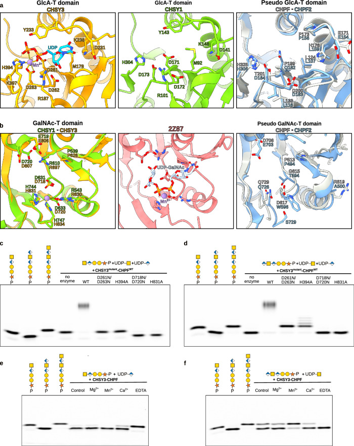
Fig. 5 Identification of catalytic residues in the N- and C-terminal GT domains of CS synthases. a, b Structural comparisons of the putative catalytic sites of the N-terminal GT31 domains and (b) the C-terminal GT7 domains of CHSY3 (colored in orange), CHSY1 (colored in green), CHPF (colored in gray), and CHPF2 (colored in blue). CHSY3 and CHPF structures were determined using cryo-EM, while CHSY1 and CHPF2 were predicted using AlphaFold 233,34. a A UDP molecule, representing a substrate analog or reaction product, bound to CHSY3, is shown in stick representation and colored in cyan, and the Mn2+ ion is shown as a purple sphere. Residues in CHSY3 important for UDP binding are highlighted in stick representation. b In the C-terminal GT domain, a structural superimposition with the chondroitin polymerase from Escherichia coli strain K4 in complex with UDP-GalNAc (colored in light blue) (PDB-ID: 2Z87)41 allowed us to propose the interactions with the donor substrate. All the residues identified to interact with the substrate donors are represented in sticks. c–f In vitro glycosylation assays for the CHSY3-CHPF complexes, followed by FACE analysis. Images shown are representative of three independent experiments. Source data is provided. c Activity of wild-type and CHSY3-mutant complexes, in which mutations are localized in the N-terminal GT domain. The pentasaccharide peptide Penta-CSF1 was used as the acceptor substrate, while both UDP-GalNAc and UDP-GlcA were used as donor substrates. d Activity of wild-type and CHSY3-mutant complexes with mutations in the C-terminal GT domain. The hexasaccharide peptide Hexa-CSF1 was used as the acceptor and UDP-GalNAc and UDP-GlcA were used as substrate donors. e Effect of different metal ion species on the GlcA and f GalNAc transferase reaction. For all the FACE gels, penta-, hexa-, and heptasaccharide peptides were loaded alongside as weight markers. Monosaccharide symbols follow the SNFG (Symbol Nomenclature for Glycans) system67. Source data are provided as a Source Data file.
Fig. 5 Identification of catalytic residues in the N- and C-terminal GT domains of CS synthases. a, b Structural comparisons of the putative catalytic sites of the N-terminal GT31 domains and (b) the C-terminal GT7 domains of CHSY3 (colored in orange), CHSY1 (colored in green), CHPF (colored in gray), and CHPF2 (colored in blue). CHSY3 and CHPF structures were determined using cryo-EM, while CHSY1 and CHPF2 were predicted using AlphaFold 233,34. a A UDP molecule, representing a substrate analog or reaction product, bound to CHSY3, is shown in stick representation and colored in cyan, and the Mn2+ ion is shown as a purple sphere. Residues in CHSY3 important for UDP binding are highlighted in stick representation. b In the C-terminal GT domain, a structural superimposition with the chondroitin polymerase from Escherichia coli strain K4 in complex with UDP-GalNAc (colored in light blue) (PDB-ID: 2Z87)41 allowed us to propose the interactions with the donor substrate. All the residues identified to interact with the substrate donors are represented in sticks. c–f In vitro glycosylation assays for the CHSY3-CHPF complexes, followed by FACE analysis. Images shown are representative of three independent experiments. Source data is provided. c Activity of wild-type and CHSY3-mutant complexes, in which mutations are localized in the N-terminal GT domain. The pentasaccharide peptide Penta-CSF1 was used as the acceptor substrate, while both UDP-GalNAc and UDP-GlcA were used as donor substrates. d Activity of wild-type and CHSY3-mutant complexes with mutations in the C-terminal GT domain. The hexasaccharide peptide Hexa-CSF1 was used as the acceptor and UDP-GalNAc and UDP-GlcA were used as substrate donors. e Effect of different metal ion species on the GlcA and f GalNAc transferase reaction. For all the FACE gels, penta-, hexa-, and heptasaccharide peptides were loaded alongside as weight markers. Monosaccharide symbols follow the SNFG (Symbol Nomenclature for Glycans) system67. Source data are provided as a Source Data file.CC BY · PubMed Central
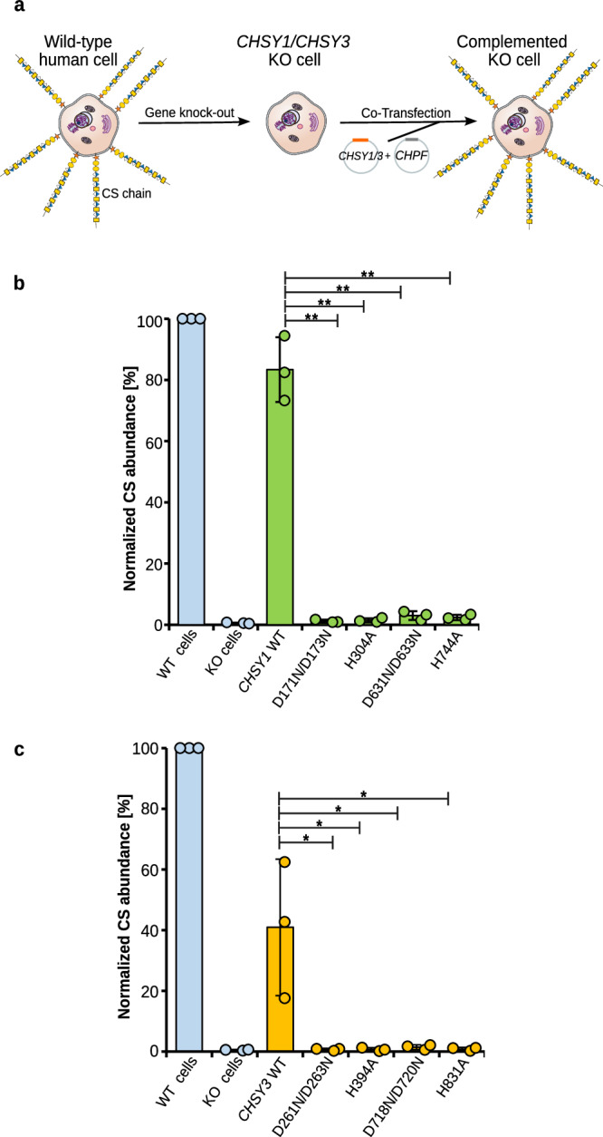
Fig. 6 Studying the effects of mutations in the CS polymerase complex on cellular CS biosynthesis. a Schematic illustration of the in-cellulo complementation assay. Knockout (KO) of the CHSY1 and CHSY3 genes via CRISPR/Cas eliminates the production of CS chains. KO cells are then co-transfected with either CHSY1 and CHPF or CHSY3 and CHPF encoding plasmids, and cell surface CS levels quantified. b Complementation of CHSY1/CHSY3 KO cells with CHPF wild-type and CHSY1 wild-type or mutant constructs. Cell surface CS levels in non-transfected cells (blue bars) and co-transfected cells (green bars) were quantified by flow cytometry using a primary anti-CS antibody and an Alexa Fluor 488-labeled secondary antibody. Results were compared to wild-type HEK293-6E cells. Experiments were performed in triplicate (n = 3). Error bars indicate standard deviation from the mean values. A one-sided paired Student’s t-test was used to determine p-values (*<0.05 and **<0.01) or more precisely CHSY1 WT vs D171N/D173N (0.0030), H304A (0.0030), D631N/D633N (0.0026) and H744A (0.0031). c Complementation assay as described in (b), but with CHSY3-CHPF (orange bars). Experiments were performed in triplicate (n = 3). Error bars indicate standard deviation from the mean values. A one-sided paired Student’s t test was used to determine p-values (*<0.05 and **<0.01) or more precisely CHSY3 WT vs D261N/D263N (0.0439), H394A (0.0442), D718N/D720N (0.0438) and H831A (0.0437). Source data is provided. Source data are provided as a Source Data file.
Fig. 6 Studying the effects of mutations in the CS polymerase complex on cellular CS biosynthesis. a Schematic illustration of the in-cellulo complementation assay. Knockout (KO) of the CHSY1 and CHSY3 genes via CRISPR/Cas eliminates the production of CS chains. KO cells are then co-transfected with either CHSY1 and CHPF or CHSY3 and CHPF encoding plasmids, and cell surface CS levels quantified. b Complementation of CHSY1/CHSY3 KO cells with CHPF wild-type and CHSY1 wild-type or mutant constructs. Cell surface CS levels in non-transfected cells (blue bars) and co-transfected cells (green bars) were quantified by flow cytometry using a primary anti-CS antibody and an Alexa Fluor 488-labeled secondary antibody. Results were compared to wild-type HEK293-6E cells. Experiments were performed in triplicate (n = 3). Error bars indicate standard deviation from the mean values. A one-sided paired Student’s t-test was used to determine p-values (*<0.05 and **<0.01) or more precisely CHSY1 WT vs D171N/D173N (0.0030), H304A (0.0030), D631N/D633N (0.0026) and H744A (0.0031). c Complementation assay as described in (b), but with CHSY3-CHPF (orange bars). Experiments were performed in triplicate (n = 3). Error bars indicate standard deviation from the mean values. A one-sided paired Student’s t test was used to determine p-values (*<0.05 and **<0.01) or more precisely CHSY3 WT vs D261N/D263N (0.0439), H394A (0.0442), D718N/D720N (0.0438) and H831A (0.0437). Source data is provided. Source data are provided as a Source Data file.CC BY · PubMed Central
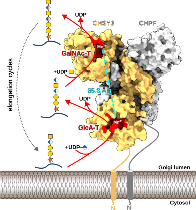
Fig. 7 Molecular mechanism of CS chain polymerization. Illustration of the proposed mechanism for chain elongation by the CS polymerase complex CHSY3-CHPF. In a first reaction step, a GlcA residue is added onto the proteoglycan carrying a pentasaccharide-linker by the N-terminal GT domain of CHSY3. The generated hexasaccharide product is released and binds to the C-terminal GT domain of CHSY3, exhibiting GalNAc transferase activity. Upon GalNAc addition, the proteoglycan dissociates, and a new reaction cycle starts. Repetitive addition of GlcA and GalNAc residues leads to the generation of the long chondroitin sulfate backbone.
Fig. 7 Molecular mechanism of CS chain polymerization. Illustration of the proposed mechanism for chain elongation by the CS polymerase complex CHSY3-CHPF. In a first reaction step, a GlcA residue is added onto the proteoglycan carrying a pentasaccharide-linker by the N-terminal GT domain of CHSY3. The generated hexasaccharide product is released and binds to the C-terminal GT domain of CHSY3, exhibiting GalNAc transferase activity. Upon GalNAc addition, the proteoglycan dissociates, and a new reaction cycle starts. Repetitive addition of GlcA and GalNAc residues leads to the generation of the long chondroitin sulfate backbone.CC BY · PubMed Central

Verification

Claims the paper makes

The claims and statistics of this paper haven’t been checked yet.

Run the check — free, no account needed. It reads the open-access text and checks the sources it cites.

A few free checks a day · papers already verified open instantly · results are public

Self-check

Percentages, sums and reported differences re-computed from the paper’s own numbers.

The self-check needs the paper’s full text.

Citations

Cites

Show all 1 references

Research works printed but not linked: 61

  1. 10.1016/j.bbagen.2013.06.006 · DOI
  2. 10.1111/febs.17107 · DOI
  3. 10.1007/s10719-017-9761-z · DOI · Paper page
  4. 10.1042/BCJ20170820 · DOI
  5. 10.1038/s41583-019-0196-3 · DOI · Paper page
  6. 10.3389/fcell.2022.745372 · DOI
  7. 10.3389/fnint.2018.00003 · DOI
  8. 10.1152/ajpcell.00095.2022 · DOI
  9. 10.3892/ol.2018.9385 · DOI
  10. 10.1002/2211-5463.13062 · DOI
  11. 10.3389/fcell.2021.693563 · DOI
  12. 10.1016/j.gene.2022.146466 · DOI
  13. 10.1038/s41467-023-42236-z · DOI
  14. 10.1093/glycob/cwae016 · DOI
  15. 10.1074/jbc.M111434200 · DOI
  16. 10.1074/jbc.M208886200 · DOI
  17. 10.1093/glycob/cwr172 · DOI
  18. 10.1074/jbc.274.4.2456 · DOI
  19. 10.1016/j.sbi.2003.08.002 · DOI
  20. 10.1074/jbc.M106871200 · DOI
  21. 10.1074/jbc.M304421200 · DOI
  22. 10.1074/jbc.M302493200 · DOI
  23. 10.1074/jbc.M303657200 · DOI
  24. 10.1074/jbc.M202601200 · DOI
  25. 10.1074/jbc.M707549200 · DOI
  26. 10.1093/nar/gkae1010 · DOI · Paper page
  27. 10.1038/s41467-022-34882-6 · DOI · Paper page
  28. 10.1038/s41589-022-01220-2 · DOI
  29. 10.1042/BJ20061876 · DOI
  30. 10.1038/S41592-022-01488-1 · DOI · Paper page
  31. 10.1002/(SICI)1098-2795(199701)46:1<4::AID-MRD2>3.0.CO;2-V · DOI
  32. Essentials of Glycobiology. (Cold Spring Harbor Laboratory Press, Cold Spring Harbor (NY), 2022).
  33. 10.1016/j.jmb.2007.05.022 · DOI
  34. 10.1093/nar/gkab1045 · DOI · Paper page
  35. 10.1126/science.aar5140 · DOI
  36. 10.7554/eLife.54532 · DOI
  37. 10.1007/978-1-0716-0270-6_3 · DOI
  38. 10.1016/j.bbrc.2008.08.121 · DOI
  39. 10.1016/j.molcel.2019.05.017 · DOI
  40. 10.1016/j.jmb.2010.03.059 · DOI
  41. 10.1007/s00018-015-2066-0 · DOI
  42. 10.1074/jbc.RA120.015305 · DOI
  43. 10.1074/jbc.M113.509984 · DOI
  44. 10.1093/glycob/cwaa086 · DOI
  45. 10.1016/j.ejmg.2013.11.001 · DOI
  46. 2013, A chondroitin synthase-1 (ChSy-1) missense mutation in a patient with neuropathy impairs the elongation of chondroitin sulfate chains initiated by chondroitin N-acetylgalactosaminyltransferase-1, Biochim Biophys Acta, 1830, 4806, 10.1016/j.bbagen.2013.06.017 · DOI
  47. 10.1038/s41467-017-01747-2 · DOI
  48. 10.1371/journal.pone.0043806 · DOI
  49. 10.1042/BJ20101456 · DOI
  50. 10.1093/nar/gkw1081 · DOI
  51. 10.1093/bioinformatics/bty1057 · DOI · Paper page
  52. 10.1093/nar/gkz1035 · DOI · Paper page
  53. 10.1002/pro.3235 · DOI · Paper page
  54. 10.1007/978-1-0716-1406-8_4 · DOI
  55. 10.3791/50635 · DOI
  56. 2009, From polymer to size-defined oligomers: a step economy process for the efficient and stereocontrolled construction of chondroitin oligosaccharides and biotinylated conjugates thereof: part 1, Chemistry, 15, 9561, 10.1002/chem.200900740 · DOI
  57. 10.1016/j.jsb.2006.05.009 · DOI
  58. 10.1107/S0907444910007493 · DOI · Paper page
  59. 10.1107/S2059798318009324 · DOI
  60. 10.1002/pro.3330 · DOI · Paper page
  61. 10.1002/PRO.4792 · DOI

Metadata

Authors
Dutta, P., Lorizolla Cordeiro, R., Friedel-Arboleas, M., Bourgeais, M., Vallet, S. D., Weber, M., Molinas, M., Boeri-Erba, E., & Wild, R.
Affiliations
  • Centre National de la Recherche Scientifique (FR)
  • Institut de Biologie Structurale, UMR 5075, University Grenoble Alpes, CNRS, CEA, 38000 Grenoble, France
Corresponding author
Wild, Rebekka
Venue
Nature Communications
Publisher
openRxiv
Published
2025-03-21
Volume
16
Issue
1
Pages
11663-11663
ISSN
2041-1723 (ISSN-L)
OpenAlex
W4416723479
Cited by
5
References
1
Citations by year
2026: 5
FWCI
2.48
Type
journal article
Language
en
Open access
Locations
  • Nature Communications (publishedVersion, cc-by)
  • bioRxiv (Cold Spring Harbor Laboratory) (acceptedVersion, cc-by-nc)
  • PubMed (publishedVersion)
  • PubMed Central (publishedVersion, cc-by)
  • Research at the University of Copenhagen (University of Copenhagen) (publishedVersion, cc-by)
  • HAL (submittedVersion, cc-by)
  • DOAJ (DOAJ: Directory of Open Access Journals) (submittedVersion, cc-by-sa)
Funders
  • LABoratoires d’EXcellence ARCANE
  • Infrastructures en Biologie Santé et Agronomie (ANR-17-EURE-0003)
  • Equipex
  • Agence Nationale de la Recherche (UAR 3518 CNRS-CEA-UGA-EMBL, ANR-23-CE44-0011, ANR-17-EURE-0003, IDEX-02, ANR-17-EURE-, ANR‐15‐IDEX‐02, ANR-15-IDEX-0002, ANR-17, ANR-10, ANR-21-ESRE-0046, ANR-10-INBS-0005, ANR-10-LABX-0049)
  • European Synchrotron Radiation Facility
  • Danmarks Grundforskningsfond (DNRF196)
  • Fondation pour la Recherche Médicale (ANR-17-EURE-0003)
  • Novo Nordisk (NNF22OC0073736)
  • Centre National de la Recherche Scientifique (UAR 3518 CNRS-CEA-UGA-EMBL)
  • Fondation Bettencourt Schueller
  • Novo Nordisk Fonden (NNF22OC0073736)
  • Région Auvergne-Rhône-Alpes (ANR-17-EURE-0003)
  • French Infrastructure for Integrated Structural Biology (ANR-17-EURE-0003, ANR-10-INBS-0005, ANR-10-INBS-000502)
Topics
  • Life Sciences › Biochemistry, Genetics and Molecular Biology › Cell Biology › Proteoglycans and glycosaminoglycans research
  • Life Sciences › Biochemistry, Genetics and Molecular Biology › Molecular Biology › Glycosylation and Glycoproteins Research
  • Life Sciences › Agricultural and Biological Sciences › Plant Science › Polysaccharides and Plant Cell Walls
Keywords
chondroitin sulfate, glycosyltransferases, cryo-EM structure, extracellular matrix, glycosaminoglycans
MeSH terms
Chondroitin Sulfates, Glycosylation, Humans, Glycosyltransferases, Catalytic Domain, Cryoelectron Microscopy, Polymerization

About these checks

Registry, retraction, Cites and statistics results are computed, not judged, with one exception: a printed reference that no index lists is read once by a model before it counts as a citation error. Claims are extracted by a model and kept only when their quote appears verbatim in the text; each cited source is then checked, with a model’s help, for whether it says what the claim relies on. Those verdicts can be wrong — read the claim’s quote and the cited source before relying on one.

Statistics and arithmetic are recomputed, not judged.

Confidence bands are shown only for fields calibrated on our annotated test set. None is calibrated yet, so no band is shown.

What counts as a claim ›How we measure confidence ›How we check this ›

Are you an author, or do you see a problem with this page? Request a review

How to cite

Cite CoChat for the checks on this page, and the paper for its own findings.

This page
CoChat. (2026). Citation check: Structural basis for human chondroitin sulfate chain polymerization. https://app.cochat.ai/papers/10.1101/2025.03.21.644485
The paper
Dutta, P., Lorizolla Cordeiro, R., Friedel-Arboleas, M., Bourgeais, M., Vallet, S. D., Weber, M., Molinas, M., Boeri-Erba, E., & Wild, R. (2025). Structural basis for human chondroitin sulfate chain polymerization. Nature Communications. https://doi.org/10.1101/2025.03.21.644485