

The DOI matches its Crossref record; 42 of the 42 references it cites were found in a registry or an index.
Protein aggregation mediates stoichiometry of protein complexes in aneuploid cells
Open accessAbstract
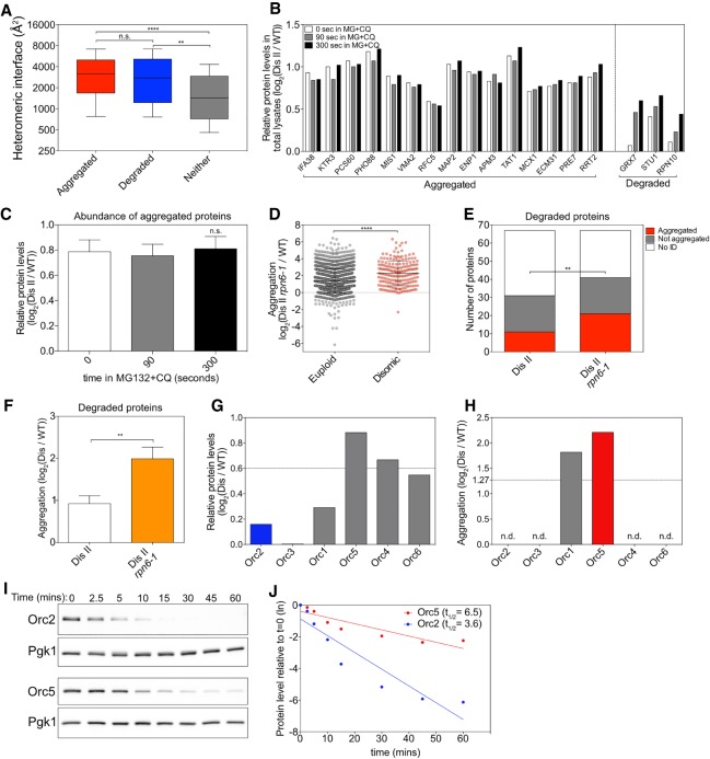
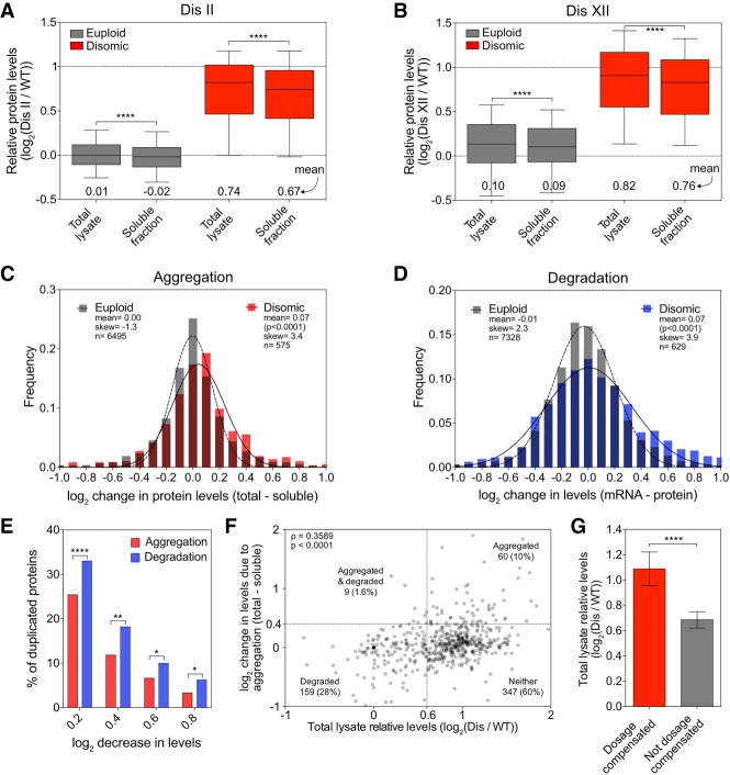
Aneuploidy, a condition characterized by chromosome gains and losses, causes reduced fitness and numerous cellular stresses, including increased protein aggregation. Here, we identify protein complex stoichiometry imbalances as a major cause of protein aggregation in aneuploid cells. Subunits of protein complexes encoded on excess chromosomes aggregate in aneuploid cells, which is suppressed when expression of other subunits is coordinately altered. We further show that excess subunits are either degraded or aggregate and that protein aggregation is nearly as effective as protein degradation at lowering levels of excess proteins. Our study explains why proteotoxic stress is a universal feature of the aneuploid state and reveals protein aggregation as a form of dosage compensation to cope with disproportionate expression of protein complex subunits.
Overview
The claims and statistics of this paper haven’t been checked yet.
Run the check — free, no account needed. It reads the open-access text and checks the sources it cites.
Integrity signals
Registry
Cites
Statistics
Figures
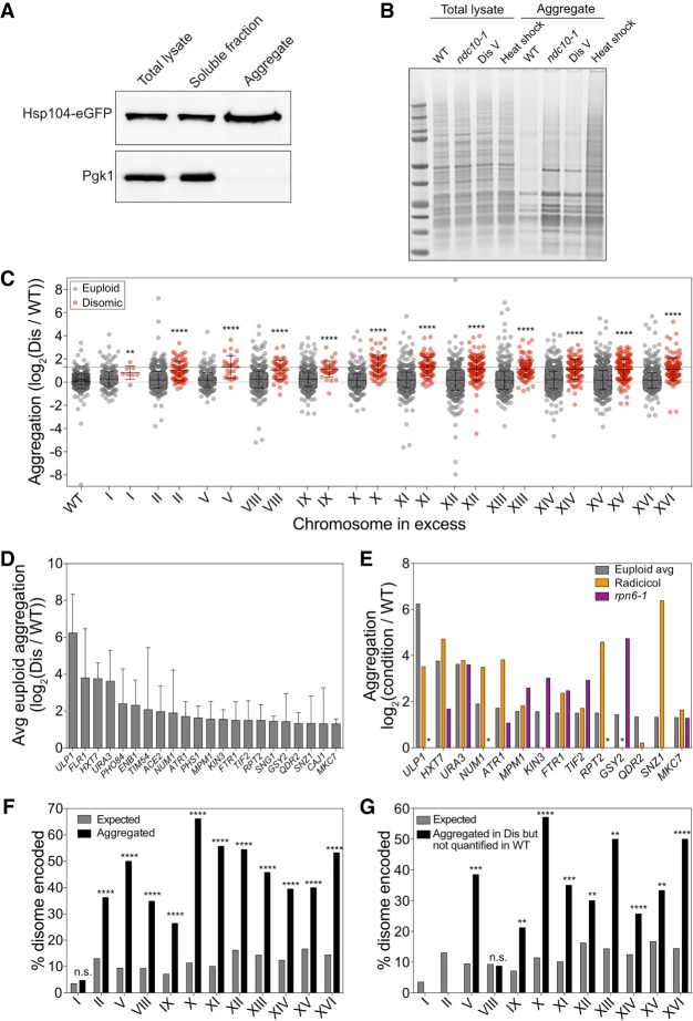
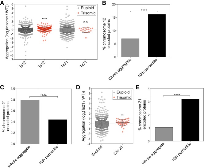
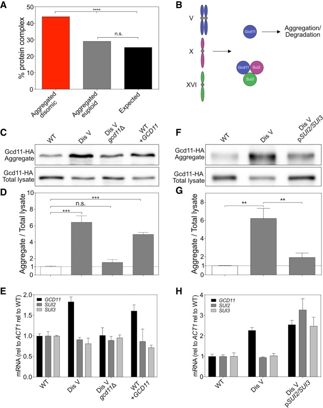
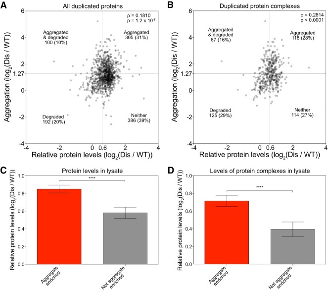
Verification
Claims the paper makes
The claims and statistics of this paper haven’t been checked yet.
Run the check — free, no account needed. It reads the open-access text and checks the sources it cites.
Claims other papers make about this one
Each is checked against this paper’s own text: does it say what the citing paper says it does?
The source backs part of the claim, not all of it — or backs it for a different population, condition or comparison.
In the citing paper
“We conclude that the reduction of Exo9 and EXOSC10 protein abundance across all subunits is caused by orphan protein decay because of the increased production of the unstable, and likely unusable, short EXOSC3 proteoform107–109.”
From EXOSC3 G191 Variants Trigger System-Wide Recalibration of RNA Processing Machinery (2026) · cites this paper as [108]
Experimental — accuracy not yet measured
Self-check
Percentages, sums and reported differences re-computed from the paper’s own numbers.
The self-check needs the paper’s full text.
Citations
Cites
Fiji: an open-source platform for biological-image analysis
Schindelin, J., Arganda-Carreras, I., Frise, E., Kaynig, V., Longair, M., Pietzsch, T., Preibisch, S., Rueden, C., Saalfeld, S., Schmid, B., et al. (2012) · Nature Methods · doi:10.1038/nmeth.2019 · Cited by 66,890
Overview of the CCP 4 suite and current developments
Winn, M. D., Ballard, C. C., Cowtan, K. D., Dodson, E. J., Emsley, P., Evans, P. R., Keegan, R. M., Krissinel, E. B., Leslie, A. G. W., McCoy, A., et al. (2011) · Acta Crystallographica Section D Biological Crystallography · doi:10.1107/s0907444910045749 · Cited by 12,353
Enzymatic assembly of DNA molecules up to several hundred kilobases
Gibson, D. G., Young, L., Chuang, R.-Y., Venter, J. C., Hutchison, C. A., & Smith, H. O. (2009) · Nature Methods · doi:10.1038/nmeth.1318 · Cited by 10,088
Biomolecular condensates: organizers of cellular biochemistry
Banani, S. F., Lee, H. O., Hyman, A. A., & Rosen, M. K. (2017) · Nature Reviews Molecular Cell Biology · doi:10.1038/nrm.2017.7 · Cited by 6,058
Ong, S.-E., Blagoev, B., Kratchmarova, I., Kristensen, D. B., Steen, H., Pandey, A., & Mann, M. (2002) · Molecular & Cellular Proteomics · doi:10.1074/mcp.m200025-mcp200 · Cited by 5,574
In-gel digestion for mass spectrometric characterization of proteins and proteomes
Shevchenko, A., Tomas, H., Havli, J., Olsen, J. V., & Mann, M. (2006) · Nature Protocols · doi:10.1038/nprot.2006.468 · Cited by 4,947
Elias, J. E., & Gygi, S. P. (2007) · Nature Methods · doi:10.1038/nmeth1019 · Cited by 3,925
2016 update of the PRIDE database and its related tools
Vizcaíno, J. A., Csordás, A., del‐Toro, N., Dianes, J. A., Griss, J., Lavidas, I., Mayer, G., Pérez‐Riverol, Y., Reisinger, F., Ternent, T., et al. (2015) · Nucleic Acids Research · doi:10.1093/nar/gkv1145 · Cited by 3,466
Bähler, J., Wu, J., Longtine, M. S., Shah, N. G., Mckenzie, A., Steever, A. B., Wach, A., Philippsen, P., & Pringle, J. R. (1998) · Yeast · doi:10.1002/(sici)1097-0061(199807)14:10 · Cited by 2,208
A tissue-specific atlas of mouse protein phosphorylation and expression
Huttlin, E. L., Jedrychowski, M. P., Elias, J. E., Goswami, T., Rad, R., Beausoleil, S. A., Villén, J., Haas, W., Sowa, M. E., & Gygi, S. P. (2010) · Cell · doi:10.1016/j.cell.2010.12.001 · Cited by 1,778
Research works printed but not linked: 1
- Guthrie C , Fink GR . 1991. Methods in enzymology: molecular biology of Saccharomyces cerevisiae. Academic Press, San Diego.
Cited by
Sorted by citations
mRNAs, proteins and the emerging principles of gene expression control
Buccitelli, C., & Selbach, M. (2020) · Nature Reviews Genetics · doi:10.1038/s41576-020-0258-4 · Cited by 1,243
Karagöz, G. E., Acosta‐Alvear, D., & Walter, P. (2019) · Cold Spring Harbor Perspectives in Biology · doi:10.1101/cshperspect.a033886 · Cited by 306
DNA Replication Stress and Chromosomal Instability: Dangerous Liaisons
Wilhelm, T., Said, M., & Naim, V. (2020) · Genes · doi:10.3390/genes11060642 · Cited by 161
Effects of aneuploidy on cell behaviour and function
Li, R., & Zhu, J. (2022) · Nature Reviews Molecular Cell Biology · doi:10.1038/s41580-021-00436-9 · Cited by 147
The epithelial–mesenchymal plasticity landscape: principles of design and mechanisms of regulation
Haerinck, J., Goossens, S., & Berx, G. (2023) · Nature Reviews Genetics · doi:10.1038/s41576-023-00601-0 · Cited by 142
Metadata
- Authors
- Brennan, C. M., Vaites, L. P., Wells, J. N., Santaguida, S., Paulo, J. A., Storchova, Z., Harper, J. W., Marsh, J. A., & Amon, A.
- Affiliations
- Howard Hughes Medical Institute (US)
- Harvard University (US)
- Western General Hospital (GB)
- ORCID
- Venue
- Genes & Development
- Publisher
- Cold Spring Harbor Laboratory
- Published
- 2019-06-13
- Volume
- 33
- Issue
- 15-16
- Pages
- 1031-1047
- ISSN
- 0890-9369 (ISSN-L)
- 1549-5477
- PMC
- PMC6672052
- OpenAlex
- W2953356987
- Cited by
- 109
- References
- 42
- Citations by year
- 2026: 10 · 2025: 15 · 2024: 16 · 2023: 14 · 2022: 14 · 2021: 18 · 2020: 19 · 2019: 3
- Type
- posted content
- Language
- en
- Open access
- hybrid
- doi.org
- Licence
- cc-by-nc
- Locations
- Genes & Development (publishedVersion, cc-by-nc)
- PubMed Central (publishedVersion, cc-by-nc)
- PubMed (publishedVersion)
- Edinburgh Research Explorer (University of Edinburgh) (acceptedVersion, other-oa)
- Archivio Istituzionale della Ricerca (Universita Degli Studi Di Milano) (submittedVersion, other-oa)
- DSpace@MIT (Massachusetts Institute of Technology) (submittedVersion, cc-by-nc)
- HighWire Press Open Archive (submittedVersion)
- Funders
- National Institutes of Health (CA206157, GM118066)
- Medical Research Council Career Development Award (MR/M02122X/1)
- American Italian Cancer Foundation
- Marie Curie Actions
- Italian Association for Cancer Research
- Koch Institute Quinquennial Cancer Research Fellowship
- Topics
- Life Sciences › Biochemistry, Genetics and Molecular Biology › Cell Biology › Microtubule and mitosis dynamics
- Life Sciences › Biochemistry, Genetics and Molecular Biology › Molecular Biology › Heat shock proteins research
- Life Sciences › Biochemistry, Genetics and Molecular Biology › Molecular Biology › Ubiquitin and proteasome pathways
- Keywords
- aneuploidy, protein aggregation, protein complex stoichiometry, proteotoxic stress, dosage compensation, protein degradation
- MeSH terms
- Aneuploidy, Cytosol, Dosage Compensation, Genetic, Humans, Saccharomyces cerevisiae, Protein Subunits, Saccharomyces cerevisiae Proteins, Multiprotein Complexes, Proteolysis, Protein Aggregation, Pathological, Protein Aggregates
About these checks
Registry, retraction, Cites and statistics results are computed, not judged, with one exception: a printed reference that no index lists is read once by a model before it counts as a citation error. Claims are extracted by a model and kept only when their quote appears verbatim in the text; each cited source is then checked, with a model’s help, for whether it says what the claim relies on. Those verdicts can be wrong — read the claim’s quote and the cited source before relying on one.
Statistics and arithmetic are recomputed, not judged.
Confidence bands are shown only for fields calibrated on our annotated test set. None is calibrated yet, so no band is shown.
What counts as a claim ›How we measure confidence ›How we check this ›
Are you an author, or do you see a problem with this page? Request a review
