

The DOI matches its Crossref record; 168 of the 168 references it cites were found in a registry or an index.
Early Archean origin of Photosystem II
Open accessPublished version of a preprint
Abstract
Photosystem II is a photochemical reaction center that catalyzes the light-driven oxidation of water to molecular oxygen. Water oxidation is the distinctive photochemical reaction that permitted the evolution of oxygenic photosynthesis and the eventual rise of eukaryotes. At what point during the history of life an ancestral photosystem evolved the capacity to oxidize water still remains unknown. Here, we study the evolution of the core reaction center proteins of Photosystem II using sequence and structural comparisons in combination with Bayesian relaxed molecular clocks. Our results indicate that a homodimeric photosystem with sufficient oxidizing power to split water had already appeared in the early Archean about a billion years before the most recent common ancestor of all described Cyanobacteria capable of oxygenic photosynthesis, and well before the diversification of some of the known groups of anoxygenic photosynthetic bacteria. Based on a structural and functional rationale, we hypothesize that this early Archean photosystem was capable of water oxidation to oxygen and had already evolved protection mechanisms against the formation of reactive oxygen species. This would place primordial forms of oxygenic photosynthesis at a very early stage in the evolutionary history of life.
Overview
The claims and statistics of this paper haven’t been checked yet.
Run the check — free, no account needed. It reads the open-access text and checks the sources it cites.
Integrity signals
Registry
Cites
Statistics
Figures
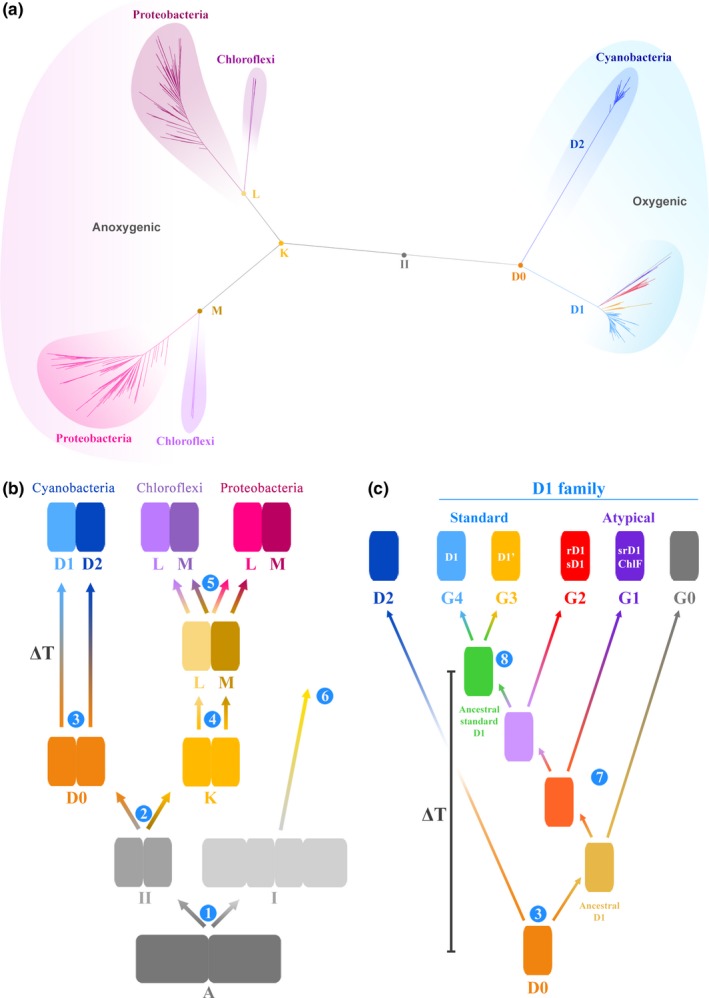
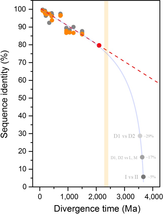
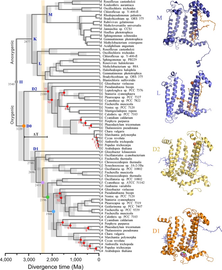
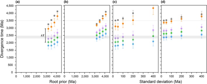
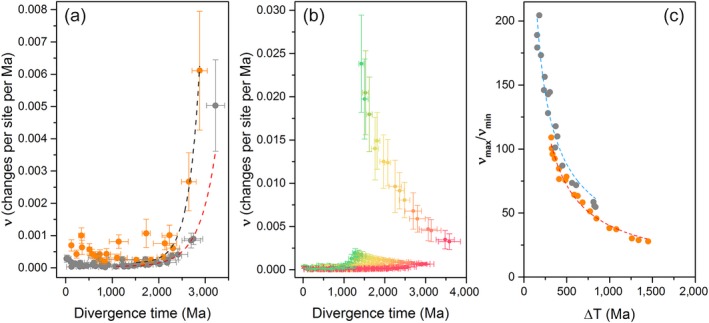
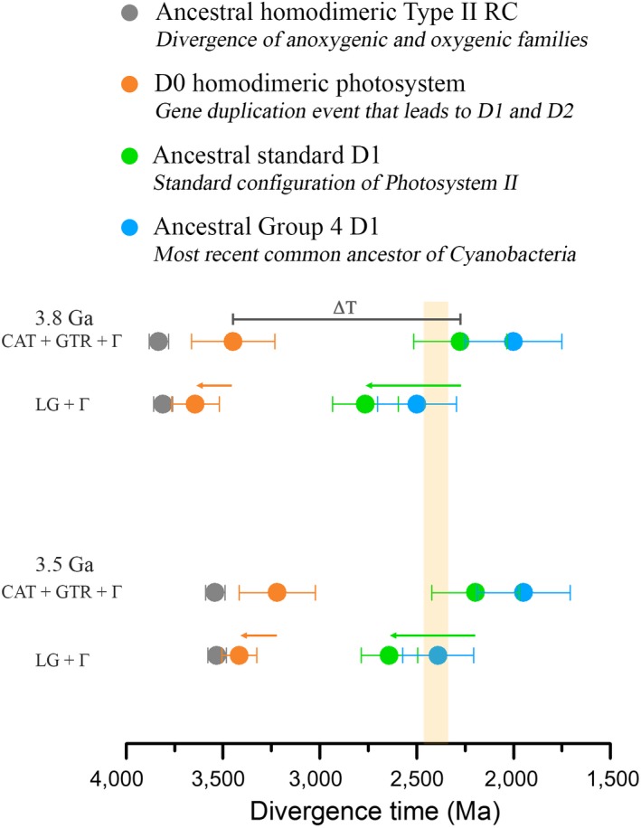
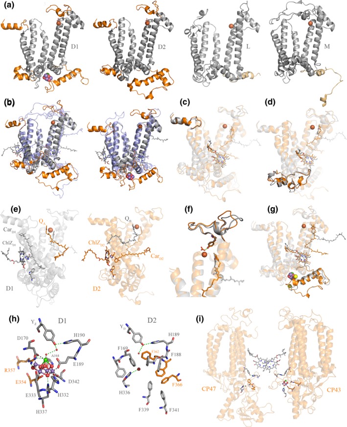
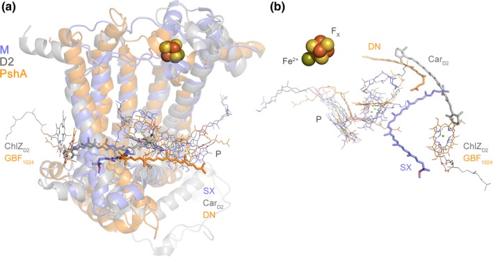
Verification
Claims the paper makes
The claims and statistics of this paper haven’t been checked yet.
Run the check — free, no account needed. It reads the open-access text and checks the sources it cites.
Claims other papers make about this one
Each is checked against this paper’s own text: does it say what the citing paper says it does?
The source backs part of the claim, not all of it — or backs it for a different population, condition or comparison.
In the citing paper
“While some argue that oxygenic photosynthesis evolved only 50 to 100 Ma before the GOE (115), most studies suggest an earlier Paleoarchean or Mesoarchean age [e.g., (3, 118, 119)].”
From The Archean atmosphere (2020) · cites this paper as [118]
Experimental — accuracy not yet measured
Self-check
Percentages, sums and reported differences re-computed from the paper’s own numbers.
The self-check needs the paper’s full text.
Citations
Cites
Guindon, S., Dufayard, J.-F., Lefort, V., Anisimova, M., Hordijk, W., & Gascuel, O. (2010) · Systematic Biology · doi:10.1093/sysbio/syq010 · Cited by 18,376
Fast, scalable generation of high-quality protein multiple sequence alignments using Clustal Omega
Sievers, F., Wilm, A., Dineen, D., Gibson, T., Karplus, K., Li, W.-Z., Lopez, R., McWilliam, H., Remmert, M., Söding, J., et al. (2011) · Molecular Systems Biology · doi:10.1038/msb.2011.75 · Cited by 15,576
The Evolutionary Fate and Consequences of Duplicate Genes
Lynch, M., & Conery, J. S. (2000) · Science · doi:10.1126/science.290.5494.1151 · Cited by 4,988
Crystal structure of oxygen-evolving photosystem II at a resolution of 1.9 Å
Umena, Y., Kawakami, K., Shen, J., & Kamiya, N. (2011) · Nature · doi:10.1038/nature09913 · Cited by 3,920
Architecture of the Photosynthetic Oxygen-Evolving Center
Ferreira, K. N., Iverson, T. M., Maghlaoui, K., Barber, J. P., & Iwata, S. (2004) · Science · doi:10.1126/science.1093087 · Cited by 3,330
The rise of oxygen in Earth's early ocean and atmosphere
Lyons, T. W., Reinhard, C. T., & Planavsky, N. J. (2014) · Nature · doi:10.1038/nature13068 · Cited by 2,810
Photoinhibition of Photosystem II. Inactivation, protein damage and turnover
Aro, E., Virgin, I., & Andersson, B. (1993) · Biochimica et Biophysica Acta (BBA) - Bioenergetics · doi:10.1016/0005-2728(93)90134-2 · Cited by 2,387
Artificial photosynthesis for solar water-splitting
Tachibana, Y., Vayssières, L., & Durrant, J. R. (2012) · Nature Photonics · doi:10.1038/nphoton.2012.175 · Cited by 2,096
Towards complete cofactor arrangement in the 3.0 Å resolution structure of photosystem II
Loll, B., Kern, J. F., Saenger, W., Zouni, A., & Biesiadka, J. (2005) · Nature · doi:10.1038/nature04224 · Cited by 1,867
A Bayesian Mixture Model for Across-Site Heterogeneities in the Amino-Acid Replacement Process
Lartillot, N., & Philippe, H. (2004) · Molecular Biology and Evolution · doi:10.1093/molbev/msh112 · Cited by 1,516
Research works printed but not linked: 1
- 10.1104/pp.010245 · DOI
Cited by
Sorted by citations
Items 101–144 of 144
Kim, T. D., Pretorius, D., Murray, J. W., & Cardona, T. (2025) · Physiologia Plantarum · doi:10.1111/ppl.70284 · Cited by 4
Self-citationTamanna, S. S., Boden, J. S., Kaiser, K. M., Wannicke, N., Hoering, J., Sánchez‐Baracaldo, P., Deponte, M., Frankenberg‐Dinkel, N., & Gehringer, M. M. (2024) · Geobiology · doi:10.1111/gbi.70005 · Cited by 5
Self-citationRolczynski, B. S., Dı́az, S. A., Goldman, E. R., Medintz, I. L., & Melinger, J. S. (2024) · The Journal of Chemical Physics · doi:10.1063/5.0181034 · Cited by 4
Cellular oxidative stress stimulated by microcystin: review
de Oliveira, I. B., & Silva, H. A. (2021) · Research Society and Development · doi:10.33448/rsd-v10i11.19765 · Cited by 4
Mondragón‐Solórzano, G., Sandoval‐Lira, J., Nochebuena, J., Cisneros, G. A., & Barroso‐Flores, J. (2022) · Journal of Chemical Theory and Computation · doi:10.1021/acs.jctc.2c00497 · Cited by 4
Hohl, S. V., Viehmann, S., Gleißner, P., Martin, A., & Kranendonk, M. J. V. (2026) · Communications Earth & Environment · doi:10.1038/s43247-026-03291-8 · Cited by 3
Рудольфовна, Л. Е., & Semin, B. К. (2022) · Origins of Life and Evolution of Biospheres · doi:10.1007/s11084-022-09625-8 · Cited by 3
Phylogeny and Evolutionary History of Respiratory Complex I Proteins in Melainabacteria
Grettenberger, C. L., Sumner, D. Y., Eisen, J. A., Jungblut, A. D., & Mackey, T. (2021) · Genes · doi:10.3390/genes12060929 · Cited by 3
Charge Transport through Self‐Assembled Monolayers of Monoterpenoids
Cafferty, B. J., Li, Y., Baghbanzadeh, M., Rappoport, D., Beyzavi, M. H., & Whitesides, G. M. (2019) · Angewandte Chemie · doi:10.1002/ange.201902997 · Cited by 3
Gray, C., Chitnavis, S., Buja, T. L., & Duffy, C. D. P. (2025) · PLoS Computational Biology · doi:10.1371/journal.pcbi.1012845 · Cited by 2
Siahi, M., & Tsikos, H. (2025) · Sedimentology · doi:10.1111/sed.70015 · Cited by 2
On the non-oxygenic origins of thylakoids
Cornet, L. (2025) · Communications Biology · doi:10.1038/s42003-025-09100-w · Cited by 2
The structure of intact and active Photosystem II from Arabidopsis thaliana at 2.44 Å resolution
Forsman, J. A., Graça, A. T., Aydin, A. O., Hall, M., Hussein, R., Schröder, W. P., & Messinger, J. (2026) · New Phytologist · doi:10.1111/nph.71085 · Cited by 2
Cyanotoxin pollution in water bodies and soils imposes potential risks to the surrounding flora
Sahu, G., Thingujam, U., Mohanty, S., Dash, B., & Bhuyan, B. (2024) · Elsevier eBooks · doi:10.1016/b978-0-443-18985-2.00017-1 · Cited by 2
Zeng, Z., Li, L., Wang, H., Tao, Y., Lv, Z., Wang, F., & Wang, Y. (2025) · PNAS Nexus · doi:10.1093/pnasnexus/pgaf035 · Cited by 3
Thinking Twice about the Evolution of PhotosynthesisPreprint
Cardona, T. (2018) · Preprints.org · doi:10.20944/preprints201812.0087.v1 · Cited by 3
Self-citationCyanobacterial redox carriers support photosynthesis in a purple phototrophic bacterium
Bowie, A. G. M., Hitchcock, A., Proctor, M. S., Martin, E. C., Swainsbury, D. J. K., & Hunter, C. N. (2025) · Biochemical Journal · doi:10.1042/bcj20253114 · Cited by 1
Ebadirad, S., Lyons, T. W., & Fournier, G. P. (2025) · Elsevier eBooks · doi:10.1016/b978-0-323-95547-8.00030-6 · Cited by 3
Tracing ancient life through the genomic record
Sobol, M. S., & Martínez-Gutiérrez, C. A. (2026) · Journal of Applied Microbiology · doi:10.1093/jambio/lxag056 · Cited by 1
Mizue, H., Suzuki, T., Matsubara, T., Kitajima‐Ihara, T., Hirano, M., Shimada, Y., Kato, Y., Dohmae, N., & Noguchi, T. (2026) · Proceedings of the National Academy of Sciences · doi:10.1073/pnas.2610028123 · Cited by 1
Walker, J. R., Varona, N. S., Wallace, B. A., Aguilar-Cámara, A., O’Beirne, M. D., Werne, J. P., Luque, A., Gilhooly, W., Bosco‐Santos, A., & Silveira, C. B. (2026) · The ISME Journal · doi:10.1093/ismejo/wrag197 · Cited by 1
Lambreva, M. D., Zobnina, V., Antal, T. K., Peeva, V. N., Giardi, M. T., Bertalan, I., Johanningmeier, U., Virtanen, O., Ray, M., Mulo, P., et al. (2024) · Physiologia Plantarum · doi:10.1111/ppl.70008 · Cited by 1
Donald Ashley Bryant (1950–2024): An Extraordinary Cyano-Bacteriologist
Govindjee, G., Larkum, A. W. D., & Soulier, N. T. (2025) · LS International Journal of Life Sciences · doi:10.5958/2319-1198.2025.00001.8 · Cited by 1
Self-citationRecent Advances in the Photosynthesis of Cyanobacteria and Eukaryotic Algae
Larkum, A. W. D., Grossman, A. R., & Raven, J. A. (2020) · Advances in photosynthesis and respiration · doi:10.1007/978-3-030-33397-3_1 · Cited by 1
Self-citationRangan, P. (2022) · Plant Physiology Reports · doi:10.1007/s40502-022-00700-w · Cited by 1
Thinking Twice about the Evolution of PhotosynthesisPreprint
Cardona, T. (2019) · Preprints.org · doi:10.20944/preprints201812.0087.v2
Self-citationMeasurement of photosynthetic linear and cyclic electron flows, CO2 capture in green microalgae
Mathiot, C. (2020) · theses.fr (ABES) · doi:10.70675/69045d56zed37z49d7zb7f7z712e6cc2a9a2 · Cited by 1
Oxygenic origin of photosynthesis
Cardona, T. (2021) · Goldschmidt Abstracts · doi:10.7185/gold2021.6298
Self-citationPhotoferrotrophy, BIF deposition, and iron-free oxygen oases in the Archean surface oceanPreprint
Crowe, S. A., Ding, A., & Zhao, G. (2023) · Research Square · doi:10.21203/rs.3.rs-3565535/v1 · Cited by 1
Tamanna, S. S., Boden, J. S., Kaiser, K. M., Wannicke, N., Hoering, J., Sánchez‐Baracaldo, P., Deponte, M., Frankenberg‐Dinkel, N., & Gehringer, M. M. (2024) · bioRxiv (Cold Spring Harbor Laboratory) · doi:10.1101/2024.03.05.583491
Self-citationEnvironmental Changes Impacting on, and Caused by, the Evolution of Photosynthetic Organisms
Raven, J. A. (2024) · Cambridge University Press eBooks · doi:10.1017/9781139049979.002
Photosynthesis and primary plastid endosymbiosis
Adl, S. M. (2024) · Elsevier eBooks · doi:10.1016/b978-0-323-95299-6.00008-1
Messinger, J., Forsman, J. A., Graça, A. T., Aydin, A. O., Hall, M., Hussein, R., & Schröder, W. P. (2025) · Research Square · doi:10.21203/rs.3.rs-5656066/v1
Cryo-EM Structure of Photosystem I from mangroves in their natural environmentPreprint
Malavath, T., Heilmann, E., Charras, Q., Yuan, X., Al‐Amoudi, A., Naschberger, A., & Gholami, A. (2025) · bioRxiv (Cold Spring Harbor Laboratory) · doi:10.1101/2025.06.10.658896
Polystyrene microparticles suppressed Pb 2+ uptake potential of Leptolyngbya boryana
Ghaffar, I., Hasan, A., Sherzada, S., & Hussain, A. (2025) · Bioremediation Journal · doi:10.1080/10889868.2025.2544600
Metagenomic Discoveries and the Expanding Horizons of Cyanobacterial Research
Nawaz, T., Gu, L., Fahad, S., Saud, S., & Zhou, R. (2026) · Springer eBooks · doi:10.1007/978-3-032-06042-6_13
The hydrogen, methane and ammonia biosphere on early Earth
Ohmoto, H., & Ferry, J. G. (2026) · Scientific Reports · doi:10.1038/s41598-026-43917-7
Kumar, A., Chuphal, M., Singh, S., Maura, J., Garg, S., Gangola, S., & Singh, A. V. (2026) · Elsevier eBooks · doi:10.1016/b978-0-443-41696-5.00012-1
Wu, Y., Aboueldahab, M., & Liu, C. (2026) · Acta Geochimica · doi:10.1007/s11631-026-00881-3
Consistent carbon isotope ordering of cyanobacterial lipids
Husain, F., Uveges, B. T. I., Izon, G., Shen, G., & Summons, R. E. (2026) · Organic Geochemistry · doi:10.1016/j.orggeochem.2026.105244
Shukla, Y., Ansari, A. H., & SHARMA, M. (2026) · Proceedings of the Indian National Science Academy · doi:10.1007/s43538-026-00829-4
Oxygen in photosymbiosis evolution
Quevarec, L., Car, C., Brasseur, G., & Robaglia, C. (2026) · Current Biology · doi:10.1016/j.cub.2026.07.060
Metadata
- Authors
- Cardona, T., Sánchez‐Baracaldo, P., Rutherford, A. W., & Larkum, A. W.
- Affiliations
- Imperial College London (GB)
- University of Bristol (GB)
- University of Technology Sydney (AU)
- Corresponding author
- Cardona, Tanai
- Venue
- Geobiology
- Publisher
- Wiley
- Published
- 2018-11-09
- Volume
- 17
- Issue
- 2
- Pages
- 127-150
- ISSN
- 1472-4669 (ISSN-L)
- 1472-4677
- PMC
- PMC6492235
- OpenAlex
- W2900344826
- Cited by
- 144
- References
- 168
- Citations by year
- 2026: 12 · 2025: 21 · 2024: 23 · 2023: 13 · 2022: 22 · 2021: 17 · 2020: 14 · 2019: 18 · 2018: 4
- FWCI
- 4.69
- Type
- journal article
- Language
- en
- Open access
- hybrid
- doi.org
- Licence
- Locations
- Geobiology (publishedVersion, cc-by)
- bioRxiv (Cold Spring Harbor Laboratory) (acceptedVersion, cc-by)
- Europe PMC (PubMed Central) (publishedVersion, cc-by)
- PubMed Central (publishedVersion, cc-by)
- PubMed (publishedVersion)
- Bristol Research (University of Bristol) (publishedVersion, cc-by)
- UTS ePRESS (University of Technology Sydney) (submittedVersion)
- Spiral (Imperial College London) (submittedVersion, other-oa)
- Funders
- Imperial College London
- Leverhulme Trust (RPG‐2017‐223)
- Royal Society
- Biotechnology and Biological Sciences Research Council (BB/K002627/1, BB/L011206/1)
- Topics
- Life Sciences › Biochemistry, Genetics and Molecular Biology › Molecular Biology › Photosynthetic Processes and Mechanisms
- Life Sciences › Agricultural and Biological Sciences › Ecology, Evolution, Behavior and Systematics › Protist diversity and phylogeny
- Life Sciences › Biochemistry, Genetics and Molecular Biology › Biochemistry › Metalloenzymes and iron-sulfur proteins
- Keywords
- photosystem II, water oxidation, oxygenic photosynthesis, Archean, evolution of photosynthesis, cyanobacteria, anoxygenic photosynthetic bacteria, reaction center proteins, reactive oxygen species, eukaryotic origin
- MeSH terms
- Cyanobacteria, Bacterial Proteins, Bayes Theorem, Photosynthesis, Phylogeny, Evolution, Molecular, Photosystem II Protein Complex
About these checks
Registry, retraction, Cites and statistics results are computed, not judged, with one exception: a printed reference that no index lists is read once by a model before it counts as a citation error. Claims are extracted by a model and kept only when their quote appears verbatim in the text; each cited source is then checked, with a model’s help, for whether it says what the claim relies on. Those verdicts can be wrong — read the claim’s quote and the cited source before relying on one.
Statistics and arithmetic are recomputed, not judged.
Confidence bands are shown only for fields calibrated on our annotated test set. None is calibrated yet, so no band is shown.
What counts as a claim ›How we measure confidence ›How we check this ›
Are you an author, or do you see a problem with this page? Request a review
