Student or educator? Sign up with your school email and get 6 months of Pro free.
Cited by 57

The DOI matches its Crossref record; 1 of the 1 references it cites were found in a registry or an index.

Isorhamnetin attenuates osteoarthritis by inhibiting osteoclastogenesis and protecting chondrocytes through modulating reactive oxygen species homeostasis

Zhou, Feng, Mei, Jingtian, Yuan, Kai, Han, Xiuguo, Qiao, Han, Tang, Tingting·2019·Journal of Cellular and Molecular Medicine

Abstract

Increasing evidence indicates that osteoarthritis (OA) is a musculoskeletal disease affecting the whole joint, including both cartilage and subchondral bone. Reactive oxygen species (ROS) have been demonstrated to be one of the important destructive factors during early‐stage OA development. The objective of this study was to investigate isorhamnetin (Iso) treatment on osteoclast formation and chondrocyte protection to attenuate OA by modulating ROS. Receptor activator of nuclear factor‐kappa B ligand (RANKL) was used to establish the osteoclast differentiation model in bone marrow macrophages (BMMs) in vivo. H 2 O 2 was used to induce ROS, which could further cause chondrocyte apoptosis. We demonstrated that Iso suppressed RANKL‐induced ROS generation, which could mediate osteoclastogenesis. Moreover, we found that Iso inhibited osteoclast formation and function by suppressing the expression of osteoclastogenesis‐related genes and proteins. We proved that Iso inhibited RANKL‐induced activation of mitogen‐activated protein kinase activation of mitogen‐activated protein kinase (MAPK), nuclear factor‐kappa B (NF‐κB) and AKT signalling pathways in BMMs. In addition, Iso inhibited ROS‐induced chondrocyte apoptosis by regulating apoptosis‐related proteins. Moreover, Iso was administered to an anterior cruciate ligament transection (ACLT)‐induced OA mouse model. The results indicated that Iso exerted beneficial effects on inhibiting excessive osteoclast activity and chondrocyte apoptosis, which further remedied cartilage damage. Overall, our data showed that Iso is an effective candidate for treating OA.

Overview

The claims and statistics of this paper haven’t been checked yet.

Run the check — free, no account needed. It reads the open-access text and checks the sources it cites.

A few free checks a day · papers already verified open instantly · results are public

Integrity signals

Registry

Crossref · 10.1111/jcmm.14333

Cites

1 of 1 references verified0 citation errors · 0 not checkable

Statistics

Figures

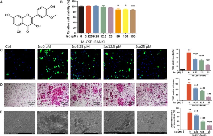
Figure 1 Iso inhibits receptor activator of nuclear factor‐kappa B ligand (RANKL)‐induced reactive oxygen species (ROS) production and osteoclastogenesis in vitro. (A) The chemical structure of Iso. (B) Bone marrow macrophages (BMMs) were treated with various concentrations of Iso for 24 h and analysed using a CCK‐8 assay. (C) Intracellular ROS levels of BMMs detected by DCFH‐DA probe. (D) Tartrate‐resistant acid phosphatase staining of osteoclasts treated with different concentrations of Iso. (E) Representative SEM images of bone resorption pits. *Compared with the control group and #compared with the RANKL and M‐CSF without Iso group
Figure 1 Iso inhibits receptor activator of nuclear factor‐kappa B ligand (RANKL)‐induced reactive oxygen species (ROS) production and osteoclastogenesis in vitro. (A) The chemical structure of Iso. (B) Bone marrow macrophages (BMMs) were treated with various concentrations of Iso for 24 h and analysed using a CCK‐8 assay. (C) Intracellular ROS levels of BMMs detected by DCFH‐DA probe. (D) Tartrate‐resistant acid phosphatase staining of osteoclasts treated with different concentrations of Iso. (E) Representative SEM images of bone resorption pits. *Compared with the control group and #compared with the RANKL and M‐CSF without Iso groupCC BY · PubMed Central
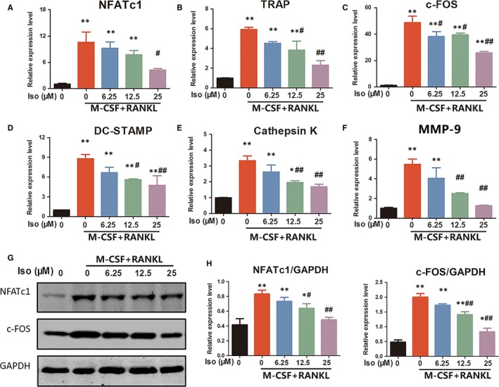
Figure 2 Iso suppresses receptor activator of nuclear factor‐kappa B ligand (RANKL)‐induced expression of osteoclastogenesis‐related genes and proteins. The expression of osteoclast‐specific mRNA, including (A) NFATc1, (B) tartrate‐resistant acid phosphatase, (C) c‐FOS, (D) DC‐STAMP, (E) cathepsin K, and (F) MMP‐9, was analysed using qRT‐PCR. (G) Western blotting was performed to analyse the expression of osteoclastogenesis‐related transcription factors including NFATc1 and c‐FOS. (H) Quantification of NFATc1 and c‐FOS expression levels. *Compared with the control group, and #compared with the RANKL and M‐CSF without Iso group
Figure 2 Iso suppresses receptor activator of nuclear factor‐kappa B ligand (RANKL)‐induced expression of osteoclastogenesis‐related genes and proteins. The expression of osteoclast‐specific mRNA, including (A) NFATc1, (B) tartrate‐resistant acid phosphatase, (C) c‐FOS, (D) DC‐STAMP, (E) cathepsin K, and (F) MMP‐9, was analysed using qRT‐PCR. (G) Western blotting was performed to analyse the expression of osteoclastogenesis‐related transcription factors including NFATc1 and c‐FOS. (H) Quantification of NFATc1 and c‐FOS expression levels. *Compared with the control group, and #compared with the RANKL and M‐CSF without Iso groupCC BY · PubMed Central
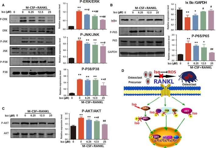
Figure 3 Iso treatment prevents osteoclastogenesis by regulating mitogen‐activated protein kinase (MAPK), NF‐κB and AKT signalling pathways. (A) The effects of Iso on MAPK signalling pathway proteins, including p‐ERK/ERK, p‐JNK/JNK and p‐p38/p38. (B) Western blotting showed that Iso affected the NF‐κB pathway‐related proteins IκBα, p65 and p‐p65. (C) The effects of Iso on AKT and the phosphorylation of AKT. *Compared with the control group and #compared with the receptor activator of nuclear factor‐kappa B ligand and M‐CSF without Iso group. (D) A schematic illustration showing signalling pathways involved in osteoclast differentiation regulated by Iso
Figure 3 Iso treatment prevents osteoclastogenesis by regulating mitogen‐activated protein kinase (MAPK), NF‐κB and AKT signalling pathways. (A) The effects of Iso on MAPK signalling pathway proteins, including p‐ERK/ERK, p‐JNK/JNK and p‐p38/p38. (B) Western blotting showed that Iso affected the NF‐κB pathway‐related proteins IκBα, p65 and p‐p65. (C) The effects of Iso on AKT and the phosphorylation of AKT. *Compared with the control group and #compared with the receptor activator of nuclear factor‐kappa B ligand and M‐CSF without Iso group. (D) A schematic illustration showing signalling pathways involved in osteoclast differentiation regulated by IsoCC BY · PubMed Central
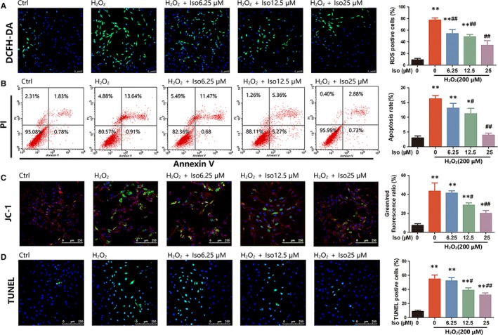
Figure 4 Iso inhibits reactive oxygen species (ROS)‐induced chondrocyte apoptosis. (A) The production of intracellular ROS detected by DCFH‐DA probe. (B) Primary chondrocyte apoptosis assessed using flow cytometry analysis. (C) Measurement of mitochondrial membrane potential by JC‐1. (D) TUNEL staining to assess cell apoptosis. *Compared with the control group and #compared with the H2O2 without Iso group
Figure 4 Iso inhibits reactive oxygen species (ROS)‐induced chondrocyte apoptosis. (A) The production of intracellular ROS detected by DCFH‐DA probe. (B) Primary chondrocyte apoptosis assessed using flow cytometry analysis. (C) Measurement of mitochondrial membrane potential by JC‐1. (D) TUNEL staining to assess cell apoptosis. *Compared with the control group and #compared with the H2O2 without Iso groupCC BY · PubMed Central
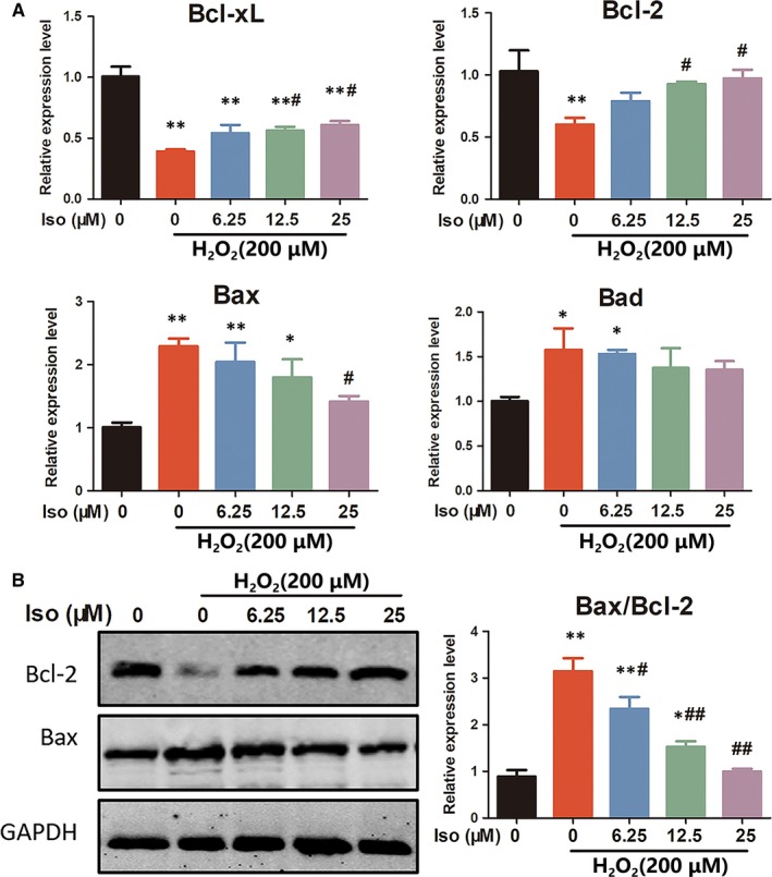
Figure 5 Iso inhibits chondrocyte apoptosis by modulating apoptosis‐related genes and proteins. (A) The expression of apoptosis‐related genes, including Bcl‐xL/Bcl‐2 (anti‐apoptotic) and Bax/Bad (pro‐apoptotic), was evaluated using qRT‐PCR. (B) Western blotting showed the effects of Iso on apoptosis‐related proteins Bcl‐2 and Bax. *Compared with the control group and # compared with the H2O2 without Iso group
Figure 5 Iso inhibits chondrocyte apoptosis by modulating apoptosis‐related genes and proteins. (A) The expression of apoptosis‐related genes, including Bcl‐xL/Bcl‐2 (anti‐apoptotic) and Bax/Bad (pro‐apoptotic), was evaluated using qRT‐PCR. (B) Western blotting showed the effects of Iso on apoptosis‐related proteins Bcl‐2 and Bax. *Compared with the control group and # compared with the H2O2 without Iso groupCC BY · PubMed Central
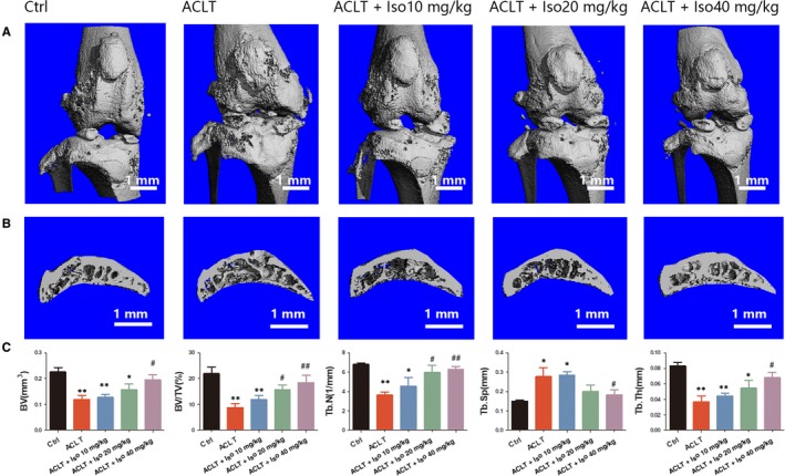
Figure 6 Micro‐CT analysis of Iso‐treated OA induced by anterior cruciate ligament transection (ACLT). (A) Three‐dimensional μCT images of frontal views of the knee joints at 4 wk after the sham operation or the ACLT operation. (B) Sagittal views of the medial compartment of subchondral bone. (C) Quantitative analysis of BV, BV/TV, Tb.N, Tb.Sp and Tb.Th. *Compared with the control group and #compared with the ACLT group
Figure 6 Micro‐CT analysis of Iso‐treated OA induced by anterior cruciate ligament transection (ACLT). (A) Three‐dimensional μCT images of frontal views of the knee joints at 4 wk after the sham operation or the ACLT operation. (B) Sagittal views of the medial compartment of subchondral bone. (C) Quantitative analysis of BV, BV/TV, Tb.N, Tb.Sp and Tb.Th. *Compared with the control group and #compared with the ACLT groupCC BY · PubMed Central
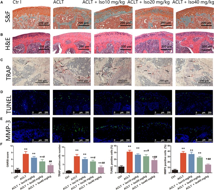
Figure 7 Staining analysis of Iso treatment in anterior cruciate ligament transection (ACLT)‐induced osteoarthritis. Knee joint medial compartment cartilage and subchondral bone were stained by (A) S&F and (B) H&E. (C) Osteoclasts in subchondral bone were stained by tartrate‐resistant acid phosphatase. (D‐E) TUNEL and MMP‐3 staining of chondrocytes in articular cartilage. (F) Quantitative analysis of the Osteoarthritis Research Society International score and positively stained cells in articular cartilage. *Compared with the control group and #compared with the ACLT group
Figure 7 Staining analysis of Iso treatment in anterior cruciate ligament transection (ACLT)‐induced osteoarthritis. Knee joint medial compartment cartilage and subchondral bone were stained by (A) S&F and (B) H&E. (C) Osteoclasts in subchondral bone were stained by tartrate‐resistant acid phosphatase. (D‐E) TUNEL and MMP‐3 staining of chondrocytes in articular cartilage. (F) Quantitative analysis of the Osteoarthritis Research Society International score and positively stained cells in articular cartilage. *Compared with the control group and #compared with the ACLT groupCC BY · PubMed Central

Verification

Claims the paper makes

The claims and statistics of this paper haven’t been checked yet.

Run the check — free, no account needed. It reads the open-access text and checks the sources it cites.

A few free checks a day · papers already verified open instantly · results are public

Claims other papers make about this one

Each is checked against this paper’s own text: does it say what the citing paper says it does?

Self-check

Percentages, sums and reported differences re-computed from the paper’s own numbers.

The self-check needs the paper’s full text.

Citations

Cites

Show all 1 references

Research works printed but not linked: 49

  1. 10.1016/j.jot.2018.03.001 · DOI
  2. 10.1038/boneres.2016.44 · DOI
  3. 10.1016/j.rdc.2012.10.004 · DOI
  4. 10.2174/1874325001206010558 · DOI
  5. 10.1016/j.joca.2014.01.003 · DOI
  6. 10.1016/S0140-6736(11)60243-2 · DOI
  7. 10.1016/j.jot.2017.12.004 · DOI
  8. 10.1016/j.jot.2015.02.002 · DOI
  9. 10.1053/joca.2001.0507 · DOI
  10. 10.1016/j.joca.2004.07.009 · DOI
  11. 10.1016/j.bone.2014.06.009 · DOI
  12. 10.1016/j.joca.2015.06.020 · DOI
  13. 10.1038/nrrheum.2016.65 · DOI
  14. 10.1007/s11010-014-2187-z · DOI
  15. 10.1002/jbmr.2974 · DOI
  16. 10.1016/j.joca.2017.07.020 · DOI
  17. 10.1016/j.joca.2007.01.008 · DOI
  18. 10.2174/138945007779940025 · DOI
  19. 10.1074/jbc.M804178200 · DOI
  20. 10.1016/j.gene.2018.05.009 · DOI
  21. Zhang BY, 2015, Isorhamnetin flavonoid synergistically enhances the anticancer activity and apoptosis induction by cis‐platin and carboplatin in non‐small cell lung carcinoma (NSCLC), Int J Clin Exp Pathol, 8, 25
  22. 10.3892/mmr.2017.7041 · DOI
  23. 10.3389/fphar.2017.00627 · DOI
  24. 10.1002/jcp.27852 · DOI
  25. 10.3389/fphar.2018.00174 · DOI
  26. 10.1002/jcb.27028 · DOI
  27. 10.1016/j.biopha.2017.09.011 · DOI
  28. 10.1007/978-1-4939-1215-5_23 · DOI
  29. 10.1016/j.bone.2012.03.025 · DOI
  30. 10.1038/cddis.2016.11 · DOI
  31. 10.1016/j.joca.2005.07.014 · DOI
  32. 10.1038/nature01658 · DOI · Paper page
  33. 10.1016/j.tem.2012.05.005 · DOI
  34. 10.1039/C7FO01497J · DOI
  35. 10.1186/1471-2474-9-20 · DOI
  36. 10.1016/j.joca.2007.03.006 · DOI
  37. 10.1182/blood-2004-09-3662 · DOI
  38. 10.1074/jbc.M109.051557 · DOI
  39. 10.1038/nrm2256 · DOI · Paper page
  40. 10.1159/000480416 · DOI
  41. 10.1002/jor.1100180306 · DOI
  42. 10.1007/s00198-013-2313-x · DOI
  43. 10.4049/jimmunol.1000885 · DOI
  44. 10.1016/j.febslet.2009.06.047 · DOI
  45. 10.1002/art.30646 · DOI
  46. 10.1007/s00204-015-1472-2 · DOI
  47. 10.1021/acs.jnatprod.7b00927 · DOI
  48. 10.7150/ijbs.12296 · DOI
  49. 10.1371/journal.pone.0023199 · DOI

Cited by

Metadata

Authors
Zhou, F., Mei, J., Yuan, K., Han, X., Qiao, H., & Tang, T.
Affiliations
Shanghai Key Laboratory of Orthopaedic Implants, Department of Orthopaedic Surgery Shanghai Ninth People's Hospital, Shanghai Jiao Tong University School of Medicine Shanghai P. R. of China
Venue
Journal of Cellular and Molecular Medicine
Publisher
Wiley
Published
2019-04-14
Volume
23
Issue
6
Pages
4395-4407
ISSN
  • 1582-1838
  • 1582-4934
Cited by
57
References
1
Language
en
Funders
  • National Natural Science Foundation of China (81672205)
  • Shanghai Science and Technology Development Foundation (18DZ2291200, 18441902700)

About these checks

Registry, retraction, Cites and statistics results are computed, not judged, with one exception: a printed reference that no index lists is read once by a model before it counts as a citation error. Claims are extracted by a model and kept only when their quote appears verbatim in the text; each cited source is then checked, with a model’s help, for whether it says what the claim relies on. Those verdicts can be wrong — read the claim’s quote and the cited source before relying on one.

Statistics and arithmetic are recomputed, not judged.

Confidence bands are shown only for fields calibrated on our annotated test set. None is calibrated yet, so no band is shown.

What counts as a claim ›How we measure confidence ›How we check this ›

Are you an author, or do you see a problem with this page? Request a review

Data sources

Bibliographic data: Semantic Scholar, OpenAlex, Crossref.

Contains information from the Semantic Scholar Academic Graph Datasets, made available under the ODC Attribution License (ODC-BY 1.0).

Semantic Scholar’s data is described in: Kinney, R., et al. (2023). The Semantic Scholar Open Data Platform. arXiv:2301.10140.