

The DOI matches its Crossref record; 4 of the 4 references it cites were found in a registry or an index.
Bone marrow stromal and anterior cruciate ligament remnant cell co‐culture‐derived extracellular vesicles promote cell activity in both cell types
Open accessAbstract
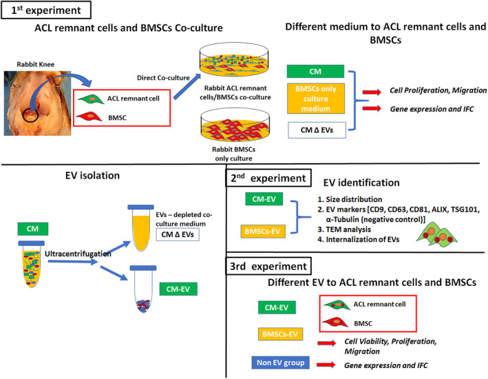
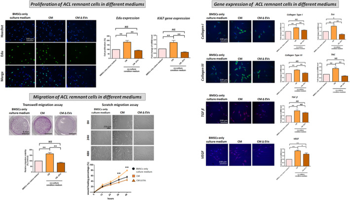
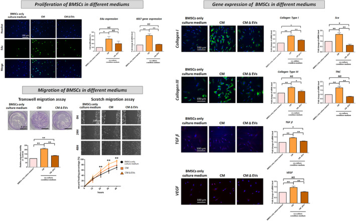
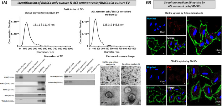
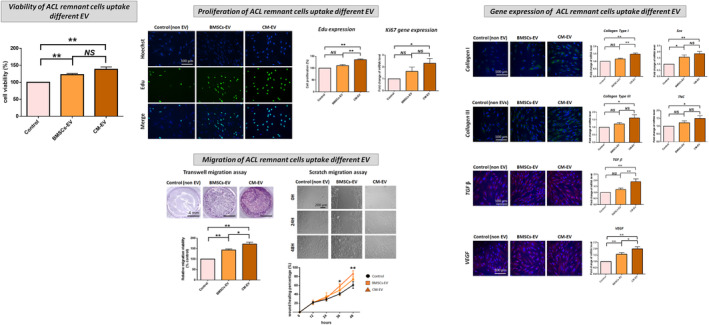
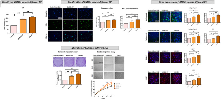
The significance of anterior cruciate ligament (ACL) remnants during reconstruction remains unclear. Co-culturing ACL remnant cells and bone marrow stromal cells (BMSCs) may reduce apoptosis and enhance hamstring tendon activity. This study investigated whether extracellular vesicles (EVs), which facilitate cell-cell interactions, act as the active components, improving graft maturation in this co-culture. The effects of EVs on cell viability, proliferation, migration and gene expression in the rabbit ACL remnant cells and BMSCs were assessed using control (BMSC-only culture), co-culture (ACL remnant cells and BMSCs, CM) and co-culture without EVs (CM ∆ EVs) media. EVs were isolated from control (BMSC-EV) and co-culture (CM-EV) media and characterized. CM significantly enhanced the proliferation, migration and expression of transforming growth factor (TGF-β)-, vascular endothelial growth factor (VEGF)-, collagen synthesis- and tenogenesis-related genes. However, CM-induced effects were reversed by the CM ∆ EVs treatment. CM-EV treatment exhibited higher potential to enhance proliferation, migration and gene expression in the ACL remnant cells and BMSCs than BMSC-EV and non-EV treatments. In conclusion, EVs, secreted under the coexistence of ACL remnant cells and BMSCs, primarily increase the cell viability, proliferation, migration and gene expression of collagen synthesis-, TGF-β-, VEGF- and tenogenesis-related genes in both cell types.
Overview
The claims and statistics of this paper haven’t been checked yet.
Run the check — free, no account needed. It reads the open-access text and checks the sources it cites.
Integrity signals
Registry
Cites
Statistics
Figures
Verification
Claims the paper makes
The claims and statistics of this paper haven’t been checked yet.
Run the check — free, no account needed. It reads the open-access text and checks the sources it cites.
Claims other papers make about this one
Each is checked against this paper’s own text: does it say what the citing paper says it does?
The source backs part of the claim, not all of it — or backs it for a different population, condition or comparison.
In the citing paper
“Besides ligamentous differentiation of ACL-derived cells, an unidentified paracrine action between transplanted ACL-derived cells and the host cells may have enhanced ligament-specific ECM synthesis [22, 62].”
From A new biological enhancement therapy for anterior cruciate ligament reconstruction: the preclinical proof of anterior cruciate ligament reconstruction with tendon graft reseeded with autologous anterior cruciate ligament-derived cells (2025) · cites this paper as [62]
Experimental — accuracy not yet measured
Self-check
Percentages, sums and reported differences re-computed from the paper’s own numbers.
The self-check needs the paper’s full text.
Citations
Cites
Incidence of Anterior Cruciate Ligament Tears and Reconstruction: A 21-Year Population-Based Study
Sanders, T. L., Maradit Kremers, H., Bryan, A. J., Larson, D. R., Dahm, D. L., Levy, B. A., Stuart, M. J., & Krych, A. J. (2016) · The American Journal of Sports Medicine · doi:10.1177/0363546516629944 · Cited by 1,325
Mesenchymal Stem Cell-Derived Exosomes Promote Fracture Healing in a Mouse Model
Furuta, T., Miyaki, S., Ishitobi, H., Ogura, T., Kato, Y., Kamei, N., Miyado, K., Higashi, Y., & Ochi, M. (2016) · Stem Cells Translational Medicine · doi:10.5966/sctm.2015-0285 · Cited by 444
Extracellular vesicles: lipids as key components of their biogenesis and functions
Record, M., Silvente-Poirot, S., Poirot, M., & Wakelam, M. O. (2018) · Journal of Lipid Research · doi:10.1194/jlr.e086173 · Cited by 328
Mifune, Y., Matsumoto, T., Ota, S., Nishimori, M., Usas, A., Kopf, S., Kuroda, R., Kurosaka, M., Fu, F. H., & Huard, J. (2012) · Cell Transplantation · doi:10.3727/096368912x647234 · Cited by 107
Research works printed but not linked: 33
- Bradley JP, 2002, Anterior cruciate ligament injuries in the National Football League: epidemiology and current treatment trends among team physicians, Art Ther, 18, 502
- 10.1016/j.jsams.2008.07.005 · DOI
- 10.1007/s00167-011-1419-y · DOI · Paper page
- 10.1007/s00167-013-2634-5 · DOI · Paper page
- 10.1177/0363546512461481 · DOI
- 10.1007/s00167-013-2749-8 · DOI
- Song GY, 2016, Biomechanical and biological findings between acute anterior cruciate ligament reconstruction with and without an augmented remnant repair: a comparative in vivo animal study, Art Ther, 32, 307
- 10.1089/scd.2010.0528 · DOI
- 10.1177/0363546512439026 · DOI
- 10.1302/2046-3758.98.BJR-2019-0365.R1 · DOI
- 10.1302/2046-3758.121.BJR-2021-0434.R2 · DOI
- 10.1042/BST20120192 · DOI
- 10.1042/BST20180079 · DOI
- 10.1186/s13287-018-0791-7 · DOI
- 10.1186/s13287-020-1562-9 · DOI
- 10.1039/C7NR00352H · DOI
- Liu WC, 2022, Extracorporeal shock wave therapy shows superiority over injections for pain relief and grip strength recovery in lateral epicondylitis: a systematic review and network meta‐analysis, Art Ther, 38, 2018
- 10.1074/jbc.M114.630558 · DOI
- 10.1016/j.gene.2014.08.041 · DOI
- 10.3389/fbioe.2020.00911 · DOI
- 10.1177/2041731417724640 · DOI
- 10.1155/2012/971907 · DOI
- 10.1016/j.semcdb.2016.11.008 · DOI
- 10.1002/stem.2575 · DOI · Paper page
- 10.1016/j.joca.2016.06.022 · DOI
- 10.1002/jor.24587 · DOI
- 10.1002/jor.24859 · DOI
- 10.1186/s12967-019-1960-x · DOI
- 10.3389/fimmu.2018.02837 · DOI · Paper page
- 10.1186/s13287-019-1484-6 · DOI
- 10.3390/ph14111093 · DOI
- 10.1186/s12964-023-01103-6 · DOI
- 10.1007/s13770-022-00490-0 · DOI
Cited by
Sorted by citations
Park, J., Soh, H., Jang, Y.-K., Lee, Y. S., Lee, M.-K., Kim, H., Kim, T.-H., Alkhelaifi, K., Kim, T.-J., & Lee, J. K. (2025) · Stem Cell Research & Therapy · doi:10.1186/s13287-025-04760-1 · Cited by 3
Metadata
- Authors
- Lin, S., Wu, S. C., Liu, Z., Chou, P. P., Zhao, C., Ho, M., & Lu, C.
- Affiliations
- Kaohsiung Medical University (TW)
- Asia University (TW)
- Kaohsiung Medical University Chung-Ho Memorial Hospital (TW)
- Mayo Clinic (US)
- Corresponding author
- Lu, Cheng‐Chang
- Venue
- Journal of Cellular and Molecular Medicine
- Publisher
- Wiley
- Published
- 2024-09
- Volume
- 28
- Issue
- 17
- Pages
- e70049-e70049
- ISSN
- 1582-1838 (ISSN-L)
- 1582-4934
- PMC
- PMC11366498
- OpenAlex
- W4402141916
- Cited by
- 5
- References
- 4
- Citations by year
- 2026: 3 · 2025: 1 · 2024: 1
- FWCI
- 0.68
- Type
- journal article
- Language
- en
- Open access
- gold
- doi.org
- Licence
- Locations
- Journal of Cellular and Molecular Medicine (publishedVersion, cc-by)
- PubMed Central (publishedVersion, cc-by)
- PubMed (publishedVersion)
- Topics
- Life Sciences › Biochemistry, Genetics and Molecular Biology › Molecular Biology › Extracellular vesicles in disease
- Health Sciences › Medicine › Orthopedics and Sports Medicine › Tendon Structure and Treatment
- Health Sciences › Medicine › Surgery › Knee injuries and reconstruction techniques
- Keywords
- extracellular vesicles, mesenchymal stem cells, co-culture, anterior cruciate ligament reconstruction, graft maturation, tenogenesis, collagen synthesis, TGF-beta, vascular endothelial growth factor, cell proliferation, cell migration, apoptosis, intercellular communication
- MeSH terms
- Extracellular Vesicles, Animals, Cell Communication, Cell Movement, Cell Survival, Cells, Cultured, Gene Expression Regulation, Male, Rabbits, Anterior Cruciate Ligament, Transforming Growth Factor beta, Coculture Techniques, Vascular Endothelial Growth Factor A, Cell Proliferation, Mesenchymal Stem Cells
About these checks
Registry, retraction, Cites and statistics results are computed, not judged, with one exception: a printed reference that no index lists is read once by a model before it counts as a citation error. Claims are extracted by a model and kept only when their quote appears verbatim in the text; each cited source is then checked, with a model’s help, for whether it says what the claim relies on. Those verdicts can be wrong — read the claim’s quote and the cited source before relying on one.
Statistics and arithmetic are recomputed, not judged.
Confidence bands are shown only for fields calibrated on our annotated test set. None is calibrated yet, so no band is shown.
What counts as a claim ›How we measure confidence ›How we check this ›
Are you an author, or do you see a problem with this page? Request a review
How to cite
Cite CoChat for the checks on this page, and the paper for its own findings.
- This page
- CoChat. (2026). Citation check: Bone marrow stromal and anterior cruciate ligament remnant cell co‐culture‐derived extracellular vesicles promote cell activity in both cell types. https://app.cochat.ai/papers/10.1111/jcmm.70049
- The paper
- Lin, S., Wu, S. C., Liu, Z., Chou, P. P., Zhao, C., Ho, M., & Lu, C. (2024). Bone marrow stromal and anterior cruciate ligament remnant cell co‐culture‐derived extracellular vesicles promote cell activity in both cell types. Journal of Cellular and Molecular Medicine. https://doi.org/10.1111/jcmm.70049
