Student or educator? Sign up with your school email and get 6 months of Pro free.
Cited by 2Enzyme Structure and FunctionAdvanced Electron Microscopy Techniques and ApplicationsMetal-Catalyzed Oxygenation Mechanisms

The DOI matches its Crossref record; 58 of the 58 references it cites were found in a registry or an index.

Visualizing the mechanism of quinol oxidation and inhibition of a bd -type oxidase using cryo-EM

Open access

Published version of a preprint

Abstract

Cytochrome bd is a prokaryotic terminal oxidase recognized as an antibiotic target against various pathogens. Despite its critical role in respiration, failure to capture the mechanism of quinol oxidation and inhibition prohibits structure guided drug discovery. Here, we present cryo–electron microscopy structures of Escherichia coli cytochrome bd -I in monomeric and dimeric forms, in several quinone and inhibitor-bound states. We identify a dynamic Q-loop lid that undergoes a disorder-to-order transition upon substrate binding to the dimer, completing the active site and enabling catalysis. Structure-guided mutagenesis confirms Tyr243 CydA and Arg298 CydA as conserved catalytic residues only found in long Q-loop oxidases, highlighting evolutionary divergence from other subfamilies. Inhibition by Aurachin D triggers refolding of the active site, occluding substrate access via an Asp239 CydA -mediated mechanism. The structural and mechanistic insights presented here establish a comprehensive framework, opening paths for drug discovery against bd oxidases.

Overview

The claims and statistics of this paper haven’t been checked yet.

Run the check — free, no account needed. It reads the open-access text and checks the sources it cites.

A few free checks a day · papers already verified open instantly · results are public

Integrity signals

Registry

Crossref · 10.1126/sciadv.aec9946

Cites

58 of 58 references verified0 citation errors · 0 not checkable

Statistics

Figures

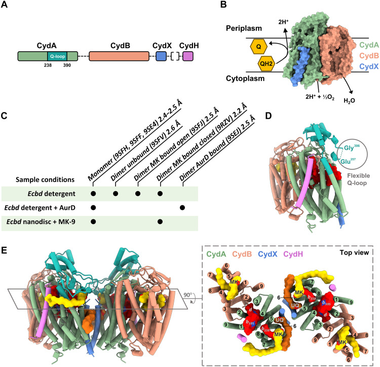
Fig. 1. Overview of the structures and function of Ecbd. (A) Genomic organization of the Ecbd subunits. Subunit CydH originates from a different operon than the other Ecbd subunits. (B) Schematic oxidase activity of Ecbd. (C) Overview of the different sample conditions used to yield the presented monomer and dimeric states, including their state and resolution. Protein Data Bank: 9RZV was resolved from the nanodisc sample. (D) Structural overview of the Ecbd monomer. CydA is shown in green, with the Q-loop in cyan, CydB in orange, CydX in blue, and CydH in purple. (E) Structural overview of the Ecbd dimer in the MK bound state with the Q-loop highlighted in cyan. MK is shown yellow and UQ in dark orange. The right shows a cross section of the Ecbd dimer with a total of 40 transmembrane helices, highlighting all identified MK (yellow) and UQ (dark orange), as well as heme groups (red).
Fig. 1. Overview of the structures and function of Ecbd. (A) Genomic organization of the Ecbd subunits. Subunit CydH originates from a different operon than the other Ecbd subunits. (B) Schematic oxidase activity of Ecbd. (C) Overview of the different sample conditions used to yield the presented monomer and dimeric states, including their state and resolution. Protein Data Bank: 9RZV was resolved from the nanodisc sample. (D) Structural overview of the Ecbd monomer. CydA is shown in green, with the Q-loop in cyan, CydB in orange, CydX in blue, and CydH in purple. (E) Structural overview of the Ecbd dimer in the MK bound state with the Q-loop highlighted in cyan. MK is shown yellow and UQ in dark orange. The right shows a cross section of the Ecbd dimer with a total of 40 transmembrane helices, highlighting all identified MK (yellow) and UQ (dark orange), as well as heme groups (red).CC BY · PubMed Central
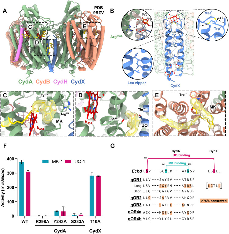
Fig. 2. The Ecbd dimer contains three distinct quinone binding sites per protomer. (A) Overview of the Ecbd dimer and the quinone binding pockets. (B) The Ecbd dimer interface is formed by CydX via Met-Trp interactions, and a leucine zipper. In addition, a PG phospholipid forms a hydrogen bond with Arg298CydA further stabilizing the interface. (C) Q- binding site near heme b558, showing a bound MK stabilized by Tyr243CydA and Arg298CydA. (D) The UQ binding site wedged between subunit CydA and CydX. The UQ is stabilized by interactions with Ser233CydA and Thr10CydX. (E) Quinone-binding pocket in CydB with a bound MK, stabilized by π-π stacking with Trp12CydB. The quinone headgroup is situated outside of electron transfer distance from the heme chain. (F) Oxidase activity of monomeric Ecbd WT and quinone binding site mutants (n = 3) (G) Consensus sequence alignment of the qOR1–4 subfamilies as defined by Murali et al. (15) The qOR1 family was subdivided in long and short Q-loop Cyt bd variants indicating conservation of Y243CydA and R298CydA in the long Q-loop family (extended sequence variation in table S2). The CydX subunit is only present in a subset of qOR1-long Q-loop family. The consensus sequence and conservation of CydX defined by Allen et al. (34)
Fig. 2. The Ecbd dimer contains three distinct quinone binding sites per protomer. (A) Overview of the Ecbd dimer and the quinone binding pockets. (B) The Ecbd dimer interface is formed by CydX via Met-Trp interactions, and a leucine zipper. In addition, a PG phospholipid forms a hydrogen bond with Arg298CydA further stabilizing the interface. (C) Q- binding site near heme b558, showing a bound MK stabilized by Tyr243CydA and Arg298CydA. (D) The UQ binding site wedged between subunit CydA and CydX. The UQ is stabilized by interactions with Ser233CydA and Thr10CydX. (E) Quinone-binding pocket in CydB with a bound MK, stabilized by π-π stacking with Trp12CydB. The quinone headgroup is situated outside of electron transfer distance from the heme chain. (F) Oxidase activity of monomeric Ecbd WT and quinone binding site mutants (n = 3) (G) Consensus sequence alignment of the qOR1–4 subfamilies as defined by Murali et al. (15) The qOR1 family was subdivided in long and short Q-loop Cyt bd variants indicating conservation of Y243CydA and R298CydA in the long Q-loop family (extended sequence variation in table S2). The CydX subunit is only present in a subset of qOR1-long Q-loop family. The consensus sequence and conservation of CydX defined by Allen et al. (34)CC BY · PubMed Central
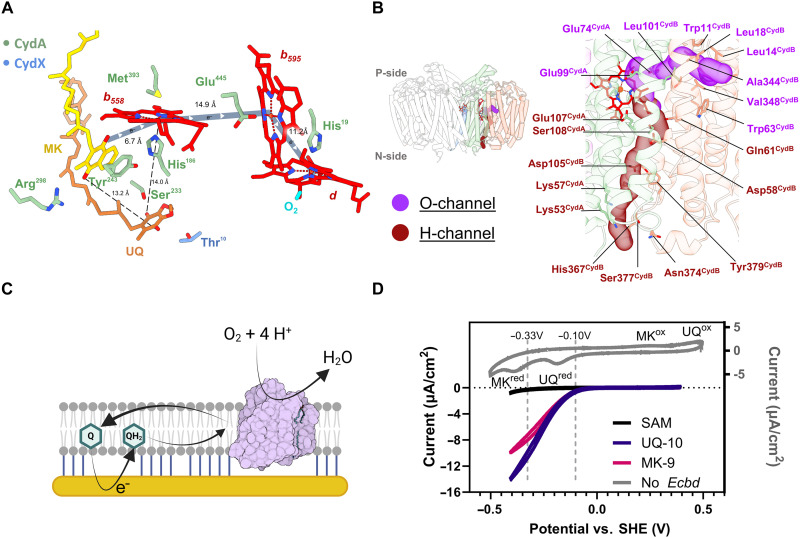
Fig. 3. The electron transfer, proton, and oxygen pathways. (A) Proposed electron transfer routes from the MK (yellow) depicted in dark blue. Distances from UQ (orange) to MK and heme b558 are indicated. (B) Residues lining of the O- and H-channel leading toward the heme d oxygen reduction center. (C) Overview of the tBLM electrochemical system, where the quinone pool is directly reduced at the gold electrode to initiate Ecbd turnover. (D) Cyclic voltammograms of the tBLM system. Inset in gray shows a model membrane containing both MK (1 mol %) and UQ (1 mol %) without Ecbd (scan rate of 100 mV/s). The reduction and oxidation peaks of UQ and MK are indicated. The SAM-negative control (black), Ecbd tBLMs containing only UQ-10 (purple), or MK-9 (pink) (scan rate of 10 mV/s) showing the same onset potential at the reduction of UQ.
Fig. 3. The electron transfer, proton, and oxygen pathways. (A) Proposed electron transfer routes from the MK (yellow) depicted in dark blue. Distances from UQ (orange) to MK and heme b558 are indicated. (B) Residues lining of the O- and H-channel leading toward the heme d oxygen reduction center. (C) Overview of the tBLM electrochemical system, where the quinone pool is directly reduced at the gold electrode to initiate Ecbd turnover. (D) Cyclic voltammograms of the tBLM system. Inset in gray shows a model membrane containing both MK (1 mol %) and UQ (1 mol %) without Ecbd (scan rate of 100 mV/s). The reduction and oxidation peaks of UQ and MK are indicated. The SAM-negative control (black), Ecbd tBLMs containing only UQ-10 (purple), or MK-9 (pink) (scan rate of 10 mV/s) showing the same onset potential at the reduction of UQ.CC BY · PubMed Central
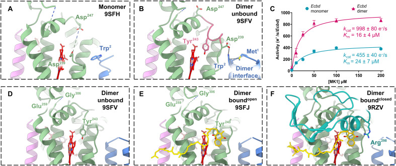
Fig. 4. Q-loop stabilization increases the turnover rate in the Ecbd dimer. (A) Selected view of the quinone binding pocket in the Ecbd monomer (B) and in the Ecbd dimer. The pink stretch is stabilized by hydrogen bonding between Asp239CydA and Trp2CydX, positioning Tyr243CydA for quinol capture. (C) Michaelis Menten kinetics of the Ecbd monomer and dimer (n = 3). (D) Unbound Ecbd dimer. (E) Ecbd dimer in the MK-boundopen state showing initial interaction with the quinone. (F) Ecbd dimer in the boundclosed state showing the closing of the Q-loop lid. CydA is represented in green, with the Q-loop lid in cyan. CydH is represented in violet, with CydX in blue.
Fig. 4. Q-loop stabilization increases the turnover rate in the Ecbd dimer. (A) Selected view of the quinone binding pocket in the Ecbd monomer (B) and in the Ecbd dimer. The pink stretch is stabilized by hydrogen bonding between Asp239CydA and Trp2CydX, positioning Tyr243CydA for quinol capture. (C) Michaelis Menten kinetics of the Ecbd monomer and dimer (n = 3). (D) Unbound Ecbd dimer. (E) Ecbd dimer in the MK-boundopen state showing initial interaction with the quinone. (F) Ecbd dimer in the boundclosed state showing the closing of the Q-loop lid. CydA is represented in green, with the Q-loop lid in cyan. CydH is represented in violet, with CydX in blue.CC BY · PubMed Central
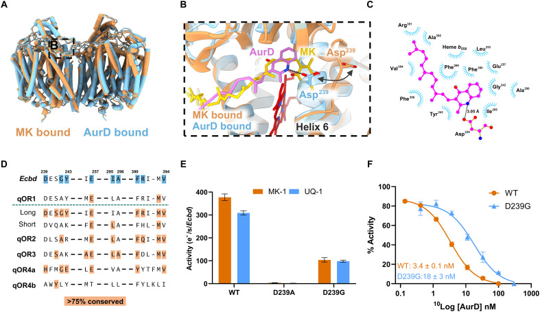
Fig. 5. Inhibitory mechanism of AurD. (A) Overview of the Ecbd dimer in the MK and AurD bound state. (B) Refolding of the Ecbd active site by AurD by the attraction of Asp239CydA. (C) Two dimensional representations of the interactions in the AurD binding pocket. (D) Conservation of the AurD binding pocket residues (extended sequence variation in table S3). (E) Activity of the monomeric Ecbd WT and D239ACydA and D239GCydA mutants (n = 3). (F) IC50 (median inhibitory concentration) of AurD against the monomeric Ecbd WT and D239GCydA mutant (n = 3), P < 0.001.
Fig. 5. Inhibitory mechanism of AurD. (A) Overview of the Ecbd dimer in the MK and AurD bound state. (B) Refolding of the Ecbd active site by AurD by the attraction of Asp239CydA. (C) Two dimensional representations of the interactions in the AurD binding pocket. (D) Conservation of the AurD binding pocket residues (extended sequence variation in table S3). (E) Activity of the monomeric Ecbd WT and D239ACydA and D239GCydA mutants (n = 3). (F) IC50 (median inhibitory concentration) of AurD against the monomeric Ecbd WT and D239GCydA mutant (n = 3), P < 0.001.CC BY · PubMed Central
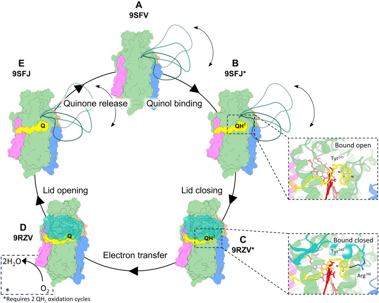
Fig. 6. Schematic overview of the proposed turnover mechanism of Ecbd. (A to D) Overall schematic view of Ecbd turnover. States with an asterix are presumed to be structurally the same in the quinol and quinone bound states. (A) Unbound Ecbd dimer has a flexible Q-loop lid and sits ready for quinol binding. (B) Ecbd dimer undergoes quinol binding. (C) From the quinol bound state (B), the Q-loop lid closes, finishing the formation of the quinol oxidation site by coordinating Arg298CydA to the quinol head (C). (D) Electron transfer occurs via the heme chain onto molecular oxygen. (E) Q-loop lid opens to release the quinone and start another catalytic cycle.
Fig. 6. Schematic overview of the proposed turnover mechanism of Ecbd. (A to D) Overall schematic view of Ecbd turnover. States with an asterix are presumed to be structurally the same in the quinol and quinone bound states. (A) Unbound Ecbd dimer has a flexible Q-loop lid and sits ready for quinol binding. (B) Ecbd dimer undergoes quinol binding. (C) From the quinol bound state (B), the Q-loop lid closes, finishing the formation of the quinol oxidation site by coordinating Arg298CydA to the quinol head (C). (D) Electron transfer occurs via the heme chain onto molecular oxygen. (E) Q-loop lid opens to release the quinone and start another catalytic cycle.CC BY · PubMed Central

Verification

Claims the paper makes

The claims and statistics of this paper haven’t been checked yet.

Run the check — free, no account needed. It reads the open-access text and checks the sources it cites.

A few free checks a day · papers already verified open instantly · results are public

Self-check

Percentages, sums and reported differences re-computed from the paper’s own numbers.

The self-check needs the paper’s full text.

Citations

Cites

Show all 58 references

Research works printed but not linked: 1

  1. 10.1021/bi061946 · DOI

Metadata

Authors
van der Velden, T. T., Kayastha, K., Pelser, F., Brünle, S., & Jeuken, L. J. C.
Affiliations
Leiden University (NL)
Corresponding author
Jeuken, Lars J. C.
Venue
Science Advances
Published
2026-05-22
Volume
12
Issue
21
Pages
eaec9946-eaec9946
ISSN
2375-2548 (ISSN-L)
OpenAlex
W7161809970
Cited by
2
References
58
Citations by year
2026: 2
FWCI
2.71
Type
journal article
Language
en
Open access
Licence
cc-by
Locations
  • Science Advances (publishedVersion, cc-by)
  • bioRxiv (Cold Spring Harbor Laboratory) (acceptedVersion, cc-by)
  • PubMed (publishedVersion)
  • PubMed Central (publishedVersion, cc-by)
  • Leiden Repository (Leiden University) (submittedVersion, cc-by)
  • Leiden Repository (Leiden University) (submittedVersion)
Funders
  • Nederlandse Organisatie voor Wetenschappelijk Onderzoek (Netherlands Electron Microscopy Infrastructure (NEMI; project number 184.034.014))
  • Nederlands Nationaal Groeifonds (Oncode (grant number NGFOP2201))
Topics
  • Life Sciences › Biochemistry, Genetics and Molecular Biology › Structural Biology › Enzyme Structure and Function
  • Life Sciences › Biochemistry, Genetics and Molecular Biology › Structural Biology › Advanced Electron Microscopy Techniques and Applications
  • Physical Sciences › Chemistry › Inorganic Chemistry › Metal-Catalyzed Oxygenation Mechanisms
Keywords
cytochrome bd oxidase, quinol oxidation, disorder-to-order transition, catalytic residues, terminal oxidase, antibiotic targets, structure-based drug design, protein dimerization
MeSH terms
Cytochrome b Group, Cytochromes, Enzyme Inhibitors, Escherichia coli, Hydroquinones, Models, Molecular, Oxidation-Reduction, Oxidoreductases, Protein Conformation, Benzoquinones, Catalytic Domain, Cryoelectron Microscopy, Escherichia coli Proteins, Electron Transport Chain Complex Proteins

About these checks

Registry, retraction, Cites and statistics results are computed, not judged, with one exception: a printed reference that no index lists is read once by a model before it counts as a citation error. Claims are extracted by a model and kept only when their quote appears verbatim in the text; each cited source is then checked, with a model’s help, for whether it says what the claim relies on. Those verdicts can be wrong — read the claim’s quote and the cited source before relying on one.

Statistics and arithmetic are recomputed, not judged.

Confidence bands are shown only for fields calibrated on our annotated test set. None is calibrated yet, so no band is shown.

What counts as a claim ›How we measure confidence ›How we check this ›

Are you an author, or do you see a problem with this page? Request a review

How to cite

Cite CoChat for the checks on this page, and the paper for its own findings.

This page
CoChat. (2026). Citation check: Visualizing the mechanism of quinol oxidation and inhibition of a bd -type oxidase using cryo-EM. https://app.cochat.ai/papers/10.1126/sciadv.aec9946
The paper
van der Velden, T. T., Kayastha, K., Pelser, F., Brünle, S., & Jeuken, L. J. C. (2026). Visualizing the mechanism of quinol oxidation and inhibition of a bd -type oxidase using cryo-EM. Science Advances. https://doi.org/10.1126/sciadv.aec9946

Data sources

Bibliographic data: Semantic Scholar, OpenAlex, Crossref.

Contains information from the Semantic Scholar Academic Graph Datasets, made available under the ODC Attribution License (ODC-BY 1.0).

Semantic Scholar’s data is described in: Kinney, R., et al. (2023). The Semantic Scholar Open Data Platform. arXiv:2301.10140.