

The DOI matches its Crossref record; 48 of the 48 references it cites were found in a registry or an index.
SUMOylation of the m6A reader YTHDF2 by PIAS1 promotes viral RNA decay to restrict EBV replication
Open accessPublished version of a preprint
Abstract
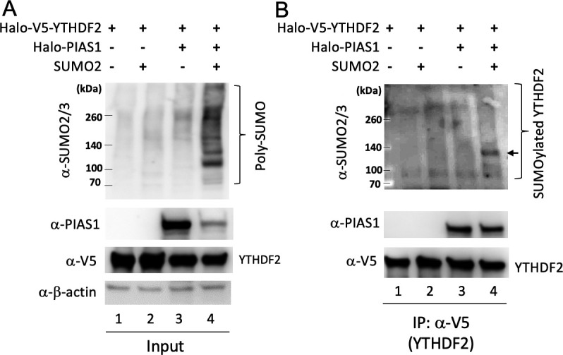
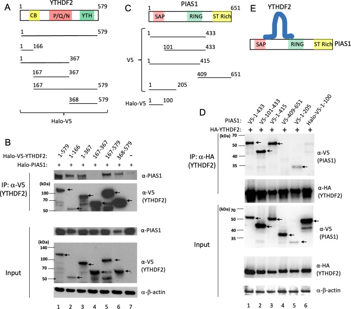
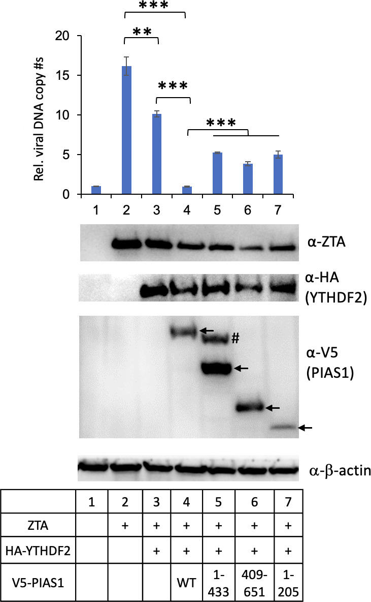
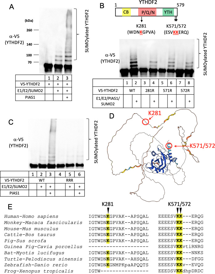
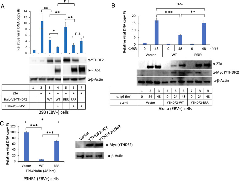
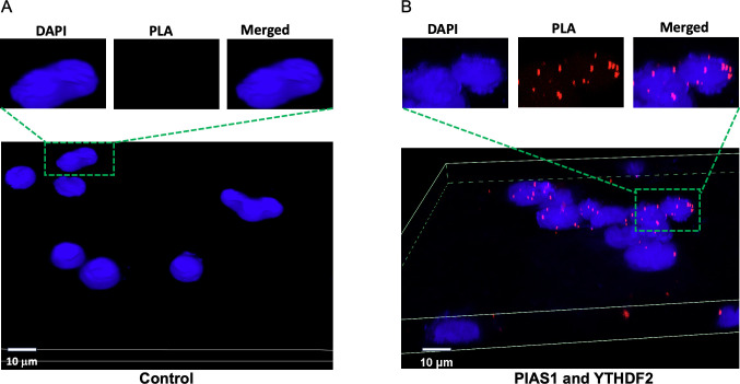
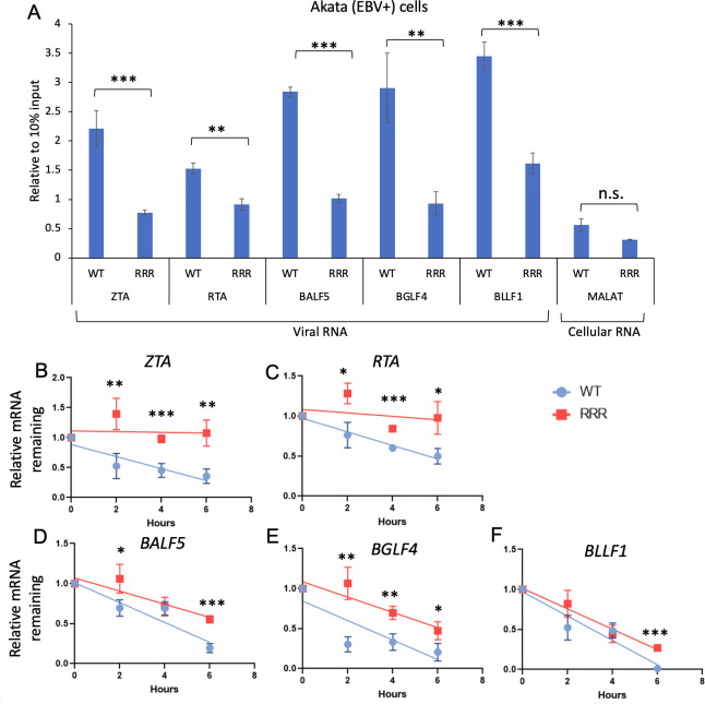
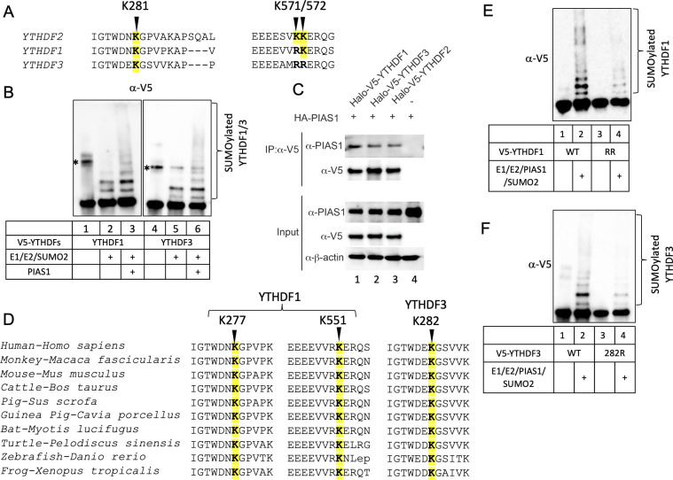
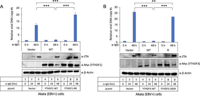
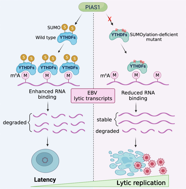
m6A RNA modification pathway plays important roles in diverse cellular processes and viral life cycle. Here, we investigated the relationship between PIAS1 and the m6A reader protein YTHDF2, which is involved in regulating RNA stability by binding to m6A-modified RNA. We found that both the N-terminal and C-terminal regions of YTHDF2 interact with PIAS1. We showed that PIAS1 promotes the SUMOylation of YTHDF2 at three specific lysine residues. We also demonstrated that PIAS1 enhances the anti-EBV activity of YTHDF2. We further revealed that PIAS1 mediates the SUMOylation of other YTHDF family members, namely, YTHDF1 and YTHDF3, to limit EBV replication. These findings together illuminate an important regulatory mechanism of YTHDF proteins in controlling viral RNA decay and EBV replication through PIAS1-mediated SUMOylation.
Overview
The claims and statistics of this paper haven’t been checked yet.
Run the check — free, no account needed. It reads the open-access text and checks the sources it cites.
Integrity signals
Registry
Cites
Statistics
Figures
Verification
Claims the paper makes
The claims and statistics of this paper haven’t been checked yet.
Run the check — free, no account needed. It reads the open-access text and checks the sources it cites.
Self-check
Percentages, sums and reported differences re-computed from the paper’s own numbers.
The self-check needs the paper’s full text.
Citations
Cites
Highly accurate protein structure prediction with AlphaFold
Jumper, J., Evans, R., Pritzel, A., Green, T., Figurnov, M., Ronneberger, O., Tunyasuvunakool, K., Bates, R., Žídek, A., Potapenko, A., et al. (2021) · Nature · doi:10.1038/s41586-021-03819-2 · Cited by 43,764
UCSF ChimeraX: Structure visualization for researchers, educators, and developers
Pettersen, E. F., Goddard, T. D., Huang, C. C., Meng, E. C., Couch, G. S., Croll, T. I., Morris, J. H., & Ferrin, T. E. (2021) · Protein Science · doi:10.1002/pro.3943 · Cited by 9,514
Dynamic RNA Modifications in Gene Expression Regulation
Roundtree, I. A., Evans, M. E., Pan, T., & He, C. (2017) · Cell · doi:10.1016/j.cell.2017.05.045 · Cited by 3,399
Rethinking m6A Readers, Writers, and Erasers
Meyer, K. D., & Jaffrey, S. R. (2017) · Annual Review of Cell and Developmental Biology · doi:10.1146/annurev-cellbio-100616-060758 · Cited by 1,150
The SUMO pathway: emerging mechanisms that shape specificity, conjugation and recognition
Gareau, J. R., & Lima, C. D. (2010) · Nature Reviews Molecular Cell Biology · doi:10.1038/nrm3011 · Cited by 1,105
A Unified Model for the Function of YTHDF Proteins in Regulating m6A-Modified mRNA
Zaccara, S., & Jaffrey, S. R. (2020) · Cell · doi:10.1016/j.cell.2020.05.012 · Cited by 797
m6A modification controls the innate immune response to infection by targeting type I interferons
Winkler, R., Gillis, E., Lasman, L., Safra, M., Geula, S., Soyris, C., Nachshon, A., Tai-Schmiedel, J., Friedman, N., Le-Trilling, V. T. K., et al. (2019) · Nature Immunology · doi:10.1038/s41590-018-0275-z · Cited by 455
Regulation of gene-activation pathways by PIAS proteins in the immune system
Shuai, K., & Liu, B. (2005) · Nature Reviews Immunology · doi:10.1038/nri1667 · Cited by 447
Interactions, localization, and phosphorylation of the m6A generating METTL3–METTL14–WTAP complex
Schöller, E., Weichmann, F., Treiber, T., Ringle, S., Treiber, N., Flatley, A., Feederle, R., Bruckmann, A., & Meister, G. (2018) · RNA · doi:10.1261/rna.064063.117 · Cited by 439
Evolutionary conflicts between viruses and restriction factors shape immunity
Duggal, N. K., & Emerman, M. (2012) · Nature Reviews Immunology · doi:10.1038/nri3295 · Cited by 366
SUMOylation of the m6A-RNA methyltransferase METTL3 modulates its function
Du, Y., Hou, G., Zhang, H., Dou, J., He, J., Guo, Y., Li, L., Chen, R., Wang, Y., Deng, R., et al. (2018) · Nucleic Acids Research · doi:10.1093/nar/gky156 · Cited by 312
Fang, R., Chen, X., Zhang, S., Shi, H., Ye, Y., Shi, H., Zou, Z., Li, P., Guo, Q., Ma, L., et al. (2021) · Nature Communications · doi:10.1038/s41467-020-20379-7 · Cited by 312
Yu, F., Wei, J., Cui, X., Yu, C., Ni, W., Bungert, J., Wu, L., He, C., & Qian, Z. (2021) · Nucleic Acids Research · doi:10.1093/nar/gkab415 · Cited by 244
RNA m 6 A modification enzymes shape innate responses to DNA by regulating interferon βPreprint
Rubio, R. M., Depledge, D. P., Bianco, C., Thompson, L., & Mohr, I. (2018) · Genes & Development · doi:10.1101/gad.319475.118 · Cited by 241
Proteome-Wide Identification of SUMO2 Modification Sites
Tammsalu, T., Matić, I., Jaffray, E., Ibrahim, A. F. M., Tatham, M. H., & Hay, R. T. (2014) · Science Signaling · doi:10.1126/scisignal.2005146 · Cited by 223
The RNA m6A reader YTHDF2 controls NK cell antitumor and antiviral immunity
Ma, S., Yan, J., Barr, T., Zhang, J., Chen, Z., Wang, L., Sun, J. C.-Y., Chen, J., Caligiuri, M. A., & Yu, J. (2021) · The Journal of Experimental Medicine · doi:10.1084/jem.20210279 · Cited by 207
Stabilization of ERK-Phosphorylated METTL3 by USP5 Increases m6A Methylation
Sun, H., Zhu, A. C., Gao, Y., Terajima, H., Fei, Q., Liu, S., Zhang, L., Zhang, Z. S., Harada, B. T., He, Y., et al. (2020) · Molecular Cell · doi:10.1016/j.molcel.2020.10.026 · Cited by 202
Hesser, C. R., Karijolich, J. J., Dominissini, D., He, C., & Glaunsinger, B. A. (2018) · PLoS Pathogens · doi:10.1371/journal.ppat.1006995 · Cited by 194
Hou, G., Zhao, X., Li, L., Yang, Q., Liu, X., Huang, C., Lu, R., Chen, R., Wang, Y., Jiang, B., et al. (2021) · Nucleic Acids Research · doi:10.1093/nar/gkab065 · Cited by 190
Li, R., Zhu, J. K., Xie, Z., Liao, G., Liu, J., Chen, M., Hu, S., Woodard, C. L., Lin, J., Taverna, S. D., et al. (2011) · Cell Host & Microbe · doi:10.1016/j.chom.2011.08.013 · Cited by 167
Self-citationTan, B., Liu, H., Zhang, S., da Silva, S. R., Zhang, L., Meng, J., Cui, X., Yuan, H., Sorel, O., Zhang, S., et al. (2017) · Nature Microbiology · doi:10.1038/s41564-017-0056-8 · Cited by 167
Baquero-Pérez, B., Antanaviciute, A., Yonchev, I. D., Carr, I., Wilson, S. A., & Whitehouse, A. (2019) · eLife · doi:10.7554/elife.47261 · Cited by 137
Imam, H., Kim, G., Mir, S. A., Khan, M. A., & Siddiqui, A. (2020) · PLoS Pathogens · doi:10.1371/journal.ppat.1008338 · Cited by 133
Cianfrocco, M. A., Wong-Barnum, M., Youn, C., Wagner, R., & Leschziner, A. E. (2017) · Practice and Experience in Advanced Research Computing · doi:10.1145/3093338.3093390 · Cited by 129
EBV epitranscriptome reprogramming by METTL14 is critical for viral-associated tumorigenesis
Lang, F., Singh, R. K., Pei, Y., Zhang, S., Sun, K., & Robertson, E. S. (2019) · PLoS Pathogens · doi:10.1371/journal.ppat.1007796 · Cited by 121
N(6)-methyladenosine-binding protein YTHDF1 suppresses EBV replication and promotes EBV RNA decay
Xia, T., Li, X., Wang, X., Zhu, Y., Zhang, H., Cheng, W., Chen, M., Ye, Y., Li, Y., Zhang, A., et al. (2021) · EMBO Reports · doi:10.15252/embr.202050128 · Cited by 104
YTHDF2 promotes mitotic entry and is regulated by cell cycle mediators
Fei, Q., Zou, Z., Roundtree, I. A., Sun, H., & He, C. (2020) · PLoS Biology · doi:10.1371/journal.pbio.3000664 · Cited by 94
Post-transcriptional regulation of antiviral gene expression by N6-methyladenosine
McFadden, M. J., McIntyre, A. B. R., Mourelatos, H. A., Abell, N. S., Gokhale, N. S., Ipas, H., Xhemalçe, B., Mason, C. E., & Horner, S. M. (2021) · Cell Reports · doi:10.1016/j.celrep.2021.108798 · Cited by 84
Blomster, H. A., Imanishi, S. Y., Joutsen, J., Kastu, J., MORRICE, N. A., Eriksson, J., & Sistonen, L. (2010) · Journal of Biological Chemistry · doi:10.1074/jbc.m110.106955 · Cited by 80
Tan, B., & Gao, S. (2018) · Reviews in Medical Virology · doi:10.1002/rmv.1983 · Cited by 74
SUMO Binding by the Epstein-Barr Virus Protein Kinase BGLF4 Is Crucial for BGLF4 Function
Li, R., Wang, L., Liao, G., Guzzo, C. M., Matunis, M. J., Zhu, H., & Hayward, S. D. (2012) · Journal of Virology · doi:10.1128/jvi.00314-12 · Cited by 70
Self-citationConserved Herpesvirus Protein Kinases Target SAMHD1 to Facilitate Virus Replication
Zhang, K., Lv, D., & Li, R. (2019) · Cell Reports · doi:10.1016/j.celrep.2019.04.020 · Cited by 69
Self-citationLi, R., Liao, G., Nirujogi, R. S., Pinto, S. M., Shaw, P. G., Huang, T., Wan, J., Qian, J. X., Gowda, H., Wu, X., et al. (2015) · PLoS Pathogens · doi:10.1371/journal.ppat.1005346 · Cited by 64
Self-citationLv, D., Zhang, K., & Li, R. (2018) · PLoS Pathogens · doi:10.1371/journal.ppat.1006868 · Cited by 56
Self-citationN6-Methyladenosine Regulates Host Responses to Viral Infection
McFadden, M. J., & Horner, S. M. (2020) · Trends in Biochemical Sciences · doi:10.1016/j.tibs.2020.11.008 · Cited by 55
Feederle, R., Mehl-Lautscham, A. M., Bannert, H., & Delecluse, H. (2009) · Journal of Virology · doi:10.1128/jvi.00525-09 · Cited by 42
Zhang, K., Lv, D., & Li, R. (2017) · Cell Reports · doi:10.1016/j.celrep.2017.11.071 · Cited by 35
Self-citationDai, D., Li, X., Wang, L., Xie, C., Jin, Y., Zeng, M., Zuo, Z., & Xia, T. (2021) · Journal of Biological Chemistry · doi:10.1016/j.jbc.2021.100547 · Cited by 33
Zhang, K., Zhang, Y., Maharjan, Y., Sugiokto, F. G., Wan, J., & Li, R. (2021) · mBio · doi:10.1128/mbio.01706-21 · Cited by 32
Self-citationAlphaherpesvirus US3 protein-mediated inhibition of the m6A mRNA methyltransferase complex
Jansens, R. J. J., Verhamme, R., Mirza, A. H., Olarerin-George, A. O., Waesberghe, C. V., Jaffrey, S. R., & Favoreel, H. W. (2022) · Cell Reports · doi:10.1016/j.celrep.2022.111107 · Cited by 30
Winkler, R., Gillis, E., Lasman, L., Safra, M., Geula, S., Soyris, C., Nachshon, A., Tai-Schmiedel, J., Friedman, N., Le‐Trilling, V. T. K., et al. (2019) · Nature Immunology · doi:10.1038/s41590-019-0314-4 · Cited by 24
Bose, D., Lin, X., Gao, L., Wei, Z., Pei, Y., & Robertson, E. S. (2023) · Journal of Biomedical Science · doi:10.1186/s12929-023-00911-9 · Cited by 22
Feng, J. J., Meng, W., Chen, L., Zhang, X., Markazi, A. D., Yuan, W., Huang, Y., & Gao, S. (2023) · mBio · doi:10.1128/mbio.03349-22 · Cited by 18
Li, J., Walsh, A., Lam, T. T., Delecluse, H., & El-Guindy, A. S. (2019) · PLoS Pathogens · doi:10.1371/journal.ppat.1007980 · Cited by 17
Hesser, C. R., & Walsh, D. (2023) · Journal of Virology · doi:10.1128/jvi.01758-22 · Cited by 16
PIAS1 potentiates the anti-EBV activity of SAMHD1 through SUMOylation
Saiada, F., Zhang, K., & Li, R. (2021) · Cell & Bioscience · doi:10.1186/s13578-021-00636-y · Cited by 15
Self-citationZhang, K., Lv, D., & Li, R. (2019) · Virology · doi:10.1016/j.virol.2019.11.011 · Cited by 9
Self-citationDai, D., Li, X., Wang, L., Xie, C., Jin, Y., Zeng, M., Zuo, Z., & Xia, T. (2021) · Journal of Biological Chemistry · doi:10.1016/j.jbc.2021.101226 · Cited by 2
Metadata
- Authors
- Sugiokto, F. G., Saiada, F., Zhang, K., & Li, R.
- Affiliations
- University of Pittsburgh (US)
- Virginia Commonwealth University (US)
- Corresponding author
- Li, Renfeng
- Venue
- mBio
- Publisher
- American Society for Microbiology
- Published
- 2024-02-14
- Volume
- 15
- Issue
- 2
- Pages
- e0316823-e0316823
- ISSN
- 2150-7511 (ISSN-L)
- PMC
- PMC10865817
- OpenAlex
- W4390973646
- Cited by
- 23
- References
- 48
- Citations by year
- 2026: 7 · 2025: 7 · 2024: 9
- FWCI
- 3.12
- Type
- journal article
- Language
- en
- Open access
- gold
- doi.org
- Licence
- cc-by
- creativecommons.org/licenses/by/4.0/ (vor)
- journals.asm.org/non-commercial-tdm-license (tdm)
- Locations
- mBio (publishedVersion, cc-by)
- bioRxiv (Cold Spring Harbor Laboratory) (acceptedVersion, cc-by-nc-nd)
- PubMed Central (publishedVersion, cc-by)
- PubMed (publishedVersion)
- PubMed Central (submittedVersion, cc-by-nc-nd)
- DOAJ (DOAJ: Directory of Open Access Journals) (submittedVersion, cc-by-sa)
- Funders
- HHS | NIH | National Institute of Allergy and Infectious Diseases (AI104828, AI141410)
- American Cancer Society (134703-RSG-20-054-01-MPC)
- Topics
- Life Sciences › Biochemistry, Genetics and Molecular Biology › Molecular Biology › RNA modifications and cancer
- Health Sciences › Medicine › Oncology › Viral-associated cancers and disorders
- Life Sciences › Biochemistry, Genetics and Molecular Biology › Molecular Biology › RNA Research and Splicing
- Keywords
- YTHDF2, PIAS1, SUMOylation, Epstein-Barr virus, m6A RNA methylation, YTHDF1, YTHDF3, viral restriction factors, RNA-binding proteins
About these checks
Registry, retraction, Cites and statistics results are computed, not judged, with one exception: a printed reference that no index lists is read once by a model before it counts as a citation error. Claims are extracted by a model and kept only when their quote appears verbatim in the text; each cited source is then checked, with a model’s help, for whether it says what the claim relies on. Those verdicts can be wrong — read the claim’s quote and the cited source before relying on one.
Statistics and arithmetic are recomputed, not judged.
Confidence bands are shown only for fields calibrated on our annotated test set. None is calibrated yet, so no band is shown.
What counts as a claim ›How we measure confidence ›How we check this ›
Are you an author, or do you see a problem with this page? Request a review
Data sources
Bibliographic data: Semantic Scholar, OpenAlex, Crossref.
Contains information from the Semantic Scholar Academic Graph Datasets, made available under the ODC Attribution License (ODC-BY 1.0).
Semantic Scholar’s data is described in: Kinney, R., et al. (2023). The Semantic Scholar Open Data Platform. arXiv:2301.10140.
