Cited by 0Microbial Natural Products and BiosynthesisBacterial Genetics and BiotechnologyMicrobial Metabolism and Applications

The DOI matches its Crossref record; 4 of the 4 references it cites were found in a registry or an index.

Inverse stable isotope labeling (InverSIL) links predicted catecholate siderophore gene clusters to their products in diverse bacteria

Open access
Robes, Jose Miguel D., Liebergesell, Tashi C. E., Medvedeva, Victoria P., Puri, Aaron W.·2026·mBio

Published version of a preprint

Abstract

Bacteria produce high-affinity, iron-chelating secondary metabolites called siderophores to access insoluble Fe(III) in their environments. Genome mining has revealed many predicted siderophore biosynthetic gene clusters (BGCs) in bacterial genomes; however, the structures of their siderophore products remain mostly undetermined. This limits our molecular-level understanding of how bacteria acquire iron. Here, we apply inverse stable isotope labeling (InverSIL) to rapidly connect predicted siderophore BGCs to their products. With InverSIL, bacteria are grown on 13 C-substituted carbon sources and then fed predicted biosynthetic precursors at their natural isotopic abundance to identify BGC products by mass spectrometry, removing issues with the availability of isotopically substituted precursors. We use InverSIL to determine the structures of the siderophore products of predicted BGCs from the methylotrophic genera Methylophilus and Methylorubrum , as well as the siderophores produced by the opportunistic pathogen Chromobacterium violaceum , which were previously shown to be essential for virulence yet remained structurally uncharacterized. We next use this approach to reveal the unexpected production of enterobactin by the genera Kushneria and Paracoccus , which was difficult to predict from genome sequences due to the distributed nature of the biosynthetic genes within the genomes. Finally, we use InverSIL to discover new siderophores, the cellulochelins, from the cellulose-degrading plant symbiont Cellulomonas sp. strain Leaf334. These findings demonstrate the utility of InverSIL for functional BGC characterization and expand our molecular understanding of bacterial iron acquisition strategies. IMPORTANCE: Iron acquisition is important for microbial survival, and bacteria produce secondary metabolites called siderophores to scavenge iron from the environment. While bacterial genome sequences show many predicted genes for making siderophores, most remain unlinked to their metabolic products. Understanding which siderophores bacteria produce is critical for elucidating microbial iron acquisition strategies, ecological interactions, and potential roles in host-microbe interactions. Here, we demonstrate how inverse stable isotope labeling (InverSIL) can rapidly link predicted siderophore gene clusters to their corresponding metabolites. By applying InverSIL to diverse bacterial strains, we validate known siderophore products and uncover unexpected products, highlighting the limitations of current in silico predictions. This study highlights the value of combining experimental approaches with genome mining to advance our understanding of how bacteria acquire iron from their environment.

Overview

The claims and statistics of this paper haven’t been checked yet.

Run the check — free, no account needed. It reads the open-access text and checks the sources it cites.

A few free checks a day · papers already verified open instantly · results are public

Integrity signals

Registry

Crossref · 10.1128/mbio.03391-25

Cites

4 of 4 references verified0 citation errors · 0 not checkable

Statistics

Figures

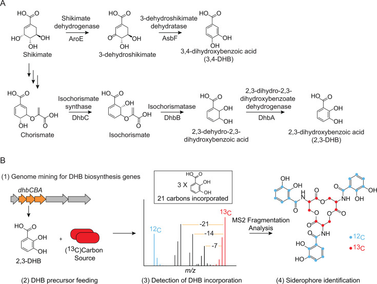
Fig 1 Inverse stable isotope labeling (InverSIL) for linking predicted catecholate siderophore BGCs with their products. (A) Enzymes responsible for the biosynthesis of catechols 3,4-DHB and 2,3-DHB. (B) Scheme of an InverSIL experiment: (1) genome mining for DHB biosynthesis genes; (2) addition of 2,3-DHB at its natural isotopic abundance to a bacterial culture grown on a 13C-substituted carbon source [indicated as (13C)]; (3) high-resolution mass spectrometry to detect DHB incorporation; and (4) identification of the siderophore aided by MS2 fragmentation. The siderophore enterobactin is shown as an example.
Fig 1 Inverse stable isotope labeling (InverSIL) for linking predicted catecholate siderophore BGCs with their products. (A) Enzymes responsible for the biosynthesis of catechols 3,4-DHB and 2,3-DHB. (B) Scheme of an InverSIL experiment: (1) genome mining for DHB biosynthesis genes; (2) addition of 2,3-DHB at its natural isotopic abundance to a bacterial culture grown on a 13C-substituted carbon source [indicated as (13C)]; (3) high-resolution mass spectrometry to detect DHB incorporation; and (4) identification of the siderophore aided by MS2 fragmentation. The siderophore enterobactin is shown as an example.CC BY · PubMed Central
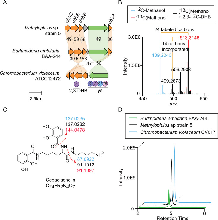
Fig 2 Using InverSIL to link an NRPS-dependent catecholate siderophore BGC with its product, cepaciachelin. (A) Comparison of an NRPS-dependent BGC in Methylophilus sp. strain 5, Burkholderia ambifaria BAA-244, and Chromobacterium violaceum CV017. Numbers indicate percent amino acid identity. Arrow colors indicate predicted 2,3-DHB biosynthesis genes (orange), predicted NRPS genes (green), and other genes (gray). Circles indicate predicted NRPS domains C (condensation domain), A (adenylation domain), CP (acyl-carrier protein), and TE (thioesterase domain), with the predicted adenylation domain substrates listed below. (B) Overlaid mass spectra of Methylophilus sp. strain 5 supernatant extract showing incorporation of two 2,3-DHB units into a metabolite with the same high-resolution mass and carbon count as cepaciachelin. (C) Structure of cepaciachelin showing MS2 fragments from different InverSIL conditions. The colors match the growth conditions indicated in panel B. (D) Extracted ion chromatograms of supernatant extracts of B. ambifaria BAA-244, Methylophilus sp. strain 5, and Chromobacterium violaceum CV017 for m/z 489.2340, corresponding to the [M+H]+ of cepaciachelin. Mass tolerance <5 ppm.
Fig 2 Using InverSIL to link an NRPS-dependent catecholate siderophore BGC with its product, cepaciachelin. (A) Comparison of an NRPS-dependent BGC in Methylophilus sp. strain 5, Burkholderia ambifaria BAA-244, and Chromobacterium violaceum CV017. Numbers indicate percent amino acid identity. Arrow colors indicate predicted 2,3-DHB biosynthesis genes (orange), predicted NRPS genes (green), and other genes (gray). Circles indicate predicted NRPS domains C (condensation domain), A (adenylation domain), CP (acyl-carrier protein), and TE (thioesterase domain), with the predicted adenylation domain substrates listed below. (B) Overlaid mass spectra of Methylophilus sp. strain 5 supernatant extract showing incorporation of two 2,3-DHB units into a metabolite with the same high-resolution mass and carbon count as cepaciachelin. (C) Structure of cepaciachelin showing MS2 fragments from different InverSIL conditions. The colors match the growth conditions indicated in panel B. (D) Extracted ion chromatograms of supernatant extracts of B. ambifaria BAA-244, Methylophilus sp. strain 5, and Chromobacterium violaceum CV017 for m/z 489.2340, corresponding to the [M+H]+ of cepaciachelin. Mass tolerance <5 ppm.CC BY · PubMed Central
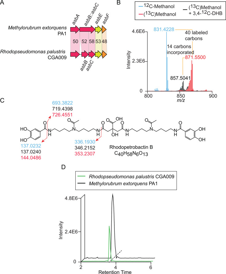
Fig 3 Using InverSIL to link a NIS catecholate siderophore BGC with its product, rhodopetrobactin B. (A) Comparison of a NIS siderophore BGC in Methylorubrum extorquens PA1 and Rhodopseudomonas palustris CGA009. Numbers indicate percent amino acid identity. Arrow colors indicate predicted iucA/iucC genes (red), a conserved hypothetical protein in petrobactin biosynthesis (yellow), and a predicted asbF gene (orange). (B) Overlaid mass spectra of M. extorquens PA1 supernatant extract showing incorporation of two 3,4-DHB units into a metabolite with the same high-resolution mass and carbon count as rhodopetrobactin B. (C) Structure of rhodopetrobactin B showing MS2 fragments from different InverSIL conditions. The colors match the growth conditions indicated in panel B. (D) Extracted ion chromatograms of supernatant extracts of R. palustris CGA009 and M. extorquens PA1 for m/z 831.4228, corresponding to the [M+H]+ of rhodopetrobactin B. Mass tolerance < 5 ppm.
Fig 3 Using InverSIL to link a NIS catecholate siderophore BGC with its product, rhodopetrobactin B. (A) Comparison of a NIS siderophore BGC in Methylorubrum extorquens PA1 and Rhodopseudomonas palustris CGA009. Numbers indicate percent amino acid identity. Arrow colors indicate predicted iucA/iucC genes (red), a conserved hypothetical protein in petrobactin biosynthesis (yellow), and a predicted asbF gene (orange). (B) Overlaid mass spectra of M. extorquens PA1 supernatant extract showing incorporation of two 3,4-DHB units into a metabolite with the same high-resolution mass and carbon count as rhodopetrobactin B. (C) Structure of rhodopetrobactin B showing MS2 fragments from different InverSIL conditions. The colors match the growth conditions indicated in panel B. (D) Extracted ion chromatograms of supernatant extracts of R. palustris CGA009 and M. extorquens PA1 for m/z 831.4228, corresponding to the [M+H]+ of rhodopetrobactin B. Mass tolerance < 5 ppm.CC BY · PubMed Central
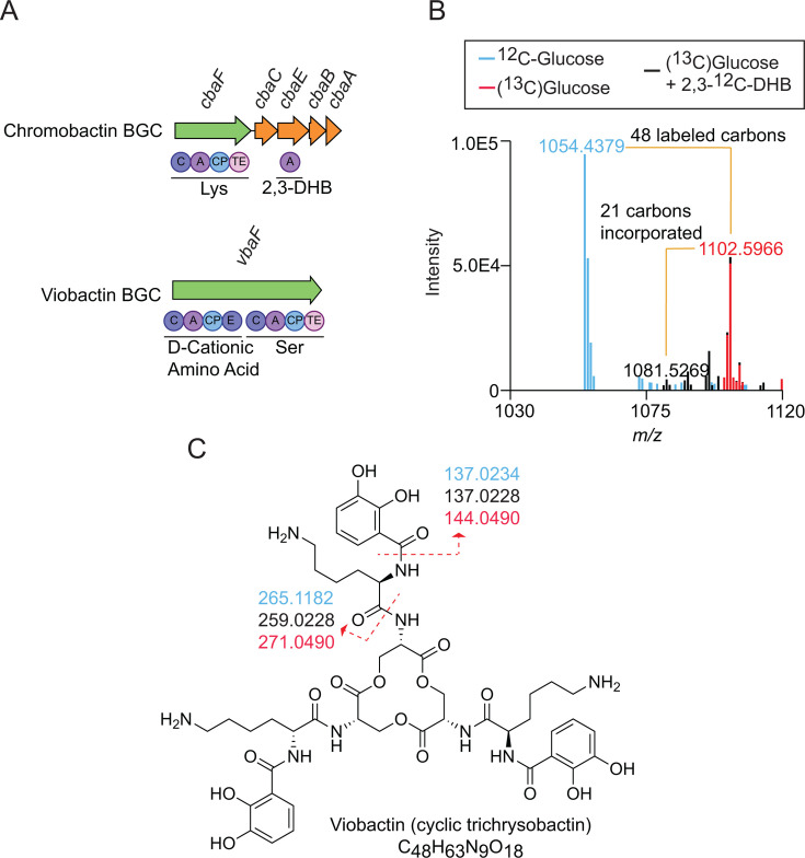
Fig 4 Using InverSIL to determine the structures of the siderophores used by the opportunistic pathogen Chromobacterium violaceum. (A) BGCs for the C. violaceum siderophores. Arrow colors indicate predicted NRPS genes (green) and 2,3-DHB biosynthesis genes (orange). Circles indicate predicted NRPS domains C (condensation domain), A (adenylation domain), CP (acyl-carrier protein), TE (thioesterase domain), and E (epimerization domain), with the predicted adenylation domain substrates listed below. (B) Overlaid mass spectra of C. violaceum CV017 supernatant extract showing incorporation of three 2,3-DHB units into a metabolite with the same high-resolution mass and carbon count as cyclic trichrysobactin. (C) Structure of viobactin (cyclic trichrysobactin) showing MS2 fragments from different InverSIL conditions. The colors match the growth conditions indicated in panel B.
Fig 4 Using InverSIL to determine the structures of the siderophores used by the opportunistic pathogen Chromobacterium violaceum. (A) BGCs for the C. violaceum siderophores. Arrow colors indicate predicted NRPS genes (green) and 2,3-DHB biosynthesis genes (orange). Circles indicate predicted NRPS domains C (condensation domain), A (adenylation domain), CP (acyl-carrier protein), TE (thioesterase domain), and E (epimerization domain), with the predicted adenylation domain substrates listed below. (B) Overlaid mass spectra of C. violaceum CV017 supernatant extract showing incorporation of three 2,3-DHB units into a metabolite with the same high-resolution mass and carbon count as cyclic trichrysobactin. (C) Structure of viobactin (cyclic trichrysobactin) showing MS2 fragments from different InverSIL conditions. The colors match the growth conditions indicated in panel B.CC BY · PubMed Central
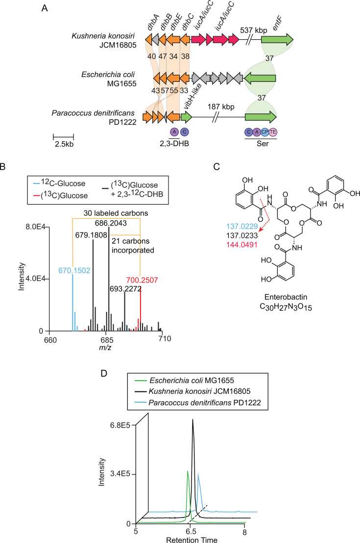
Fig 5 Non-colocalized enterobactin BGCs in Kushneria konosiri JCM16805 and Paracoccus denitrificans PD1222. (A) Comparison of a predicted dhb operon, NIS BGC, and distant entF gene in K. konosiri JCM16805 with the enterobactin BGC in E. coli MG1655 and another non-colocalized enterobactin BGC in Paracoccus denitrificans PD1222. Numbers indicate percent amino acid identity. Arrow colors indicate predicted 2,3-DHB biosynthesis genes (orange), predicted iucA/iucC genes (red), predicted NRPS genes (green), and other genes (gray). Circles indicate predicted NRPS domains C (condensation domain), A (adenylation domain), CP (acyl-carrier protein), and TE (thioesterase domain), with the predicted adenylation domain substrates listed below. (B) Overlaid mass spectra of K. konosiri JCM16805 supernatant extract showing incorporation of three 2,3-DHB units into a metabolite with the same high-resolution mass and carbon count as enterobactin. (C) Structure of enterobactin showing MS2 fragments from different InverSIL conditions. The colors match the growth conditions indicated in panel B. (D) Extracted ion chromatograms of supernatant extracts from K. konosiri JCM 16805, P. denitrificans PD1222, and E. coli MG1655 for m/z 670.1502, corresponding to the [M+H]+ of enterobactin. Mass tolerance < 5 ppm.
Fig 5 Non-colocalized enterobactin BGCs in Kushneria konosiri JCM16805 and Paracoccus denitrificans PD1222. (A) Comparison of a predicted dhb operon, NIS BGC, and distant entF gene in K. konosiri JCM16805 with the enterobactin BGC in E. coli MG1655 and another non-colocalized enterobactin BGC in Paracoccus denitrificans PD1222. Numbers indicate percent amino acid identity. Arrow colors indicate predicted 2,3-DHB biosynthesis genes (orange), predicted iucA/iucC genes (red), predicted NRPS genes (green), and other genes (gray). Circles indicate predicted NRPS domains C (condensation domain), A (adenylation domain), CP (acyl-carrier protein), and TE (thioesterase domain), with the predicted adenylation domain substrates listed below. (B) Overlaid mass spectra of K. konosiri JCM16805 supernatant extract showing incorporation of three 2,3-DHB units into a metabolite with the same high-resolution mass and carbon count as enterobactin. (C) Structure of enterobactin showing MS2 fragments from different InverSIL conditions. The colors match the growth conditions indicated in panel B. (D) Extracted ion chromatograms of supernatant extracts from K. konosiri JCM 16805, P. denitrificans PD1222, and E. coli MG1655 for m/z 670.1502, corresponding to the [M+H]+ of enterobactin. Mass tolerance < 5 ppm.CC BY · PubMed Central
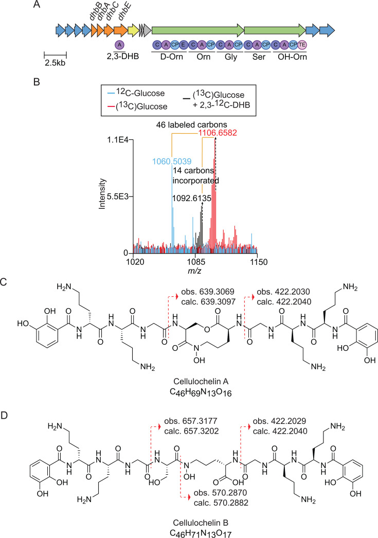
Fig 6 Characterization of a novel siderophore from Cellulomonas sp. strain Leaf334 using InverSIL. (A) The cellulochelin BGC. Arrow colors indicate predicted 2,3-DHB biosynthesis genes (orange), predicted NRPS genes (green), predicted transport-related genes (blue), and a predicted ferric reductase gene (yellow). Circles indicate predicted NRPS domains C (condensation domain), A (adenylation domain), CP (acyl-carrier protein), and TE (thioesterase domain), and E (epimerization domain), with the predicted adenylation domain substrates listed below. (B) Overlaid mass spectra of Cellulomonas sp. strain Leaf334 supernatant extract showing incorporation of two 2,3-DHB units into a new metabolite. (C and D) Structure of (C) cellulochelin A and (D) cellulochelin B showing diagnostic MS2 fragments that differentiate the two siderophores.
Fig 6 Characterization of a novel siderophore from Cellulomonas sp. strain Leaf334 using InverSIL. (A) The cellulochelin BGC. Arrow colors indicate predicted 2,3-DHB biosynthesis genes (orange), predicted NRPS genes (green), predicted transport-related genes (blue), and a predicted ferric reductase gene (yellow). Circles indicate predicted NRPS domains C (condensation domain), A (adenylation domain), CP (acyl-carrier protein), and TE (thioesterase domain), and E (epimerization domain), with the predicted adenylation domain substrates listed below. (B) Overlaid mass spectra of Cellulomonas sp. strain Leaf334 supernatant extract showing incorporation of two 2,3-DHB units into a new metabolite. (C and D) Structure of (C) cellulochelin A and (D) cellulochelin B showing diagnostic MS2 fragments that differentiate the two siderophores.CC BY · PubMed Central

Verification

Claims the paper makes

The claims and statistics of this paper haven’t been checked yet.

Run the check — free, no account needed. It reads the open-access text and checks the sources it cites.

A few free checks a day · papers already verified open instantly · results are public

Self-check

Percentages, sums and reported differences re-computed from the paper’s own numbers.

The self-check needs the paper’s full text.

Citations

Cites

Research works printed but not linked: 65

  1. 10.3389/fmicb.2012.00383 · DOI
  2. 10.1039/b906679a · DOI · Paper page
  3. 10.1038/s41579-024-01090-6 · DOI
  4. 10.1007/s00775-018-1617-x · DOI
  5. 10.1080/10409238.2025.2476476 · DOI
  6. 10.1016/bs.mie.2024.06.007 · DOI
  7. 10.1016/j.copbio.2022.102757 · DOI
  8. 10.1093/nar/gkaf334 · DOI
  9. 10.1038/s41467-020-19986-1 · DOI
  10. 10.7554/eLife.109154 · DOI
  11. 10.1021/acschembio.5b00801 · DOI
  12. 10.1016/j.chembiol.2010.01.011 · DOI
  13. 10.1021/ja1109453 · DOI
  14. 10.1038/s41557-021-00803-1 · DOI · Paper page
  15. 10.1073/pnas.1516878112 · DOI
  16. 10.1016/j.chembiol.2006.11.007 · DOI
  17. 10.1038/s41589-021-00949-6 · DOI · Paper page
  18. 10.1128/mbio.01999-23 · DOI
  19. 10.1016/bs.mie.2024.06.005 · DOI
  20. 10.1002/anie.200501942 · DOI
  21. 10.1021/np060409n · DOI
  22. 10.1039/c7mt00111h · DOI
  23. 10.1007/s00775-002-0375-x · DOI
  24. 10.1099/00221287-144-7-1747 · DOI
  25. 10.1039/D3NP00037K · DOI
  26. 10.1074/jbc.M009140200 · DOI
  27. 10.1073/pnas.0808118105 · DOI
  28. 10.1021/bi801876q · DOI
  29. 10.1021/acs.analchem.4c03528 · DOI
  30. 10.1073/pnas.2507323122 · DOI
  31. 10.1007/s00253-015-6713-z · DOI
  32. 10.1128/mBio.02430-17 · DOI
  33. 10.1002/mbo3.347 · DOI
  34. 10.1128/AEM.03160-15 · DOI
  35. 10.1371/journal.pone.0102458 · DOI
  36. 10.1134/S0026261711040047 · DOI
  37. 10.1128/JCM.03561-13 · DOI
  38. 10.1073/pnas.2322096121 · DOI
  39. 10.1111/1462-2920.14078 · DOI
  40. 10.1007/s00248-010-9725-3 · DOI
  41. 10.1128/JB.01009-12 · DOI
  42. 10.1128/IAI.00577-19 · DOI
  43. 10.1002/1873-3468.14539 · DOI
  44. 10.1021/np200126z · DOI
  45. 10.1128/JB.180.17.4435-4441.1998 · DOI · Paper page
  46. 10.1128/genomeA.00080-16 · DOI
  47. 10.1099/ijsem.0.002897 · DOI
  48. 10.3390/microorganisms11122910 · DOI
  49. 10.1186/s40793-018-0324-0 · DOI
  50. 10.1073/pnas.0630018100 · DOI · Paper page
  51. 10.1007/BF00447011 · DOI
  52. 10.1016/S0040-4039(01)86717-4 · DOI
  53. 10.1042/bj1460191 · DOI
  54. 10.1073/pnas.1401734111 · DOI
  55. 10.1099/00221287-148-2-353 · DOI
  56. 10.1038/s41564-021-00997-7 · DOI
  57. 10.1073/pnas.0805451105 · DOI
  58. 10.1039/c8mt00252e · DOI
  59. 10.1021/acschembio.7b00597 · DOI
  60. 10.1099/00221287-128-9-1959 · DOI
  61. 10.1016/j.tim.2025.05.002 · DOI
  62. 10.1128/JB.06817-11 · DOI
  63. 10.1093/nar/gkr1044 · DOI
  64. 10.1186/s12859-023-05311-2 · DOI
  65. 10.1016/0003-2697(87)90612-9 · DOI

Metadata

Authors
Robes, J. M. D., Liebergesell, T. C. E., Medvedeva, V. P., & Puri, A. W.
Affiliations
  • University of Utah (US)
  • University of California, Irvine (US)
Corresponding author
Puri, Aaron W.
Venue
mBio
Published
2026-06-10
Volume
17
Issue
6
Pages
e0339125-e0339125
ISSN
2150-7511 (ISSN-L)
OpenAlex
W4415946583
Cited by
0
References
4
FWCI
0.00
Type
journal article
Language
en
Open access
Locations
  • mBio (publishedVersion, cc-by)
  • bioRxiv (Cold Spring Harbor Laboratory) (acceptedVersion, cc-by-nc)
  • PubMed (publishedVersion)
  • PubMed Central (publishedVersion, cc-by)
  • Europe PMC (PubMed Central) (submittedVersion, cc-by-nc)
  • DOAJ (DOAJ: Directory of Open Access Journals) (submittedVersion, cc-by-sa)
Funders
  • National Institute of General Medical Sciences (R35 GM147018)
  • Simons Foundation Early Career Investigator in Aquatic Microbial Ecology and Evolution (00006628)
  • NIH training (T32 AI055434)
Topics
  • Life Sciences › Immunology and Microbiology › Applied Microbiology and Biotechnology › Microbial Natural Products and Biosynthesis
  • Life Sciences › Biochemistry, Genetics and Molecular Biology › Genetics › Bacterial Genetics and Biotechnology
  • Life Sciences › Biochemistry, Genetics and Molecular Biology › Biotechnology › Microbial Metabolism and Applications
Keywords
siderophores, iron acquisition, enterobactin, Methylobacterium, Methylophilus, Chromobacterium violaceum, Paracoccus, Cellulomonas, bacterial secondary metabolites

About these checks

Registry, retraction, Cites and statistics results are computed, not judged, with one exception: a printed reference that no index lists is read once by a model before it counts as a citation error. Claims are extracted by a model and kept only when their quote appears verbatim in the text; each cited source is then checked, with a model’s help, for whether it says what the claim relies on. Those verdicts can be wrong — read the claim’s quote and the cited source before relying on one.

Statistics and arithmetic are recomputed, not judged.

Confidence bands are shown only for fields calibrated on our annotated test set. None is calibrated yet, so no band is shown.

What counts as a claim ›How we measure confidence ›How we check this ›

Are you an author, or do you see a problem with this page? Request a review