

The DOI matches its Crossref record; 204 of the 204 references it cites were found in a registry or an index.
Artificial intelligence and deep learning in ophthalmology
Open accessAbstract
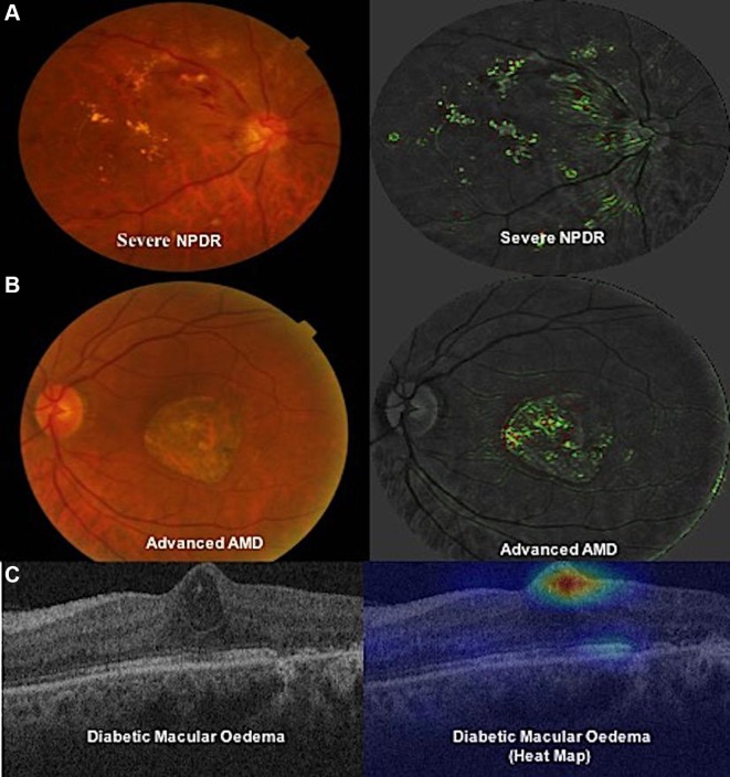
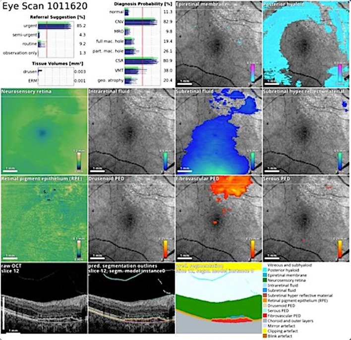
Artificial intelligence (AI) based on deep learning (DL) has sparked tremendous global interest in recent years. DL has been widely adopted in image recognition, speech recognition and natural language processing, but is only beginning to impact on healthcare. In ophthalmology, DL has been applied to fundus photographs, optical coherence tomography and visual fields, achieving robust classification performance in the detection of diabetic retinopathy and retinopathy of prematurity, the glaucoma-like disc, macular oedema and age-related macular degeneration. DL in ocular imaging may be used in conjunction with telemedicine as a possible solution to screen, diagnose and monitor major eye diseases for patients in primary care and community settings. Nonetheless, there are also potential challenges with DL application in ophthalmology, including clinical and technical challenges, explainability of the algorithm results, medicolegal issues, and physician and patient acceptance of the AI ‘black-box’ algorithms. DL could potentially revolutionise how ophthalmology is practised in the future. This review provides a summary of the state-of-the-art DL systems described for ophthalmic applications, potential challenges in clinical deployment and the path forward.
Overview
The claims and statistics of this paper haven’t been checked yet.
Run the check — free, no account needed. It reads the open-access text and checks the sources it cites.
Integrity signals
Registry
Cites
Statistics
Figures

Verification
Claims the paper makes
The claims and statistics of this paper haven’t been checked yet.
Run the check — free, no account needed. It reads the open-access text and checks the sources it cites.
Claims other papers make about this one
Each is checked against this paper’s own text: does it say what the citing paper says it does?
In the citing paper
“Indeed, AI has proven its capability to detect diabetic retinopathy, glaucoma, and age-related macular degeneration when applied to fundus photographs, optical coherence tomography, and visual fields.4”
From Artificial intelligence and endo-histo-omics: new dimensions of precision endoscopy and histology in inflammatory bowel disease (2024) · cites this paper as [4]
In the citing paper
“Ophthalmology, in recent years, has observed a significant uptick in AI efforts, with dozens of papers demonstrating clinical diagnostic and analytical capabilities that extend beyond current human capability [99][100][101] .”
From Deep learning-enabled medical computer vision (2021) · cites this paper as [99]
Self-check
Percentages, sums and reported differences re-computed from the paper’s own numbers.
The self-check needs the paper’s full text.
Citations
Cites
U-Net: Convolutional Networks for Biomedical Image Segmentation
Ronneberger, O., Fischer, P., & Brox, T. (2015) · Lecture notes in computer science · doi:10.1007/978-3-319-24574-4_28 · Cited by 99,404
LeCun, Y., Bengio, Y., & Hinton, G. (2015) · Nature · doi:10.1038/nature14539 · Cited by 84,126
Chobanian, A. V. (2003) · JAMA · doi:10.1001/jama.289.19.2560 · Cited by 25,386
Dermatologist-level classification of skin cancer with deep neural networks
Esteva, A., Kuprel, B., Novoa, R. A., Ko, J., Swetter, S. M., Blau, H. M., & Thrun, S. (2017) · Nature · doi:10.1038/nature21056 · Cited by 14,362
(2013) · European Heart Journal · doi:10.1093/eurheartj/eht151 · Cited by 13,655
Hinton, G., Deng, L., Yu, D., Dahl, G., Mohamed, A.-R., Jaitly, N., Senior, A., Vanhoucke, V., Nguyen, P., Sainath, T., et al. (2012) · IEEE Signal Processing Magazine · doi:10.1109/msp.2012.2205597 · Cited by 10,260
MIMIC-III, a freely accessible critical care database
Johnson, A. E. W., Pollard, T. J., Shen, L., Lehman, L.-W. H., Feng, M., Ghassemi, M., Moody, B., Szolovits, P., Anthony Celi, L., & Mark, R. G. (2016) · Scientific Data · doi:10.1038/sdata.2016.35 · Cited by 8,141
Gulshan, V., Peng, L., Coram, M., Stumpe, M. C., Wu, D., Narayanaswamy, A., Venugopalan, S., Widner, K., Madams, T., Cuadros, J., et al. (2016) · JAMA · doi:10.1001/jama.2016.17216 · Cited by 7,697
Self-citationGlobal Prevalence of Glaucoma and Projections of Glaucoma Burden through 2040
Tham, Y.-C., Li, X., Wong, T. Y., Quigley, H. A., Aung, T., & Cheng, C.-Y. (2014) · Ophthalmology · doi:10.1016/j.ophtha.2014.05.013 · Cited by 7,159
Self-citationShin, H.-C., Roth, H. R., Gao, M., Lu, L., Xu, Z., Nogues, I., Yao, J., Mollura, D., & Summers, R. M. (2016) · IEEE Transactions on Medical Imaging · doi:10.1109/tmi.2016.2528162 · Cited by 5,759
Cited by
Artificial intelligence in healthcare: transforming the practice of medicine
Bajwa, J., Munir, U., Nori, A., & Williams, B. (2021) · Future Healthcare Journal · doi:10.7861/fhj.2021-0095 · Cited by 1,856
Deep learning-enabled medical computer vision
Esteva, A., Chou, K., Yeung, S., Naik, N., Madani, A., Mottaghi, A., Liu, Y., Topol, E., Dean, J., & Socher, R. (2021) · npj Digital Medicine · doi:10.1038/s41746-020-00376-2 · Cited by 1,406
Diabetic retinopathy: Looking forward to 2030
Tan, T.-E., & Wong, T. Y. (2023) · Frontiers in Endocrinology · doi:10.3389/fendo.2022.1077669 · Cited by 511
Self-citationDeep learning in ophthalmology: The technical and clinical considerations
Ting, D. S. W., Peng, L., Varadarajan, A. V., Keane, P. A., Burlina, P. M., Chiang, M. F., Schmetterer, L., Pasquale, L. R., Bressler, N. M., Webster, D. R., et al. (2019) · Progress in Retinal and Eye Research · doi:10.1016/j.preteyeres.2019.04.003 · Cited by 534
Self-citationEvaluating the Performance of ChatGPT in Ophthalmology
Antaki, F., Touma, S., Milad, D., El-Khoury, J., & Duval, R. (2023) · Ophthalmology Science · doi:10.1016/j.xops.2023.100324 · Cited by 563
Metadata
- Authors
- Ting, D. S. W., Pasquale, L. R., Peng, L., Campbell, J. P., Lee, A. Y., Raman, R., Tan, G. S. W., Schmetterer, L., Keane, P. A., & Wong, T. Y.
- Affiliations
- SingHealth (SG)
- Mount Sinai Hospital (US)
- Google (United States) (US)
- Oregon Health & Science University (US)
- University of Washington (US)
- Sankara Nethralaya (IN)
- National University of Singapore (SG)
- Moorfields Eye Hospital (GB)
- Corresponding author
- Ting, Daniel Shu Wei
- ORCID
- Venue
- British Journal of Ophthalmology
- Publisher
- BMJ
- Published
- 2018-10-25
- Volume
- 103
- Issue
- 2
- Pages
- 167-175
- ISSN
- 0007-1161
- 1468-2079
- PMC
- PMC6362807
- OpenAlex
- W2898192966
- Cited by
- 1,554
- References
- 204
- Citations by year
- 2026: 245 · 2025: 280 · 2024: 229 · 2023: 247 · 2022: 181 · 2021: 184 · 2020: 132 · 2019: 52 · 2018: 1
- FWCI
- 380.44
- Type
- book chapter
- Language
- en
- Open access
- hybrid
- doi.org
- Licence
- cc-by-nc
- Locations
- Springer eBooks (publishedVersion)
- British Journal of Ophthalmology (publishedVersion, cc-by-nc)
- Europe PMC (PubMed Central) (publishedVersion, other-oa)
- PubMed Central (publishedVersion, cc-by-nc)
- PubMed (publishedVersion)
- DR-NTU (Nanyang Technological University) (submittedVersion, cc-by-nc)
- UCL Discovery (University College London) (submittedVersion)
- Minerva Access (University of Melbourne) (submittedVersion, cc-by-nc)
- HighWire Press Open Archive (submittedVersion)
- Funders
- NIH Clinical Center (K12 EY027720, R01EY019474, P30EY10572, P41EB015896)
- National Medical Research Council (NHIC-I2D-1409022, grants 0796/2003, IRG07nov013, I)
- National Science Foundation (SCH-1622542, SCH-1622536, SCH-1622679)
- National Institute for Health Research (NIHR-CS--2014-12-023)
- Topics
- Health Sciences › Medicine › Radiology, Nuclear Medicine and Imaging › Retinal Imaging and Analysis
- Health Sciences › Medicine › Health Informatics › Artificial Intelligence in Healthcare and Education
- Health Sciences › Medicine › Ophthalmology › Ophthalmology and Visual Health Research
- Keywords
- artificial intelligence, deep learning, ophthalmology, diabetic retinopathy detection, retinopathy of prematurity, glaucoma, macular edema, age-related macular degeneration, fundus images, optical coherence tomography, visual field, ocular imaging, telemedicine, medicolegal issues
- MeSH terms
- Deep Learning, Animals, Artificial Intelligence, Eye Diseases, Humans, Ophthalmology
About these checks
Registry, retraction, Cites and statistics results are computed, not judged, with one exception: a printed reference that no index lists is read once by a model before it counts as a citation error. Claims are extracted by a model and kept only when their quote appears verbatim in the text; each cited source is then checked, with a model’s help, for whether it says what the claim relies on. Those verdicts can be wrong — read the claim’s quote and the cited source before relying on one.
Statistics and arithmetic are recomputed, not judged.
Confidence bands are shown only for fields calibrated on our annotated test set. None is calibrated yet, so no band is shown.
What counts as a claim ›How we measure confidence ›How we check this ›
Are you an author, or do you see a problem with this page? Request a review
How to cite
Cite CoChat for the checks on this page, and the paper for its own findings.
- This page
- CoChat. (2026). Citation check: Artificial intelligence and deep learning in ophthalmology. https://app.cochat.ai/papers/10.1136/bjophthalmol-2018-313173
- The paper
- Ting, D. S. W., Pasquale, L. R., Peng, L., Campbell, J. P., Lee, A. Y., Raman, R., Tan, G. S. W., Schmetterer, L., Keane, P. A., & Wong, T. Y. (2019). Artificial intelligence and deep learning in ophthalmology. British Journal of Ophthalmology. https://doi.org/10.1136/bjophthalmol-2018-313173
Data sources
Bibliographic data: Semantic Scholar, OpenAlex, Crossref.
Contains information from the Semantic Scholar Academic Graph Datasets, made available under the ODC Attribution License (ODC-BY 1.0).
Semantic Scholar’s data is described in: Kinney, R., et al. (2023). The Semantic Scholar Open Data Platform. arXiv:2301.10140.
