

The DOI matches its Crossref record; 38 of the 39 references it cites were found in a registry or an index.
HNF4α is a therapeutic target that links AMPK to WNT signalling in early-stage gastric cancer
Open accessAbstract
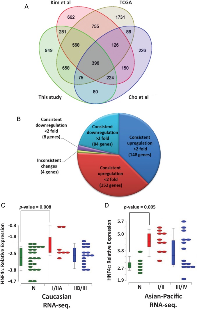
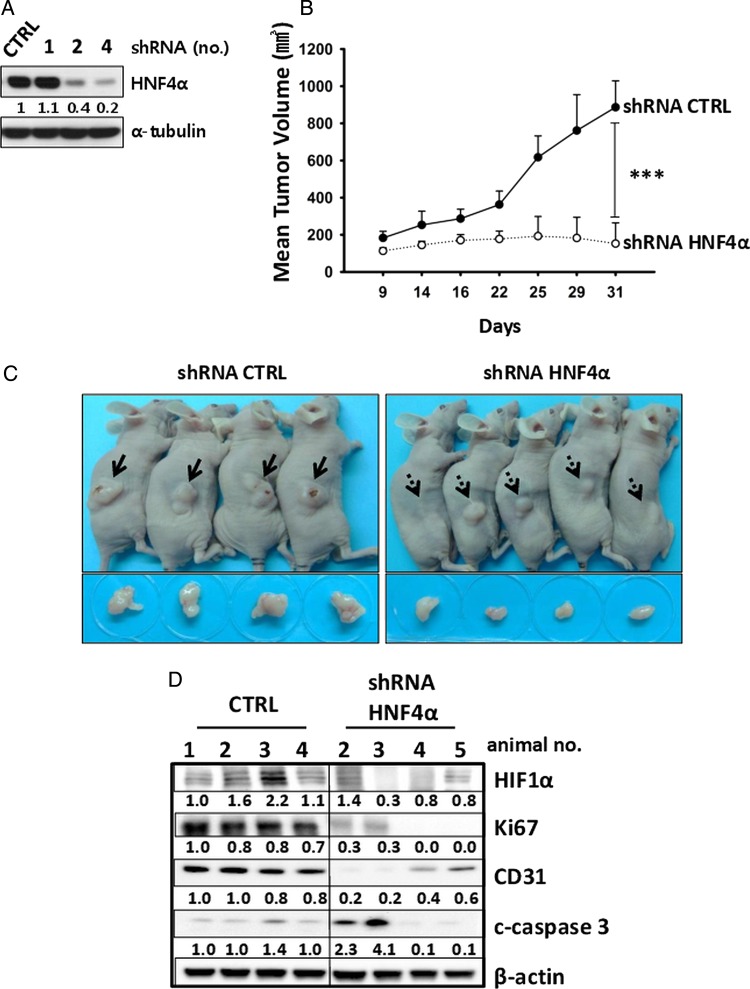
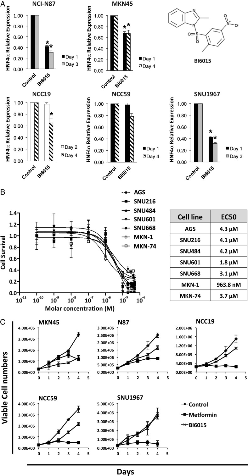
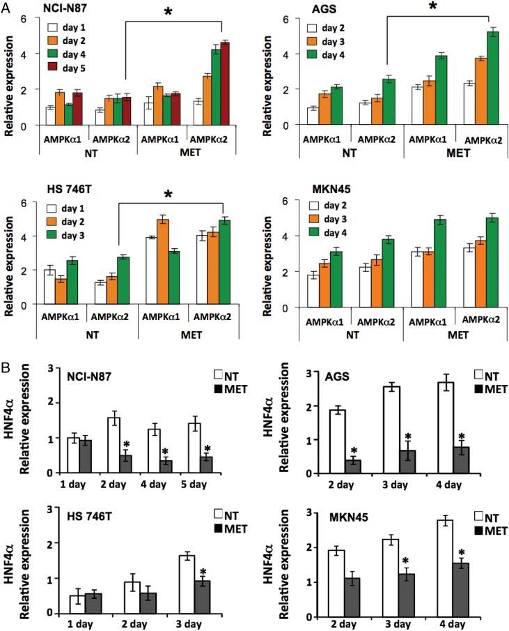
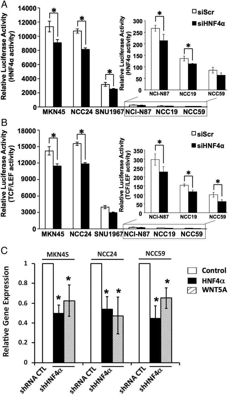
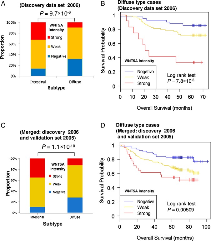
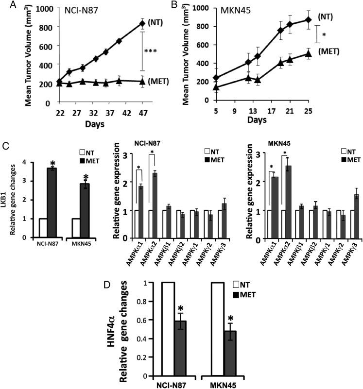
BACKGROUND: Worldwide, gastric cancer (GC) is the fourth most common malignancy and the most common cancer in East Asia. Development of targeted therapies for this disease has focused on a few known oncogenes but has had limited effects. OBJECTIVE: To determine oncogenic mechanisms and novel therapeutic targets specific for GC by identifying commonly dysregulated genes from the tumours of both Asian-Pacific and Caucasian patients. METHODS: We generated transcriptomic profiles of 22 Caucasian GC tumours and their matched non-cancerous samples and performed an integrative analysis across different GC gene expression datasets. We examined the inhibition of commonly overexpressed oncogenes and their constituent signalling pathways by RNAi and/or pharmacological inhibition. RESULTS: Hepatocyte nuclear factor-4α (HNF4α) upregulation was a key signalling event in gastric tumours from both Caucasian and Asian patients, and HNF4α antagonism was antineoplastic. Perturbation experiments in GC tumour cell lines and xenograft models further demonstrated that HNF4α is downregulated by AMPKα signalling and the AMPK agonist metformin; blockade of HNF4α activity resulted in cyclin downregulation, cell cycle arrest and tumour growth inhibition. HNF4α also regulated WNT signalling through its target gene WNT5A, a potential prognostic marker of diffuse type gastric tumours. CONCLUSIONS: Our results indicate that HNF4α is a targetable oncoprotein in GC, is regulated by AMPK signalling through AMPKα and resides upstream of WNT signalling. HNF4α may regulate 'metabolic switch' characteristic of a general malignant phenotype and its target WNT5A has potential prognostic values. The AMPKα-HNF4α-WNT5A signalling cascade represents a potentially targetable pathway for drug development.
Overview
The claims and statistics of this paper haven’t been checked yet.
Run the check — free, no account needed. It reads the open-access text and checks the sources it cites.
Integrity signals
Registry
Cites
Statistics
Figures
Verification
Claims the paper makes
The claims and statistics of this paper haven’t been checked yet.
Run the check — free, no account needed. It reads the open-access text and checks the sources it cites.
Claims other papers make about this one
Each is checked against this paper’s own text: does it say what the citing paper says it does?
The cited source says what the claim attributes to it.
In the citing paper
“Chang et al., for example, demonstrated that metformin reduced hepatocyte nuclear factor 4α (HNF4α) levels via activation of AMPKα, which subsequently modulated the WNT signaling pathway.199”
From Drug repurposing for cancer therapy (2024) · cites this paper as [199]
Self-check
Percentages, sums and reported differences re-computed from the paper’s own numbers.
The self-check needs the paper’s full text.
Citations
Cites
Jemal, A., Bray, F., Ferlay, J., Ward, E., & Forman, D. (2011) · CA A Cancer Journal for Clinicians · doi:10.3322/caac.20107 · Cited by 55,077
Analyzing real-time PCR data by the comparative CT method
Schmittgen, T. D., & Livak, K. J. (2008) · Nature Protocols · doi:10.1038/nprot.2008.73 · Cited by 26,190
Global Cancer Statistics, 2002
Parkin, D. M., Bray, F., Ferlay, J., & Pisani, P. (2005) · CA A Cancer Journal for Clinicians · doi:10.3322/canjclin.55.2.74 · Cited by 18,710
Bang, Y.-J., Van Cutsem, E., Feyereislova, A., Chung, H. C., Shen, L., Sawaki, A., Lordick, F., Ohtsu, A., Omuro, Y., Satoh, T., et al. (2010) · The Lancet · doi:10.1016/s0140-6736(10)61121-x · Cited by 7,302
Targeting HIF-1 for cancer therapy
Semenza, G. L. (2003) · Nature Reviews Cancer · doi:10.1038/nrc1187 · Cited by 6,717
Suerbaum, S., & Michetti, P. (2002) · New England Journal of Medicine · doi:10.1056/nejmra020542 · Cited by 2,984
The LKB1-AMPK pathway: metabolism and growth control in tumour suppression
Shackelford, D. B., & Shaw, R. J. (2009) · Nature Reviews Cancer · doi:10.1038/nrc2676 · Cited by 1,943
LKB1 is the upstream kinase in the AMP-activated protein kinase cascade
Woods, A., Johnstone, S. R., Dickerson, K., Leiper, F. C., Fryer, L. G. D., Neumann, D., Schlattner, U., Wallimann, T., Carlson, M., & Carling, D. (2003) · Current Biology · doi:10.1016/j.cub.2003.10.031 · Cited by 1,650
Cancer Statistics in Korea: Incidence, Mortality, Survival and Prevalence in 2010
Jung, K., Won, Y., Kong, H., Oh, C.-M., Seo, H., & Lee, J.-S. (2013) · Cancer research and treatment : official journal of Korean Cancer Association · doi:10.4143/crt.2013.45.1.1 · Cited by 1,086
Self-citationGene Expression Signature–Based Prognostic Risk Score in Gastric Cancer
Choi, Y., Lim, J. Y., Cheong, J., Park, Y., Yoon, S.-L., Kim, S. M., Kim, S.-B., Kim, H., Hong, S. W., Park, Y. N., et al. (2011) · Clinical Cancer Research · doi:10.1158/1078-0432.ccr-10-2180 · Cited by 351
Research works printed but not linked: 2
- McDonald, 2008, Pfizer facts, the burden of cancer in Asia
- Ferlay, 2010, GLOBOCAN 2008, cancer incidence and mortality worldwide, CancerBase, 2010
Cited by
Sorted by citations
Items 101–119 of 119
A review of potential mechanisms and treatments of gastric intestinal metaplasia
Wu, Y., Zhang, K., Zheng, Y., & Jin, H. (2024) · European Journal of Gastroenterology & Hepatology · doi:10.1097/meg.0000000000002903 · Cited by 2
Qu, H., Shi, R., Yang, B., Chen, X., Song, Y., He, L., Sun, Y., YANG, Y., Li, Z., & Qu, X. (2021) · Biocell · doi:10.32604/biocell.2021.015867 · Cited by 2
AMPK activation prevents hepatocellular carcinoma development through inhibition of HNF4α activityPreprint
Sun, Z., Linares, B., Urdiales, C., Al-sarmi, F., Putluri, N., & Nostrand, J. L. V. (2025) · bioRxiv (Cold Spring Harbor Laboratory) · doi:10.1101/2025.05.16.652569 · Cited by 2
HNF4A promotes tumor progression in gastric cancer by transcriptional activation of KIF2C
Lu, H., Gan, J., Zhou, L., Yuan, Y., Zhou, Q., & Lin, X. (2025) · Clinical and Experimental Medicine · doi:10.1007/s10238-025-01748-2 · Cited by 1
Iso, H., Ariyasu, R., Fujishima, S., Ito, M., Nibuya, K., Sugai, M., Tsugitomi, R., Amino, Y., Uchibori, K., Yanagitani, N., et al. (2025) · Lung Cancer · doi:10.1016/j.lungcan.2025.108631 · Cited by 1
OLI, K., & Masood, J. A. I. S. (2025) · Frontiers in Bioinformatics · doi:10.3389/fbinf.2025.1654326 · Cited by 1
Divenko, M., & Mills, J. C. (2026) · Physiological Reviews · doi:10.1152/physrev.00033.2025 · Cited by 4
Therapeutic insight into the role of nuclear protein HNF4α in liver carcinogenesis
Das, S., Ravi, H., Rajeswari, V. D., Venkatraman, G., Ramasamy, M., Dhanasekaran, S., & Ramanathan, G. (2024) · Advances in protein chemistry and structural biology · doi:10.1016/bs.apcsb.2024.05.001 · Cited by 1
Yang, W., Qin, Z., Bai, Y., Shi, W., Li, K., Song, L., Zhuang, A., & Ding, C. (2024) · SSRN Electronic Journal · doi:10.2139/ssrn.4855548 · Cited by 1
Combinational logic network for digitally coded gene expression of gastric cancer
Park, S., & Nam, S. (2016) · IEEE International Conference on Bioinformatics and Biomedicine (BIBM) · doi:10.1109/bibm.2016.7822788
Self-citationKwon, M.-S., Nam, S., Lee, S., Ahn, Y. Z., Chang, H. R., Kim, Y. H., & Park, T. (2017) · Oncotarget · doi:10.18632/oncotarget.19270
Self-citationScreening of independent prognostic long non-coding RNA for gastric cancer in TCGAPreprint
Zhang, Q., Chen, Q., Lv, Y., Dong, X., Huang, X., Yan, C., & Xu, H. (2020) · doi:10.21203/rs.3.rs-37844/v1
Therapeutic effect of metformin on reversing gastric intestinal metaplasia
Li, S., Li, X., Zhu, X., Zhao, X., Qu, X., Lin, K., Yao, N., Wang, N., Chen, M., Zhang, L., et al. (2025) · Chinese Medical Journal · doi:10.1097/cm9.0000000000003675
Zhu, H., Zhao, W., Qian, J., Zhu, X., Zhang, C., Geng, Y., Yuan, Y., Xia, Y., Huang, L., Fan, J., et al. (2026) · Biological Procedures Online · doi:10.1186/s12575-025-00320-2
Xu, X., Wu, H., Shang, J., Wang, Y., Yang, Y., Cai, T., Chen, L., Xu, X., Zhang, C., Zhang, W. N., et al. (2026) · Cell Death and Disease · doi:10.1038/s41419-026-08627-y
The role of HNF4α in adenocarcinoma
Dadzie, H. E., & Snyder, E. L. (2026) · Biochemical Society Transactions · doi:10.1042/bst20253085
Courtois, S. (2017) · theses.fr (ABES) · doi:10.70675/9cea7a12z80a2z4c42za889z55d1acdfaf97 · Cited by 1
Sun, Z., Linarés, B., Urdiales, C., Al-sarmi, F., Sang, B., Putluri, N., & Nostrand, J. L. V. (2026) · Science Advances · doi:10.1126/sciadv.aea8017 · Cited by 2
Metadata
- Authors
- Chang, H. R., Nam, S., Kook, M.-C., Kim, K.-T., Liu, X., Yao, H., Jung, H. R., Lemos, R., Seo, H. H., Park, H. S., Gim, Y., Hong, D., Huh, I., Kim, Y.-W., Tan, D., Liu, C.-G., Powis, G., Park, T., Liang, H., & Kim, Y. H.
- Affiliations
- National Cancer Center (KR)
- The University of Texas MD Anderson Cancer Center (US)
- Sanford Burnham Prebys Medical Discovery Institute (US)
- Seoul National University (KR)
- Corresponding author
- Kim, Yon Hui
- ORCID
- Venue
- Gut
- Publisher
- BMJ
- Published
- 2014-11-19
- Volume
- 65
- Issue
- 1
- Pages
- 19-32
- ISSN
- 0017-5749 (ISSN-L)
- 1468-3288
- PMC
- PMC4717359
- OpenAlex
- W1974918249
- Cited by
- 119
- References
- 39
- Citations by year
- 2026: 5 · 2025: 12 · 2024: 11 · 2023: 10 · 2022: 9 · 2021: 20 · 2020: 11 · 2019: 6 · 2018: 7 · 2017: 13 · 2016: 12 · 2015: 3
- FWCI
- 7.49
- Type
- journal article
- Language
- en
- Open access
- hybrid
- doi.org
- Licence
- cc-by-nc
- Locations
- Gut (publishedVersion, cc-by-nc)
- PubMed Central (publishedVersion, cc-by-nc)
- PubMed (publishedVersion)
- Funders
- National Research Foundation (2013M3A9C4078158, 2008–0062618, 2012R1A3A2026438)
- Ministry of Science, ICT and Future Planning (2013M3A9C4078158, 2012R1A3A2026438, 2008-0062618)
- National Research Foundation of Korea (2013M3A9C4078158, 2012R1A3A2026438, 20080062618)
- National Institutes of Health (CA- 016672, CA143883, CA95060)
- National Cancer Institute (CA 016672, P01CA017094, CA143883, R01CA098920, CA9506, CA-170-94, U24CA143883, CA98920, R01CA129616, CA129616, P50CA095060, P30CA016672)
- Topics
- Health Sciences › Medicine › Oncology › Gastric Cancer Management and Outcomes
- Life Sciences › Biochemistry, Genetics and Molecular Biology › Molecular Biology › Metabolism, Diabetes, and Cancer
- Life Sciences › Biochemistry, Genetics and Molecular Biology › Molecular Biology › Wnt/β-catenin signaling in development and cancer
- Keywords
- HNF4α, gastric cancer, AMPK signaling, Wnt signaling, Wnt5a, metformin, therapeutic target, oncogenic mechanisms, cell cycle arrest, metabolic switch, prognostic factors, diffuse-type gastric cancer, gene expression profiling, xenograft models
- MeSH terms
- Wnt-5a Protein, Adenocarcinoma, Animals, Female, Humans, Immunohistochemistry, Mice, Inbred BALB C, Neoplasm Transplantation, Proto-Oncogene Proteins, Random Allocation, Retrospective Studies, Stomach Neoplasms, Biomarkers, Tumor, Blotting, Western, Down-Regulation, Up-Regulation, Gene Expression Regulation, Neoplastic, Case-Control Studies, Reverse Transcriptase Polymerase Chain Reaction, White People, Asian People, Cell Line, Tumor, Wnt Proteins, Mice, Hepatocyte Nuclear Factor 4, AMP-Activated Protein Kinases, Wnt Signaling Pathway
About these checks
Registry, retraction, Cites and statistics results are computed, not judged, with one exception: a printed reference that no index lists is read once by a model before it counts as a citation error. Claims are extracted by a model and kept only when their quote appears verbatim in the text; each cited source is then checked, with a model’s help, for whether it says what the claim relies on. Those verdicts can be wrong — read the claim’s quote and the cited source before relying on one.
Statistics and arithmetic are recomputed, not judged.
Confidence bands are shown only for fields calibrated on our annotated test set. None is calibrated yet, so no band is shown.
What counts as a claim ›How we measure confidence ›How we check this ›
Are you an author, or do you see a problem with this page? Request a review
