

The DOI matches its Crossref record; 29 of the 29 references it cites were found in a registry or an index.
Telomere-mediated lung disease
Abstract
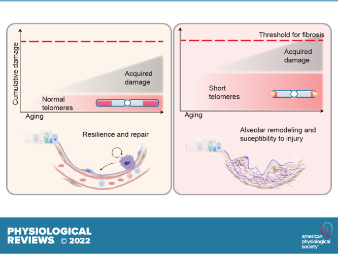
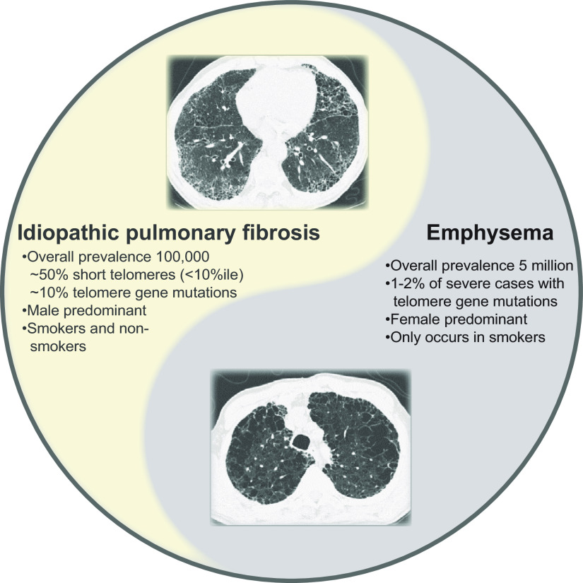
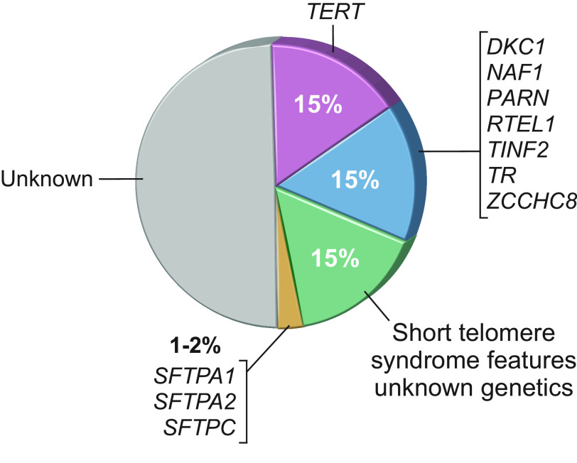
Parenchymal lung disease is the fourth leading cause of death in the United States; among the top causes, it continues on the rise. Telomeres and telomerase have historically been linked to cellular processes related to aging and cancer, but surprisingly, in the recent decade genetic discoveries have linked the most apparent manifestations of telomere and telomerase dysfunction in humans to the etiology of lung disease: both idiopathic pulmonary fibrosis (IPF) and emphysema. The short telomere defect is pervasive in a subset of IPF patients, and human IPF is the phenotype most intimately tied to germline defects in telomere maintenance. One-third of families with pulmonary fibrosis carry germline mutations in telomerase or other telomere maintenance genes, and one-half of patients with apparently sporadic IPF have short telomere length. Beyond explaining genetic susceptibility, short telomere length uncovers clinically relevant syndromic extrapulmonary disease, including a T-cell immunodeficiency and a propensity to myeloid malignancies. Recognition of this subset of patients who share a unifying molecular defect has provided a precision medicine paradigm wherein the telomere-mediated lung disease diagnosis provides more prognostic value than histopathology or multidisciplinary evaluation. Here, we critically evaluate this progress, emphasizing how the genetic findings put forth a new pathogenesis paradigm of age-related lung disease that links telomere abnormalities to alveolar stem senescence, remodeling, and defective gas exchange.
Overview
The claims and statistics of this paper haven’t been checked yet.
Run the check — free, no account needed. It reads the open-access text and checks the sources it cites.
Integrity signals
Registry
Cites
Statistics
Figures
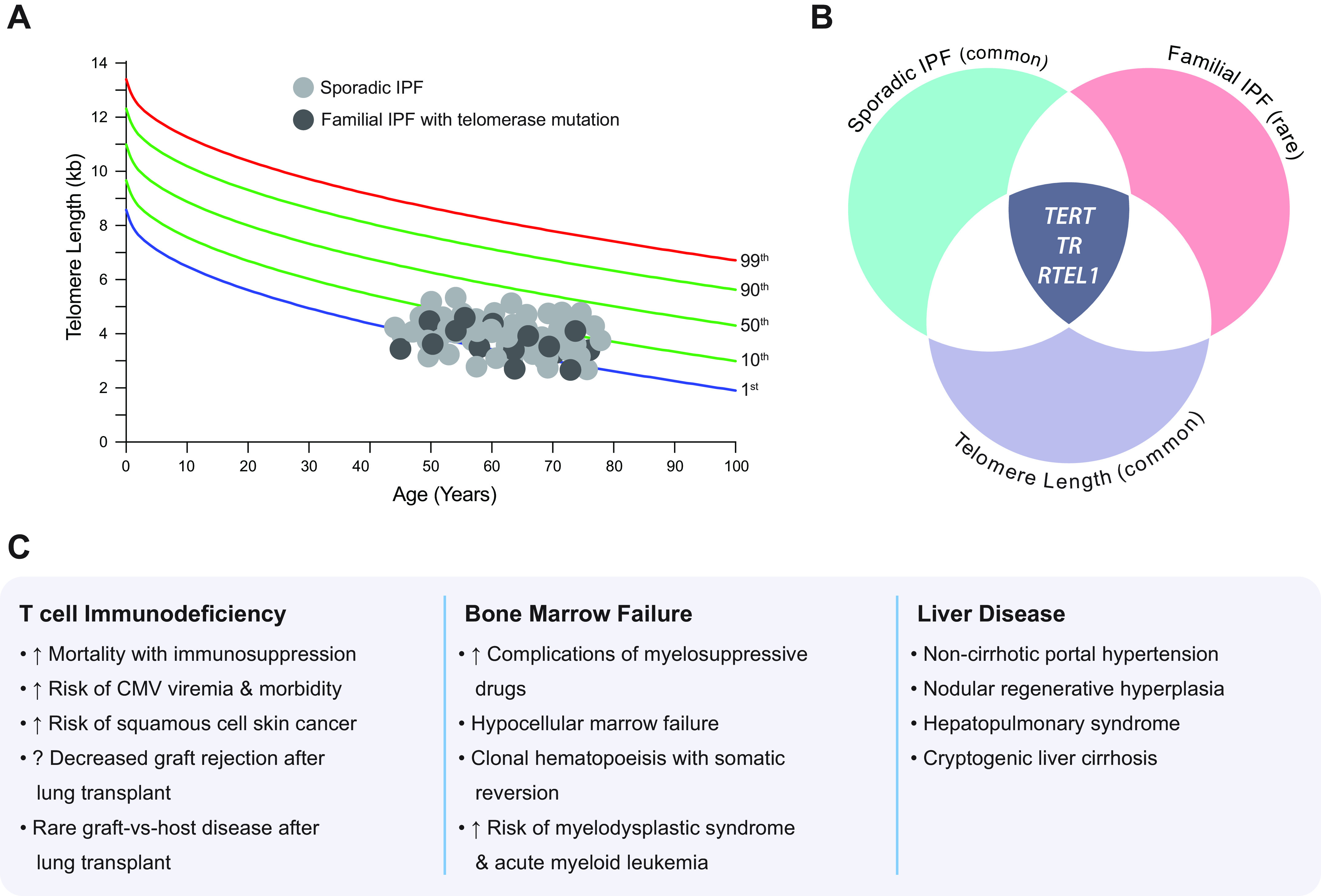
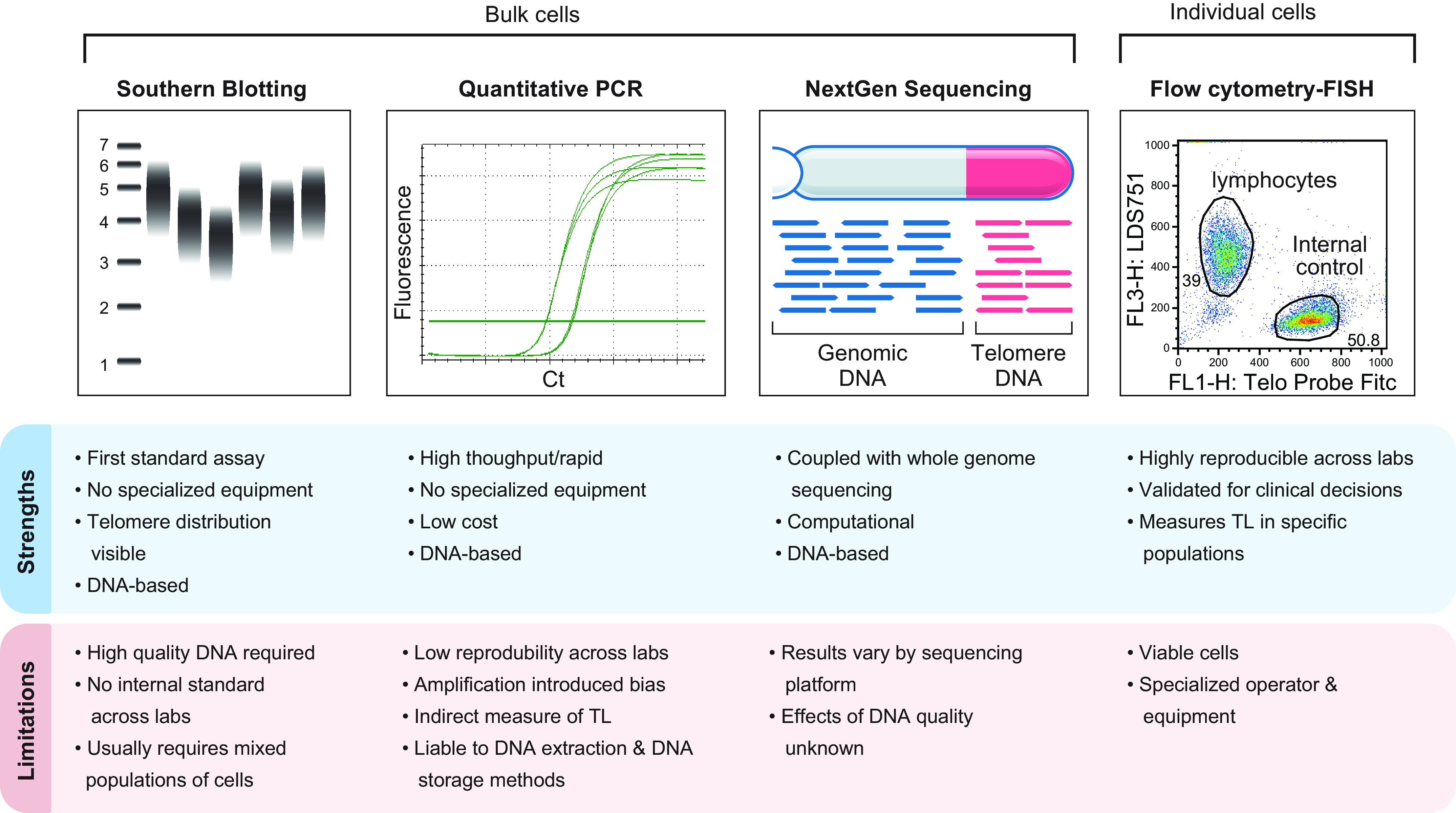
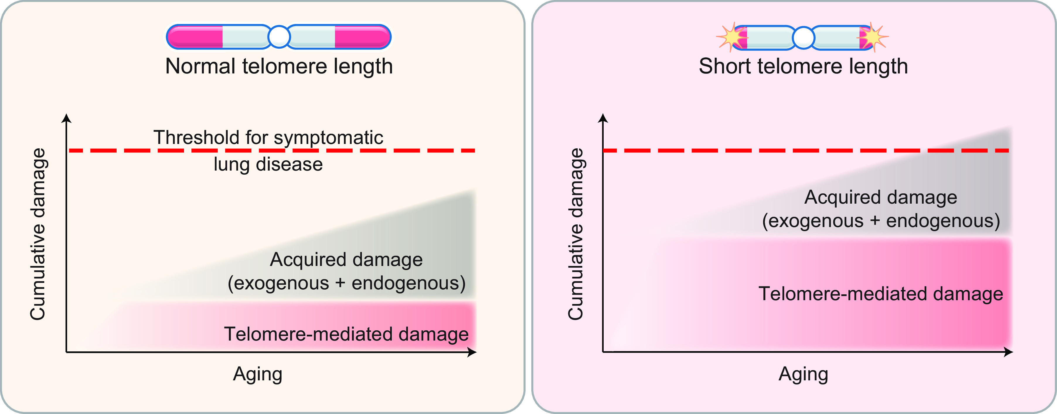
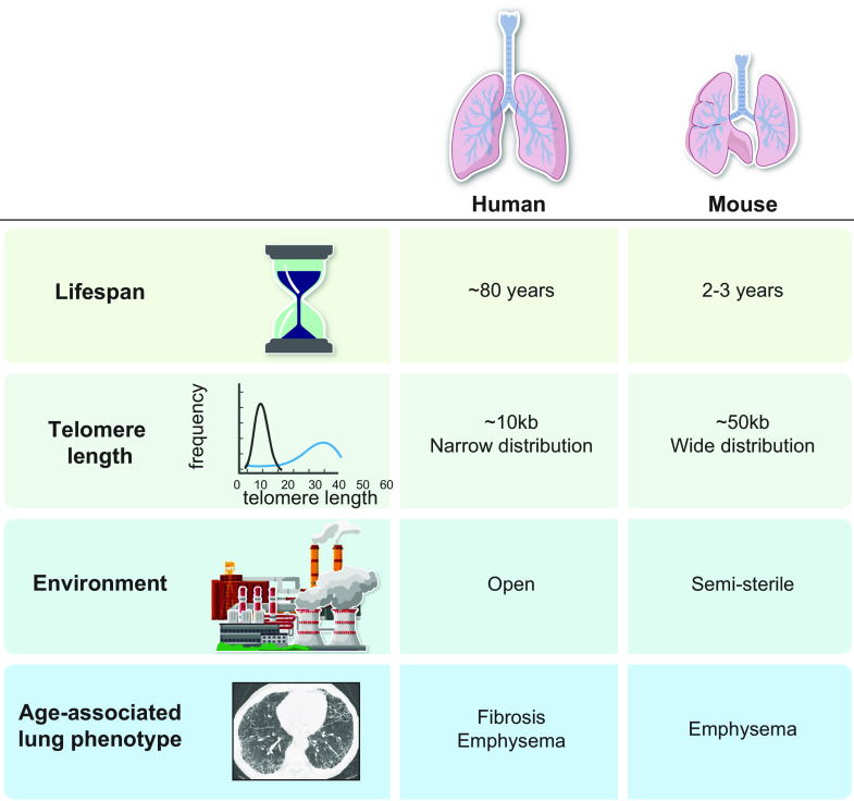
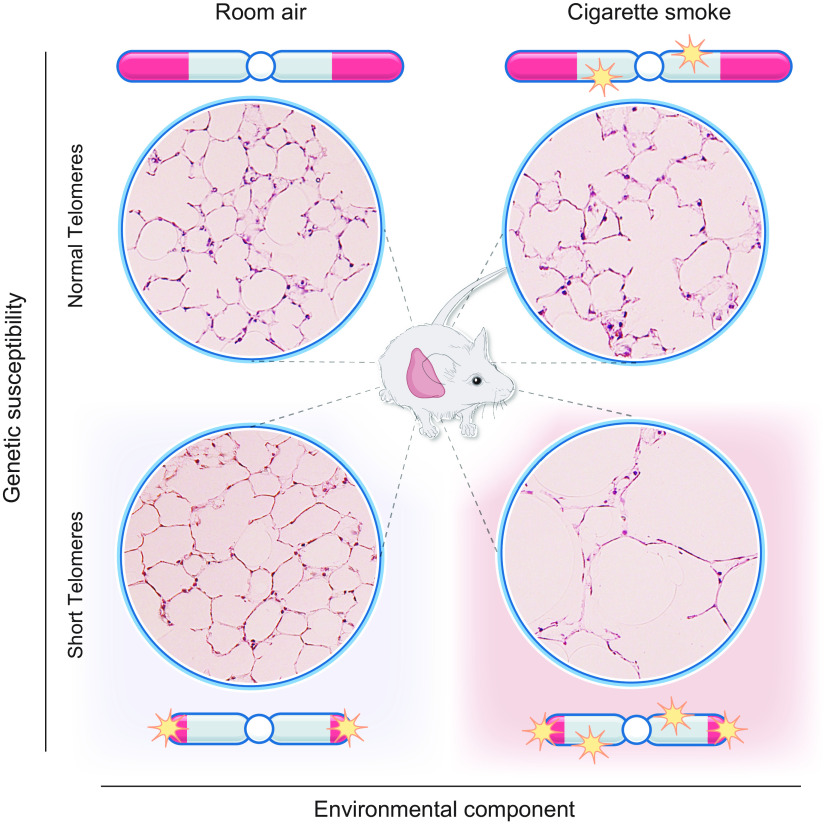
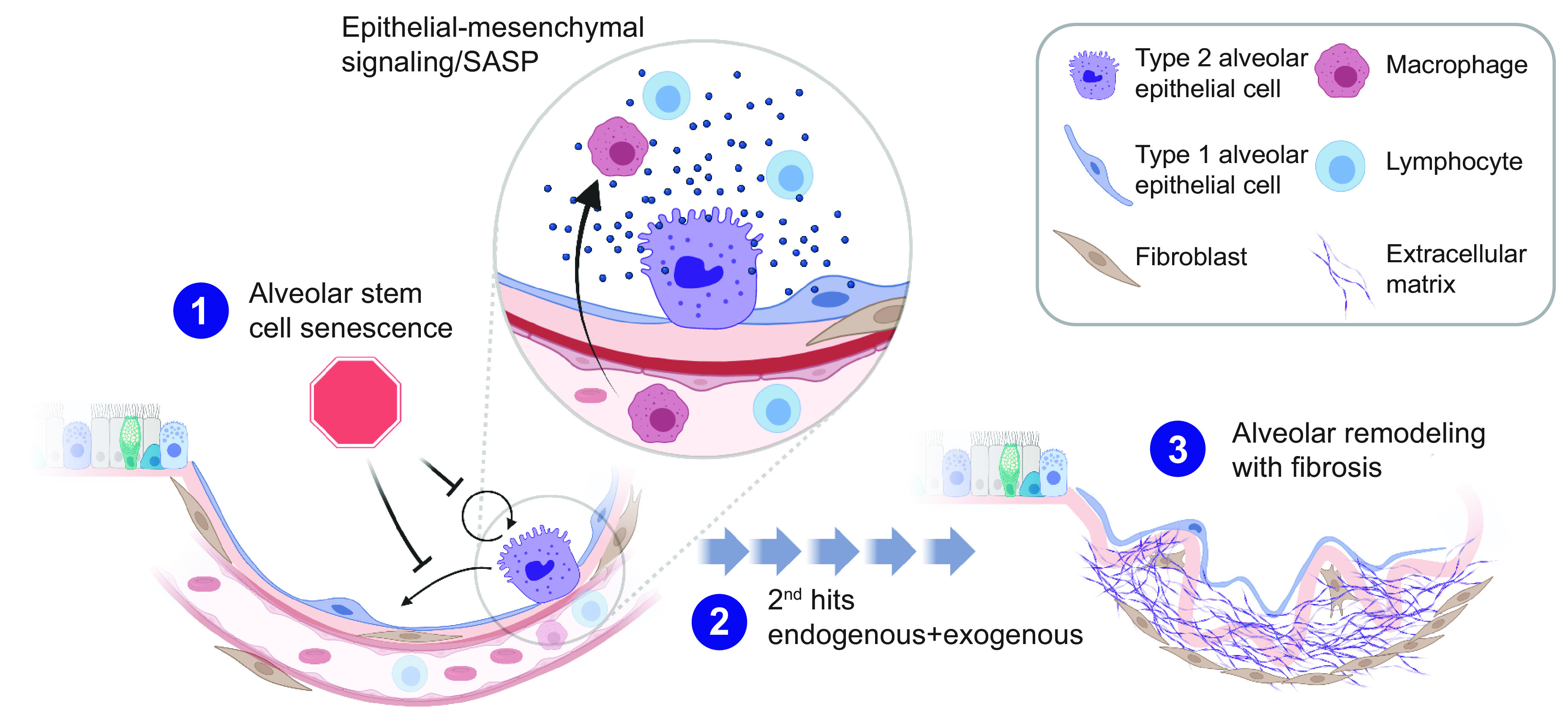
Verification
Claims the paper makes
The claims and statistics of this paper haven’t been checked yet.
Run the check — free, no account needed. It reads the open-access text and checks the sources it cites.
Claims other papers make about this one
Each is checked against this paper’s own text: does it say what the citing paper says it does?
The cited source says what the claim attributes to it.
In the citing paper
“Telomerase deficiency in humans is associated with premature development of diseases such as pulmonary fibrosis, aplastic anemia and dyskeratosis congenita, all of which hamper the regenerative capacity of the affected tissues 34 .”
From Hallmarks of aging: An expanding universe (2023) · cites this paper as [34]
Self-check
Percentages, sums and reported differences re-computed from the paper’s own numbers.
The self-check needs the paper’s full text.
Citations
Cites
Telomeres shorten during ageing of human fibroblasts
Harley, C. B., Futcher, A. B., & Greider, C. W. (1990) · Nature · doi:10.1038/345458a0 · Cited by 5,750
Telomere Shortening and Tumor Formation by Mouse Cells Lacking Telomerase RNA
Blasco, M. A., Lee, H.-W., Hande, M. P., Samper, E., Lansdorp, P. M., DePinho, R. A., & Greider, C. W. (1997) · Cell · doi:10.1016/s0092-8674(01)80006-4 · Cited by 2,108
Type 2 alveolar cells are stem cells in adult lung
Barkauskas, C. E., Cronce, M. J., Rackley, C. R., Bowie, E. J., Keene, D. R., Stripp, B. R., Randell, S. H., Noble, P. W., & Hogan, B. L. M. (2013) · Journal of Clinical Investigation · doi:10.1172/jci68782 · Cited by 1,776
A telomeric sequence in the RNA of Tetrahymena telomerase required for telomere repeat synthesis
Greider, C. W., & Blackburn, E. H. (1989) · Nature · doi:10.1038/337331a0 · Cited by 1,643
Telomerase Mutations in Families with Idiopathic Pulmonary Fibrosis
Armanios, M. Y., Chen, J. J.-L., Cogan, J. D., Alder, J. K., Ingersoll, R. G., Markin, C., Lawson, W. E., Xie, M., Vulto, I., Phillips, J. A., et al. (2007) · New England Journal of Medicine · doi:10.1056/nejmoa066157 · Cited by 1,357
Self-citationEssential role of mouse telomerase in highly proliferative organs
Lee, H.-W., Blasco, M. A., Gottlieb, G. J., Horner, J. W., Greider, C. W., & DePinho, R. A. (1998) · Nature · doi:10.1038/33345 · Cited by 1,270
Greider, C. W., & Blackburn, E. H. (1987) · Cell · doi:10.1016/0092-8674(87)90576-9 · Cited by 1,150
Evidence for a mitotic clock in human hematopoietic stem cells: loss of telomeric DNA with age.
Vaziri, H., Dragowska, W., Allsopp, R. C., Thomas, T. E., Harley, C. B., & Lansdorp, P. M. (1994) · Proceedings of the National Academy of Sciences · doi:10.1073/pnas.91.21.9857 · Cited by 1,132
Armanios, M., & Blackburn, E. H. (2012) · Nature Reviews Genetics · doi:10.1038/nrg3246 · Cited by 982
Self-citationHeiss, N. S., Knight, S. W., Vulliamy, T. J., Klauck, S. M., Wiemann, S., Mason, P. J., Poustka, A., & Dokal, I. (1998) · Nature Genetics · doi:10.1038/ng0598-32 · Cited by 976
Research works printed but not linked: 81
- 10.1016/0092-8674(85)90170-9 · DOI · Paper page
- 10.1016/0022-2836(78)90294-2 · DOI · Paper page
- 10.1038/nm1006-1133 · DOI · Paper page
- 10.1126/science.276.5312.561 · DOI · Paper page
- 10.1172/JCI120851 · DOI
- 10.1183/13993003.02115-2016 · DOI
- 10.1016/j.gde.2015.06.004 · DOI
- 10.1016/S0140-6736(11)60052-4 · DOI · Paper page
- Loyd JE, 2003, Am J Respir Cell Mol Biol, 29, S47
- 10.1164/rccm.200408-1104OC · DOI · Paper page
- 10.1002/humu.22397 · DOI
- 10.1378/chest.14-1947 · DOI
- 10.1126/scitranslmed.aaf7837 · DOI
- 10.1101/gad.326785.119 · DOI
- 10.1164/rccm.201408-1510OC · DOI
- 10.1038/ng.3278 · DOI · Paper page
- 10.1182/blood.2019003264 · DOI
- 10.1164/rccm.201610-2088OC · DOI
- 10.1016/j.healun.2021.11.008 · DOI
- 10.1164/rccm.200112-123OC · DOI · Paper page
- 10.1016/j.ajhg.2008.11.010 · DOI
- 10.1093/hmg/ddw014 · DOI
- 10.1038/ng.2528 · DOI · Paper page
- 10.1038/ng.2609 · DOI · Paper page
- 10.1007/s00439-013-1265-8 · DOI
- 10.1001/jamaoncol.2016.5945 · DOI
- 10.1164/rccm.201805-0825OC · DOI
- 10.1172/JCI120216 · DOI
- 10.1183/09031936.00060014 · DOI
- 10.1056/NEJMc1207471 · DOI
- 10.1164/rccm.201809-1646OC · DOI
- 10.1016/j.gde.2020.02.019 · DOI
- 10.1172/JCI147598 · DOI
- 10.1371/journal.pone.0007926 · DOI
- 10.1378/chest.15-0825 · DOI
- 10.1002/lt.26109 · DOI
- 10.1111/acel.12041 · DOI
- 10.1182/hematology.2020000170 · DOI
- 10.1073/pnas.1720427115 · DOI
- 10.1371/journal.pgen.1002696 · DOI
- 10.1136/annrheumdis-2018-214499 · DOI
- 10.1038/317687a0 · DOI
- 10.1093/nar/gkn1027 · DOI
- 10.1371/journal.pone.0184098 · DOI
- 10.1093/ije/dyu191 · DOI
- 10.1093/nar/gku181 · DOI
- 10.1093/nar/gkm700 · DOI
- 10.1200/PO.21.00110 · DOI
- 10.1182/blood-2017-09-806489 · DOI
- 10.1182/blood.2021011075 · DOI
- 10.1080/15476286.2016.1205768 · DOI
- 10.1378/chest.13-2224 · DOI
- 10.1016/s0092-8674(00)80687-x · DOI · Paper page
- 10.1038/ng.3423 · DOI
- 10.1128/MCB.00593-18 · DOI
- 10.1111/j.1399-0004.2011.01658.x · DOI
- 10.1067/mpd.2000.104295 · DOI
- 10.1136/jmg.2010.085100 · DOI
- 10.1016/j.ajhg.2017.04.001 · DOI
- 10.1016/j.chest.2020.07.025 · DOI
- 10.1007/s00412-005-0333-9 · DOI
- 10.1183/13993003.02115-2015 · DOI
- 10.1164/rccm.201902-0360OC · DOI
- 10.1513/AnnalsATS.201609-718AW · DOI
- 10.1136/thoraxjnl-2015-207252 · DOI
- 10.1164/rccm.200606-833OC · DOI
- 10.1007/s00408-012-9389-5 · DOI
- 10.1016/j.stem.2009.04.002 · DOI
- Kauffman SL, 1980, Int Rev Exp Pathol, 22, 131
- 10.1038/nature12930 · DOI
- 10.1172/JCI99287 · DOI
- 10.1172/JCI93560 · DOI
- 10.1038/nature14112 · DOI
- 10.1016/j.cell.2005.03.032 · DOI
- 10.1242/dev.140103 · DOI
- 10.1038/s41588-019-0346-6 · DOI
- 10.1016/j.stem.2020.03.009 · DOI · Paper page
- 10.3109/01902148.2012.658148 · DOI
- 10.1152/ajplung.00062.2019 · DOI
- 10.1172/JCI91161 · DOI
- 10.1111/j.1440-1843.2011.01955.x · DOI
Cited by
Sorted by citations
Hallmarks of aging: An expanding universe
López-Otín, C., Blasco, M. A., Partridge, L., Serrano, M., & Kroemer, G. (2023) · Cell · doi:10.1016/j.cell.2022.11.001 · Cited by 6,549
Meta-hallmarks of aging and cancer
López-Otín, C., Pietrocola, F., Roiz-Valle, D., Galluzzi, L., & Kroemer, G. (2023) · Cell Metabolism · doi:10.1016/j.cmet.2022.11.001 · Cited by 529
Idiopathic Pulmonary Fibrosis: Where do We Stand and How Far to Go?
Singh, V., Ulasov, I., Gupta, S., Singh, A., Roy, V. K., & Kharwar, R. K. (2024) · Discovery Medicine · doi:10.24976/discov.med.202436180.3 · Cited by 17
Metadata
- Authors
- Alder, J. K., & Armanios, M.
- Affiliations
- Division of Pulmonary and Critical Care Medicine, University of Pittsburgh School of Medicine, Pittsburgh, Pennsylvania
- Departments of Oncology and Genetic Medicine, Telomere Center at Johns Hopkins, Johns Hopkins University School of Medicine, Baltimore, Maryland
- Venue
- Physiological Reviews
- Publisher
- American Physiological Society
- Published
- 2022-10-01
- Volume
- 102
- Issue
- 4
- Pages
- 1703-1720
- ISSN
- 0031-9333
- 1522-1210
- PMC
- PMC9306791
- Cited by
- 130
- References
- 29
- Language
- en
- Funders
- HHS | NIH | National Cancer Institute (CA225027)
- HHS | NIH | National Heart, Lung, and Blood Institute (HL119476, HL135062)
- S&R Kuno Foundation
About these checks
Registry, retraction, Cites and statistics results are computed, not judged, with one exception: a printed reference that no index lists is read once by a model before it counts as a citation error. Claims are extracted by a model and kept only when their quote appears verbatim in the text; each cited source is then checked, with a model’s help, for whether it says what the claim relies on. Those verdicts can be wrong — read the claim’s quote and the cited source before relying on one.
Statistics and arithmetic are recomputed, not judged.
Confidence bands are shown only for fields calibrated on our annotated test set. None is calibrated yet, so no band is shown.
What counts as a claim ›How we measure confidence ›How we check this ›
Are you an author, or do you see a problem with this page? Request a review
