

The DOI matches its Crossref record; 130 of the 131 references it cites were found in a registry or an index.
Endothelial-to-Mesenchymal Transition (EndoMT): Roles in Tumorigenesis, Metastatic Extravasation and Therapy Resistance
Open accessAbstract
Cancer cells evolve in a very complex tumor microenvironment, composed of several cell types, among which the endothelial cells are the major actors of the tumor angiogenesis. Today, these cells are also characterized for their plasticity, as endothelial cells have demonstrated their potential to modify their phenotype to differentiate into mesenchymal cells through the endothelial-to-mesenchymal transition (EndoMT). This cellular plasticity is mediated by various stimuli including transforming growth factor-β (TGF-β) and is modulated dependently of experimental conditions. Recently, emerging evidences have shown that EndoMT is involved in the development and dissemination of cancer and also in cancer cell to escape from therapeutic treatment. In this review, we summarize current updates on EndoMT and its main induction pathways. In addition, we discuss the role of EndoMT in tumorigenesis, metastasis, and its potential implication in cancer therapy resistance.
Overview
The claims and statistics of this paper haven’t been checked yet.
Run the check — free, no account needed. It reads the open-access text and checks the sources it cites.
Integrity signals
Registry
Cites
Statistics
Figures
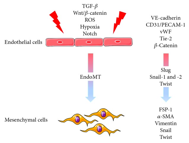
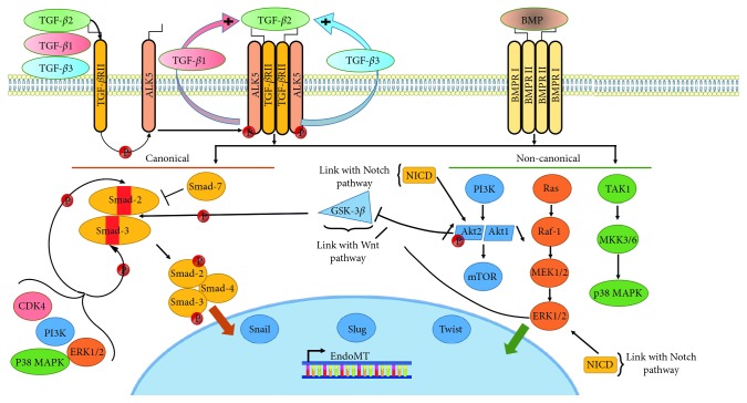
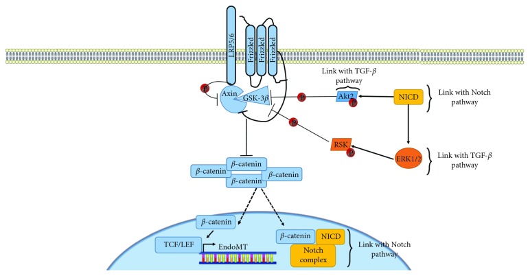
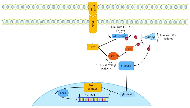
Verification
Claims the paper makes
The claims and statistics of this paper haven’t been checked yet.
Run the check — free, no account needed. It reads the open-access text and checks the sources it cites.
Claims other papers make about this one
Each is checked against this paper’s own text: does it say what the citing paper says it does?
In the citing paper
“Extravasation is known to be influenced by various environmental factors, such as ECM heterogeneity [4], immune cells [68], epithelial-to-mesenchymal transition (EMT) [69], integrins [70,71], blood flow [4,72], and biochemical gradients [73].”
From Cancer-on-chip models for metastasis: importance of the tumor microenvironment (2024) · cites this paper as [69]
Self-check
Percentages, sums and reported differences re-computed from the paper’s own numbers.
The self-check needs the paper’s full text.
Citations
Cites
1 retracted
Vetter, G., Saumet, A., Moes, M., Vallar, L., Béchec, A. L., Laurini, C., Sabbah, M., Arar, K., Theillet, C. G., Lecellier, C., et al. (2010) · Oncogene · doi:10.1038/onc.2010.181 · Cited by 122
RetractedHallmarks of Cancer: The Next Generation
Hanahan, D., & Weinberg, R. A. (2011) · Cell · doi:10.1016/j.cell.2011.02.013 · Cited by 66,590
Hanahan, D., & Weinberg, R. A. (2000) · Cell · doi:10.1016/s0092-8674(00)81683-9 · Cited by 29,219
Tumor angiogenesis: therapeutic implications
Sherwood, L. M., Parris, E. E., & Folkman, J. (1971) · New England Journal of Medicine · doi:10.1056/nejm197111182852108 · Cited by 10,187
Microenvironmental regulation of tumor progression and metastasis
Quail, D. F., & Joyce, J. A. (2013) · Nature Medicine · doi:10.1038/nm.3394 · Cited by 7,994
Accessories to the Crime: Functions of Cells Recruited to the Tumor Microenvironment
Hanahan, D., & Coussens, L. M. (2012) · Cancer Cell · doi:10.1016/j.ccr.2012.02.022 · Cited by 4,632 · cited 1×
“*is microenvironment is composed of numerous cell types: immune cells (bone marrow-derived inflammatory cells, monocytes/ macrophages, and lymphocytes), vascular cells (endothelial cells and pericytes), and stromal fibroblastic cells, and of an extracellular matrix composed of collagen and proteoglycans [3, 4].”
BackgroundKalluri, R., & Zeisberg, M. (2006) · Nature Reviews Cancer · doi:10.1038/nrc1877 · Cited by 4,538
Cano, A., Pérez‐Moreno, M., Rodrigo, I., Locascio, A., Blanco, M. J., Barrio, M. G. D., Portillo, F., & Nieto, M. A. (2000) · Nature Cell Biology · doi:10.1038/35000025 · Cited by 3,672
T cell exclusion, immune privilege, and the tumor microenvironment
Joyce, J. A., & Fearon, D. T. (2015) · Science · doi:10.1126/science.aaa6204 · Cited by 2,539
Endothelial-to-mesenchymal transition contributes to cardiac fibrosis
Zeisberg, E. M., Tarnavski, O., Zeisberg, M., Dorfman, A. L., McMullen, J. R., Gustafsson, E., Chandraker, A. K., Yuan, X., Pu, W. T., Roberts, A. B., et al. (2007) · Nature Medicine · doi:10.1038/nm1613 · Cited by 2,251
Cited by
Sorted by citations
Kumar, V., Ramnarayanan, K., Sundar, R., Padmanabhan, N., Srivastava, S., Koiwa, M., Yasuda, T., Koh, V., Huang, K. K., Tay, S. T., et al. (2022) · Cancer Discovery · doi:10.1158/2159-8290.cd-21-0683 · Cited by 573
TGF-β-Induced Endothelial to Mesenchymal Transition in Disease and Tissue Engineering
Ma, J., Sánchez‐Duffhues, G., Goumans, M., & ten Dijke, P. (2020) · Frontiers in Cell and Developmental Biology · doi:10.3389/fcell.2020.00260 · Cited by 250
Single-cell atlas of the human brain vasculature across development, adulthood and disease
Wälchli, T., Ghobrial, M., Schwab, M., Takada, S., Zhong, H., Suntharalingham, S., Vetiska, S., Rodríguez, D. G., Wu, R., Rehrauer, H., et al. (2024) · Nature · doi:10.1038/s41586-024-07493-y · Cited by 217
Endothelial cells in tumor microenvironment: insights and perspectives
Leone, P., Malerba, E., Susca, N., Favoino, E., Perosa, F., Brunori, G., Prete, M., & Racanelli, V. (2024) · Frontiers in Immunology · doi:10.3389/fimmu.2024.1367875 · Cited by 158
Endothelial-to-mesenchymal transition in anticancer therapy and normal tissue damage
Choi, K. J., Nam, J.-K., Kim, J.-H., Choi, S., & Lee, Y. (2020) · Experimental & Molecular Medicine · doi:10.1038/s12276-020-0439-4 · Cited by 116
EPISCORE: cell type deconvolution of bulk tissue DNA methylomes from single-cell RNA-Seq data
Teschendorff, A. E., Zhu, T., Breeze, C. E., & Beck, S. (2020) · Genome biology · doi:10.1186/s13059-020-02126-9 · Cited by 118
Endothelial cell plasticity at the single-cell level
Pasut, A., Becker, L. M., Cuypers, A., & Carmeliet, P. (2021) · Angiogenesis · doi:10.1007/s10456-021-09797-3 · Cited by 100
Troletti, C. D., Fontijn, R. D., Gowing, E., Charabati, M., Hof, B. V. H., Didouh, I., van der Pol, S. M. A., Geerts, D., Prat, A., van Horssen, J., et al. (2019) · Cell Death and Disease · doi:10.1038/s41419-018-1294-2 · Cited by 96
Sampson, N., Brunner, E., Weber, A., Puhr, M., Schäfer, G., Szyndralewiez, C., & Klocker, H. (2018) · International Journal of Cancer · doi:10.1002/ijc.31316 · Cited by 85
Metabolic Signatures of Distinct Endothelial Phenotypes
Dumas, S. J., García‐Caballero, M., & Carmeliet, P. (2020) · Trends in Endocrinology and Metabolism · doi:10.1016/j.tem.2020.05.009 · Cited by 82
Kang, E., Seo, J., Yoon, H., & Cho, S. (2021) · International Journal of Molecular Sciences · doi:10.3390/ijms22073591 · Cited by 81
Tumor Vessels Fuel the Fire in Glioblastoma
Rosińska, S., & Gavard, J. (2021) · International Journal of Molecular Sciences · doi:10.3390/ijms22126514 · Cited by 84
Lin, P. P. (2020) · Cells · doi:10.3390/cells9061539 · Cited by 78
Khouzam, R. A., Zaarour, R. F., Brodaczewska, K. K., Azakir, B., Venkatesh, G. H., Thiery, J., Terry, S., & Chouaı̈b, S. (2022) · Frontiers in Immunology · doi:10.3389/fimmu.2022.828875 · Cited by 80
Cancer-on-chip models for metastasis: importance of the tumor microenvironment
Jouybar, M., de Winde, C. M., Wolf, K., Friedl, P., Mebius, R. E., & den Toonder, J. M. J. (2024) · Trends in Biotechnology · doi:10.1016/j.tibtech.2023.10.001 · Cited by 66
Kanno, Y. (2023) · International Journal of Molecular Sciences · doi:10.3390/ijms24021796 · Cited by 67
The hallmarks of severe pulmonary arterial hypertension: the cancer hypothesis—ten years later
Cool, C. D., Kuebler, W. M., Bogaard, H. J., Spiekerkoetter, E. F., Nicolls, M. R., & Voelkel, N. F. (2020) · American Journal of Physiology-Lung Cellular and Molecular Physiology · doi:10.1152/ajplung.00476.2019 · Cited by 63
Watabe, T., Takahashi, K., Pietras, K., & Yoshimatsu, Y. (2023) · Seminars in Cancer Biology · doi:10.1016/j.semcancer.2023.04.007 · Cited by 64
Osman, A., Afify, S. M., Hassan, G., Fu, X., Seno, A., & Seno, M. (2020) · Cancers · doi:10.3390/cancers12040879 · Cited by 63
Extracellular Vesicles Are Key Regulators of Tumor Neovasculature
Kuriyama, N., Yoshioka, Y., Kikuchi, S., Azuma, N., & Ochiya, T. (2020) · Frontiers in Cell and Developmental Biology · doi:10.3389/fcell.2020.611039 · Cited by 58
Gu, S., Liu, Y., Zou, J., Wang, W., Wei, T., Wang, X., Zhu, L., Zhang, M., Zhu, J., Xie, T., et al. (2020) · Experimental Eye Research · doi:10.1016/j.exer.2020.108271 · Cited by 59
Transforming Growth Factor-β: An Agent of Change in the Tumor Microenvironment
Stuelten, C. H., & Zhang, Y. E. (2021) · Frontiers in Cell and Developmental Biology · doi:10.3389/fcell.2021.764727 · Cited by 57
Angiogenesis and targeted therapy in the tumour microenvironment: From basic to clinical practice
Yang, S., Fang, Y., Ma, Y., Wang, F., Wang, Y., Jia, J., Yang, Y., Sun, W., Zhou, Q., & Li, Z. (2025) · Clinical and Translational Medicine · doi:10.1002/ctm2.70313 · Cited by 61
A bi-directional dialog between vascular cells and monocytes/macrophages regulates tumor progression
Delprat, V., & Michiels, C. (2021) · Cancer and Metastasis Reviews · doi:10.1007/s10555-021-09958-2 · Cited by 51
Endothelial-to-Mesenchymal Transition: Role in Cardiac Fibrosis
Cheng, W., Li, X., Liu, D., Cui, C., & Wang, X. (2020) · Journal of Cardiovascular Pharmacology and Therapeutics · doi:10.1177/1074248420952233 · Cited by 45
Transforming growth factor-β in tumour development
Trelford, C. B., Dagnino, L., & Guglielmo, G. M. D. (2022) · Frontiers in Molecular Biosciences · doi:10.3389/fmolb.2022.991612 · Cited by 45
To be or not to be: endothelial cell plasticity in development, repair, and disease
Greenspan, L. J., & Weinstein, B. M. (2021) · Angiogenesis · doi:10.1007/s10456-020-09761-7 · Cited by 46
Sun, Z., Zhao, H., Fang, D., Davis, C. T., Shi, D., Lei, K., Rich, B. E., Winter, J. M., Guo, L., Sorensen, L. K., et al. (2022) · Neuron · doi:10.1016/j.neuron.2022.07.015 · Cited by 43
Resident vascular endothelial progenitor definition and function: the age of reckoning
Dight, J., Zhao, J., Styke, C., Khosrotehrani, K., & Patel, J. (2021) · Angiogenesis · doi:10.1007/s10456-021-09817-2 · Cited by 39
Cadherin Signaling in Cancer and Autoimmune Diseases
Sisto, M., Ribatti, D., & Lisi, S. (2021) · International Journal of Molecular Sciences · doi:10.3390/ijms222413358 · Cited by 37
Liu, J., Yu, X., Braucht, A., Smith, S., & Wang, C. (2024) · ACS Nano · doi:10.1021/acsnano.3c12281 · Cited by 34
Wawro, M. E., Chojnacka, K., Wieczorek, K., Sobierajska, K., & Niewiarowska, J. (2018) · International Journal of Molecular Sciences · doi:10.3390/ijms20010053 · Cited by 33
Zhang, T., Zhang, L., Gao, Y. J., Wang, Y., Liu, Y., Zhang, H., Wang, Q., Hu, F., Li, J., Tan, J., et al. (2021) · Molecular Oncology · doi:10.1002/1878-0261.13092 · Cited by 33
Fonta, C. M., Loustau, T., Li, C., Surendran, S. P., Hansen, U. J., Murdamoothoo, D., Benn, M. C., Velázquez-Quesada, I., Carapito, R., Orend, G., et al. (2023) · Matrix Biology · doi:10.1016/j.matbio.2023.01.002 · Cited by 29
Zhao, J., Zhao, C., Yang, F., Jiang, Z., Zhu, J., Yao, W., Pang, W., & Zhou, J. (2023) · Theranostics · doi:10.7150/thno.84427 · Cited by 27
Yin, Z., & Wang, L. (2023) · Annals of Medicine · doi:10.1080/07853890.2023.2180155 · Cited by 26
Cooke, J. P., & Lai, L. (2023) · Advanced Drug Delivery Reviews · doi:10.1016/j.addr.2023.115118 · Cited by 25
Gonzales, J., Holbert, K., Czysz, K., George, J. T., Fernandes, C., & Fraidenburg, D. R. (2022) · International Journal of Molecular Sciences · doi:10.3390/ijms23094763 · Cited by 23
Adnani, L., Spinelli, C., Tawil, N., & Rak, J. W. (2022) · Seminars in Cancer Biology · doi:10.1016/j.semcancer.2022.11.003 · Cited by 25
Bourreau, C., Treps, L., Faure, S., Fradin, D., & Clere, N. (2023) · Pharmacology & Therapeutics · doi:10.1016/j.pharmthera.2023.108347 · Cited by 20
Self-citationShifting Shapes: The Endothelial-to-Mesenchymal Transition as a Driver for Cancer Progression
Giordanengo, L., Proment, A., Botta, V., Picca, F., Munir, H., Tao, J., Olivero, M., Taulli, R., Bersani, F., Sangiolo, D., et al. (2025) · International Journal of Molecular Sciences · doi:10.3390/ijms26136353 · Cited by 20
Obesity-Related Fatty Acid and Cholesterol Metabolism in Cancer-Associated Host Cells
Ye, Y., Sun, X., & Lu, Y. (2020) · Frontiers in Cell and Developmental Biology · doi:10.3389/fcell.2020.600350 · Cited by 19
Hu, B., Gong, Y., Wang, Y., Xie, J., Cheng, J. Q., & Huang, Q. (2022) · Frontiers in Oncology · doi:10.3389/fonc.2022.821454 · Cited by 18
Molecular atlas of the human brain vasculature at the single-cell levelPreprint
Wälchli, T., Ghobrial, M., Schwab, M., Takada, S., Zhong, H., Suntharalingham, S., Vetiska, S., Rodrigues, D. R., Rehrauer, H., Wu, R., et al. (2021) · bioRxiv (Cold Spring Harbor Laboratory) · doi:10.1101/2021.10.18.464715 · Cited by 16
Ciccone, V., Filippelli, A., Bacchella, C., Monzani, E., & Morbidelli, L. (2022) · Cancers · doi:10.3390/cancers14174240 · Cited by 15
Endothelial-to-Mesenchymal Transition: Potential Target of Doxorubicin-Induced Cardiotoxicity
Feng, J., & Wu, Y. (2023) · American Journal of Cardiovascular Drugs · doi:10.1007/s40256-023-00573-w · Cited by 16
Slenders, L., Wesseling, M., Wei, S., Boltjes, A., Kapteijn, D., van de Kraak, P., Depuydt, M., Prange, K., van den Dungen, N. V. D., Benavente, E. D., et al. (2025) · Vascular pharmacology · doi:10.1016/j.vph.2025.107498 · Cited by 8
ZC3H4 mediates silica-induced EndoMT via ER stress and autophagy
Jiang, R., Han, L., Gao, Q., & Chao, J. (2021) · Environmental Toxicology and Pharmacology · doi:10.1016/j.etap.2021.103605 · Cited by 15
Deluque, A. L., Oliveira, B. M., Souza, C. S., Maciel, A. L. D., Francescato, H. D. C., da Silva, C. G. A., de Almeida, L. F., de Paula, F. J. A., Costa, R. S., Antunes‐Rodrigues, J., et al. (2022) · Nutrients · doi:10.3390/nu14245316 · Cited by 15
Alternative Strategies to Inhibit Tumor Vascularization
Brossa, A., Buono, L., Fallo, S., Fiorio, A., Munaron, L., & Bussolati, B. (2019) · International Journal of Molecular Sciences · doi:10.3390/ijms20246180 · Cited by 15
SUMOylation of Smad2 mediates TGF-β-regulated endothelial–mesenchymal transition
Su, Q., Chen, X., Ling, X., Li, D., Ren, X., Zhao, Y., Yang, Y., Liu, Y., He, A., Zhu, X., et al. (2023) · Journal of Biological Chemistry · doi:10.1016/j.jbc.2023.105244 · Cited by 14
Andreucci, E., Bugatti, K., Peppicelli, S., Ruzzolini, J., Lulli, M., Calorini, L., Battistini, L., Zanardi, F., Sartori, A. M., & Bianchini, F. (2023) · International Journal of Molecular Sciences · doi:10.3390/ijms24021475 · Cited by 13
Choi, S., Jang, J., Kim, Y., Park, C. G., Lee, S. Y., Kim, H.-J., & Kim, H. (2024) · Cell Death and Disease · doi:10.1038/s41419-024-06678-7 · Cited by 13
Metastatic Breast Cancer, Organotropism and Therapeutics: A Review
Waza, A. A., Tarfeen, N., Majid, S., Hassan, Y., Mir, R., Rather, M. Y., & Shah, N. U. D. (2021) · Current Cancer Drug Targets · doi:10.2174/1568009621666210806094410 · Cited by 13
Changchien, C., Chang, H., Dai, M., Tsai, W., Tsai, H., Wang, C., Shen, M., Cheng, L., Lee, H., Chen, Y., et al. (2020) · Cancer Science · doi:10.1111/cas.14772 · Cited by 12
Lu, Q., Ma, J., Zhao, Y., Ding, G., Wang, Y., Qiao, X., & Cheng, X. (2024) · Biomedicine & Pharmacotherapy · doi:10.1016/j.biopha.2024.117521 · Cited by 11
Verloy, R., Privat‐Maldonado, A., Audenaerde, J. R. M. V., Rovers, S., Zaryouh, H., Waele, J. D., Quatannens, D., Peeters, D. J. E., Roeyen, G., Deben, C., et al. (2025) · Cells · doi:10.3390/cells14060450 · Cited by 11
MEF2C and miR-194-5p: New Players in Triple Negative Breast Cancer Tumorigenesis
Caetano, S., Garcia, A. R., Figueira, I., & Brito, M. A. (2023) · International Journal of Molecular Sciences · doi:10.3390/ijms241814297 · Cited by 10
Metastasis prevention: How to catch metastatic seeds
Gerashchenko, T. S., Schegoleva, A. A., Khozyainova, A. A., Чойнзонов, Е. Л., & Denisov, E. V. (2023) · Biochimica et Biophysica Acta (BBA) - Reviews on Cancer · doi:10.1016/j.bbcan.2023.188867 · Cited by 9
Pham, T. P., van Bergen, A. S., Kremer, V., Glaser, S.-F., Dimmeler, S., & Boon, R. A. (2021) · International Journal of Molecular Sciences · doi:10.3390/ijms22158088 · Cited by 9
Platel, V., Lechevalier, D., Bourreau, C., Renault, S., Šoborová, I., Jeannière, C., Martin, L., Hérault, O., Corre, I., & Clere, N. (2022) · Pharmacological Research · doi:10.1016/j.phrs.2022.106097 · Cited by 11
Self-citationXie, J., Hu, B., Gong, Y., He, S., Lin, J., Huang, Q., & Cheng, J. Q. (2023) · Journal of Translational Medicine · doi:10.1186/s12967-023-04746-8 · Cited by 8
Single-cell analysis revealing the metabolic landscape of prostate cancer
Wang, J., Ding, H.-K., Xu, H., Hu, D.-K., Hankey, W., Chen, L., Xiao, J., Liang, C.-Z., Zhao, B. Q., & Xu, L. (2024) · Asian Journal of Andrology · doi:10.4103/aja20243 · Cited by 9
Smęda, M., Jasztal, A., Maleki, E. H., Bar, A., Sternak, M., Kwiatkowski, G., Suraj, J., Proniewski, B., Kieronska‐Rudek, A., Wojnar‐Lason, K., et al. (2022) · Frontiers in Molecular Biosciences · doi:10.3389/fmolb.2022.1050112 · Cited by 8
Mikula, M. W., Najjar, S. N., Jabbour, T. E., Dalvi, S., Umrau, K., Li, H., Koo, B. H., & Lee, H. (2020) · Applied immunohistochemistry & molecular morphology · doi:10.1097/pai.0000000000000888 · Cited by 7
Far-Infrared Therapy Decreases Orthotopic Allograft Transplantation Vasculopathy
Lin, Y.-W., Tsai, C., Huang, C., Tsai, Y., Shih, C.-M., Lin, S., Li, C., Lin, C., Sung, S.-Y., & Lin, F. (2022) · Biomedicines · doi:10.3390/biomedicines10051089 · Cited by 7
Crestani, M., Gerber, I., Miéville, A., Frauenknecht, K. B. M., Maragkou, T., Hortobágyi, T., & Viola Vogel, B. (2025) · Advanced Science · doi:10.1002/advs.202416526 · Cited by 6
Bourreau, C., Navarro, E., Cotinat, M., Krejbich, M., Guillonneau, F., Guette, C., Boissard, A., Henry, C., Corre, I., Treps, L., et al. (2025) · Cancer Medicine · doi:10.1002/cam4.70707 · Cited by 6
Self-citationLin, F., Lin, Y., Shih, C., Lin, S., Tung, Y., Li, C., Chen, Y., Lin, C.-Y., Tsai, Y.-T., & Huang, C. (2021) · International Journal of Molecular Sciences · doi:10.3390/ijms22105210 · Cited by 6
ADP-Ribosylation Factor 6 Pathway Acts as a Key Executor of Mesenchymal Tumor Plasticity
Hashimoto, A., & Hashimoto, S. (2023) · International Journal of Molecular Sciences · doi:10.3390/ijms241914934 · Cited by 6
New insights into the pathogenesis of cardiac papillary fibroelastomas
Matysiak, N., Mielańczyk, Ł., Kaczmarek, K., Zaba, M., Reichman-Warmusz, E., & Wojnicz, R. (2021) · Folia Histochemica et Cytobiologica · doi:10.5603/fhc.a2021.0027 · Cited by 5
Kageyama, T., Kato, M., Miyachi, S., Bao, X., Sekito, S., Sugino, Y., Higashi, S., Sasaki, T., Nishikawa, K., Murakawa, Y., et al. (2025) · Cancer Science · doi:10.1111/cas.70144 · Cited by 4
Kim, J. S., Choi, W., Kim, H. I., Chung, S. W., Choi, J., Lee, D., & Kim, K. M. (2025) · Journal of Liver Cancer · doi:10.17998/jlc.2024.12.27 · Cited by 6
Saha, D., Annadorai, R. K., Thannimangalath, S., Shroff, N. P., Kataria, S., Dam, B., Dutta, A., Hegde, A., Hiwale, A. A., Ravula, V., et al. (2025) · Journal of Clinical Investigation · doi:10.1172/jci177123 · Cited by 3
Campbell, I., Glinka, M., Shaban, F., Kirkwood, K. J., Nadalin, F., Adams, D. J., Papatheodorou, I., Burger, A., Baldock, R., Arends, M. J., et al. (2023) · Journal of Clinical Medicine · doi:10.3390/jcm12123884 · Cited by 3
Molecular and therapeutic bases of cancer
Fajardo-Orduña, G. R., & Chamorro-Plata, D. (2021) · Elsevier eBooks · doi:10.1016/b978-0-12-819828-5.00002-4 · Cited by 3
Cell-Type Heterogeneity in DNA Methylation Studies: Statistical Methods and Guidelines
Teschendorff, A. E. (2022) · Springer eBooks · doi:10.1007/978-3-030-94475-9_4 · Cited by 2
Tsai, C., Huang, C., Tsai, Y., Shih, C., Lai, Z., Liu, C., Lin, Y., & Lin, F. (2024) · Molecular Oral Microbiology · doi:10.1111/omi.12453 · Cited by 3
Chiang, K., Chang, W.-J., Liu, Y., Lin, H.-L., Lin, C., Chiou, T., Wu, H., & Tung, Y. (2025) · Sensors and Actuators Reports · doi:10.1016/j.snr.2025.100396 · Cited by 2
Zhao, S., Liu, S., Shao, W., & Liu, D. (2025) · International Journal of Molecular Sciences · doi:10.3390/ijms262211128 · Cited by 3
Verloy, R., Peeters, E., Privat‐Maldonado, A., Rovers, S., Lau, H. W., Brants, L., Hermans, C., Waele, J. D., Deben, C., Smits, E., et al. (2025) · npj Precision Oncology · doi:10.1038/s41698-025-01241-9 · Cited by 2
Francescato, R., Moretti, M., & Bersini, S. (2024) · Bioengineering & Translational Medicine · doi:10.1002/btm2.10644 · Cited by 2
Zhuo, D., Mei, Y., Lin, C., Wu, A., Luo, Y., Lu, H., & Fu, J. (2024) · Journal of Cancer · doi:10.7150/jca.91566 · Cited by 2
Mirata, S., Almonti, V., Passalacqua, M., Vernazza, S., Bassi, A. M., Giuseppe, D. D., Gualtieri, A. F., & Scarfı̀, S. (2024) · Toxicology · doi:10.1016/j.tox.2024.154032 · Cited by 2
Pericytes Relationship with Cancer Stem Cells in the Colon
Garza-Treviño, E. N., González, P. D., Salgado, C. I. V., & Garcia, J. L. O. (2020) · Current Tissue Microenvironment Reports · doi:10.1007/s43152-020-00015-8 · Cited by 2
Shahriar, S., Biswas, S., Zhao, K., Akcan, U., Tuohy, M. C., Glendinning, M. D., Kurt, A., Wayne, C. R., Prochilo, G., Price, M. Z., et al. (2022) · bioRxiv (Cold Spring Harbor Laboratory) · doi:10.1101/2022.11.15.516660 · Cited by 2
Tucureanu, M. M., & Mânduțeanu, I. (2025) · Journal of Cellular and Molecular Medicine · doi:10.1111/jcmm.70835 · Cited by 1
Endothelial Notch1 drives multicellular remodelling during hyaloid vessel regression
Khazaal, S., Sango, A.-R., zouine, K., Silva, A., Megne, A. V., Chidiac, R., Bora, K., Mawambo, G., Chen, J., & Oubaha, M. (2026) · Angiogenesis · doi:10.1007/s10456-026-10062-8 · Cited by 1
Lorente-Herraiz, L., Cuesta, Á. M., Recio-Poveda, L., Botella, L. M., & Albiñana, V. (2024) · International Journal of Molecular Sciences · doi:10.3390/ijms25116163 · Cited by 1
Kiéda, C., Wilkus, K., & Szczylik, C. A. (2022) · Onco Therapeutics · doi:10.1615/oncotherap.2022042541 · Cited by 1
Xie, J., Ruan, Z., Zheng, J., Gong, Y., Wang, Y., Hu, B., Cheng, J. Q., & Huang, Q. (2021) · Preprints.org · doi:10.20944/preprints202101.0129.v1 · Cited by 1
Verloy, R., Privat‐Maldonado, A., Audenaerde, J. R. M. V., Rovers, S., Zaryouh, H., Waele, J. D., Deben, C., Smits, E., & Bogaerts, A. (2024) · Research Square · doi:10.21203/rs.3.rs-3788739/v1
Koudstaal, T., Bogaard, H. J., & Boomars, K. A. (2024) · Elsevier eBooks · doi:10.1016/b978-0-12-822951-4.00019-9
Kosmac, K., Joshi, J. K., McDermott, M. M., Stewart, J. C., Zhang, D., Xu, S., Ho, K. J., Sufit, R. L., Ferrucci, L., Peterson, C. A., et al. (2025) · JVS Vascular Science · doi:10.1016/j.jvssci.2025.100294
Xie, J., Hu, B., Gong, Y., He, S., Lin, J., Huang, Q., & Cheng, J. (2024) · Figshare · doi:10.6084/m9.figshare.26648220
Xie, J., Hu, B., Gong, Y., He, S., Lin, J., Huang, Q., & Cheng, J. (2024) · Figshare · doi:10.6084/m9.figshare.26648217
Xie, J., Hu, B., Gong, Y., He, S., Lin, J., Huang, Q., & Cheng, J. (2024) · Figshare · doi:10.6084/m9.figshare.26648223
Rôle de NIPBL dans le développement cardiaque
Boulet, F. (2020) · theses.fr (ABES) · doi:10.70675/b2c0ee9dzbdffz4159z8a54z82a924d05ad9
Eldjoudi, A. N. (2023) · theses.fr (ABES) · doi:10.70675/690000f1zbe7dz4590zaa5fz92ff6ce464c8
Metadata
- Authors
- Platel, V., Faure, S., Corre, I., & Clere, N.
- Affiliations
- Centre National de la Recherche Scientifique (FR)
- Inserm (FR)
- Corresponding author
- Corre, Isabelle
- Clere, Nicolas
- Venue
- Journal of Oncology
- Publisher
- Wiley
- Published
- 2019-08-01
- Volume
- 2019
- Pages
- 1-13
- ISSN
- 1687-8450 (ISSN-L)
- 1687-8469
- PMC
- PMC6701373
- OpenAlex
- W2953419714
- Cited by
- 101
- References
- 131
- Citations by year
- 2026: 2 · 2025: 14 · 2024: 16 · 2023: 17 · 2022: 14 · 2021: 18 · 2020: 16 · 2019: 2
- FWCI
- 1.19
- Type
- journal article
- Language
- en
- Open access
- hybrid
- doi.org
- Locations
- Journal of Oncology (publishedVersion, cc-by)
- PubMed Central (publishedVersion, cc-by)
- PubMed (publishedVersion)
- HAL (submittedVersion, other-oa)
- DOAJ (DOAJ: Directory of Open Access Journals) (submittedVersion, cc-by-sa)
- Funders
- Région Pays de la Loire
- Comité départemental du Maine et Loire de la Ligue contre le Cancer
- Comité départemental de Loire Atlantique de la Ligue contre le Cancer
- Topics
- Health Sciences › Medicine › Oncology › Cancer Cells and Metastasis
- Life Sciences › Biochemistry, Genetics and Molecular Biology › Molecular Biology › TGF-β signaling in diseases
- Life Sciences › Biochemistry, Genetics and Molecular Biology › Molecular Biology › Angiogenesis and VEGF in Cancer
- Keywords
- endothelial-to-mesenchymal transition, endothelial cells, cellular plasticity, carcinogenesis, metastasis, cancer therapy resistance, tumor microenvironment, tumor angiogenesis, TGF-beta, mesenchymal differentiation
About these checks
Registry, retraction, Cites and statistics results are computed, not judged, with one exception: a printed reference that no index lists is read once by a model before it counts as a citation error. Claims are extracted by a model and kept only when their quote appears verbatim in the text; each cited source is then checked, with a model’s help, for whether it says what the claim relies on. Those verdicts can be wrong — read the claim’s quote and the cited source before relying on one.
Statistics and arithmetic are recomputed, not judged.
Confidence bands are shown only for fields calibrated on our annotated test set. None is calibrated yet, so no band is shown.
What counts as a claim ›How we measure confidence ›How we check this ›
Are you an author, or do you see a problem with this page? Request a review
