

Deep Learning Algorithm-Based Ultrasound Image Information in Diagnosis and Treatment of Pernicious Placenta Previa
Open accessThe DOI matches its Crossref record; 22 of the 22 references it cites were found in a registry or an index.
Abstract
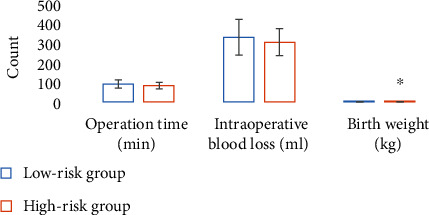
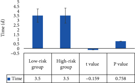
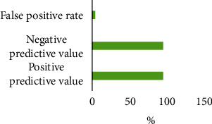
This study was to explore the value of the deep dictionary learning algorithm in constructing a B ultrasound scoring system and exploring its application in the clinical diagnosis and treatment of pernicious placenta previa (PPP). 60 patients with PPP were divided into a low-risk group (severe, implantable) and high-risk group (adhesive, penetrating) according to their clinical characteristics, B ultrasound imaging characteristics, and postpartum pathological examination results. Under PPP ultrasonic image information using the deep learning algorithm, the B ultrasound image diagnostic scoring system was established to predict the depth of various types of placenta accreta. The results showed that the cut-off values of severe, implantable, adhesive, and penetrating types were <2.3, 2.3-6.5, 6.5-9, and ≥9 points, respectively; there were significant differences in the termination of pregnancy and neonatal birth weight between the two groups ( P < 0.05 ); the positive predictive value, negative predictive value, and false positive rate of ultrasound images based on the deep dictionary learning algorithm for PPP were 95.33%, 94.89%, and 3.56%, respectively. Thus, the ultrasound image diagnostic scoring system based on the deep learning algorithm has an important predictive role for PPP, which can provide a more targeted diagnosis and treatment plan for patients in clinical practice and improve the prediction and treatment efficiency.
Overview
The claims and statistics of this paper haven’t been checked yet.
Free: it reads the open-access text and checks the first 5 claims that cite a source we can check. Create a free account to check every claim.
Integrity signals
Registry
Cites
Statistics
Figures



Verification
Claims the paper makes
The claims and statistics of this paper haven’t been checked yet.
Free: it reads the open-access text and checks the first 5 claims that cite a source we can check. Create a free account to check every claim.
Claims other papers make about this one
Each is checked against this paper’s own text: does it say what the citing paper says it does?
The source backs part of the claim, not all of it — or backs it for a different population, condition or comparison.
In the citing paper
“These results suggest that deep dictionary learning offers a viable and more accurate alternative to manual ultrasound diagnosis, which typically yields PPVs between 82% and 93% [23].”
From Machine Learning in Maternity: Early Diagnosis of Placenta Accreta Spectrum (2026) · cites this paper as [23]
Experimental — accuracy not yet measuredThe source backs part of the claim, not all of it — or backs it for a different population, condition or comparison.
In the citing paper
“The highlighted studies [20,21,23] focused on specific demographic groups, limiting the generalizability of their results to broader or underrepresented populations.”
From Machine Learning in Maternity: Early Diagnosis of Placenta Accreta Spectrum (2026) · cites this paper as [23]
Experimental — accuracy not yet measuredThe source backs part of the claim, not all of it — or backs it for a different population, condition or comparison.
In the citing paper
“Yang et al., therefore, investigated the predictive role of a scoring system for the occurrence of pernicious placenta previa [121]”
From Evolving the Era of 5D Ultrasound? A Systematic Literature Review on the Applications for Artificial Intelligence Ultrasound Imaging in Obstetrics and Gynecology (2023) · cites this paper as [121]
Experimental — accuracy not yet measuredThe cited source says what the claim attributes to it.
In the citing paper
“The positive predictive value (PPV), negative predictive value (NPV), and false positive rate (FPR) of ultrasound image information based on the deep dictionary learning algorithm were 95.33%, 94.89%, and 3.56%, respectively [23].”
From Machine Learning in Maternity: Early Diagnosis of Placenta Accreta Spectrum (2026) · cites this paper as [23]
Self-check
Percentages, sums and reported differences re-computed from the paper’s own numbers.
The self-check needs the paper’s full text.
Citations
Cites
Emerging role of deep learning‐based artificial intelligence in tumor pathology
Jiang, Y., Yang, M., Wang, S., Li, X., & Sun, Y. (2020) · Cancer Communications · doi:10.1002/cac2.12012 · Cited by 438 · cited 1×
“In order to improve the prediction of the dangers of prenatal diagnosis of PPP, foreign scholars found that ultrasound has a high positive and negative predictive value for placenta accreta; domestic scholars proposed the concept of the ultrasound scoring system for placenta accreta and pointed out that an ultrasound…”
BackgroundTang, H., Liu, H., Wei, X., & Sebe, N. (2020) · IEEE Transactions on Neural Networks and Learning Systems · doi:10.1109/tnnls.2020.2997289 · Cited by 119 · cited 1×
“As one of the algorithms in the field of machine learning, deep learning is a deep neural network with multiple hidden layers [14].”
BackgroundDeep learning in magnetic resonance image reconstruction
Chandra, S. S., Lorenzana, M. B., Liu, X., Liu, S., Bollmann, S., & Crozier, . (2021) · Journal of Medical Imaging and Radiation Oncology · doi:10.1111/1754-9485.13276 · Cited by 75 · cited 1×
“Deep dictionary learning combines the advantages of deep learning and dictionary learning, constructs a deep structure through multilevel dictionary learning, and constitutes a deep dictionary learning model to learn sample data to obtain a dictionary [15].”
BackgroundEarly development of the human placenta and pregnancy complications
Haram, K., Mortensen, J. H. S., Myking, O. L., Roald, B., Magann, E. F., & Morrison, J. C. (2019) · The Journal of Maternal-Fetal & Neonatal Medicine · doi:10.1080/14767058.2019.1578745 · Cited by 50 · cited 1×
“In PPP patients with placenta accreta, the most common maternal complication is postpartum hemorrhage, intraoperative hemorrhage will exceed 3,000mL in 90% of parturients, and hemorrhage greater than 10,000mL is not uncommon, causing a series of serious complications if not effectively controlled, such as hemorrhagic…”
BackgroundZaghal, A. A., Hussain, H. K., & Berjawi, G. (2019) · Journal of Magnetic Resonance Imaging · doi:10.1002/jmri.26764 · Cited by 45 · cited 1×
“3% of the perinatal hysterectomy rate, and the maternal mortality rate is as high as 7%, and the main complications of newborns are iatrogenic preterm delivery, stillbirth, and asphyxia [8].”
BackgroundChen, C., Liu, X., Chen, D., Huang, S. M., Yan, X., Liu, H., Chang, Q., & Liang, Z. (2019) · Annals of Palliative Medicine · doi:10.21037/apm.2019.09.04 · Cited by 39 · cited 1×
“History of previous cesarean section and placenta previa are the main risk factors for PPP with placenta accreta [4].”
BackgroundAbinader, R., Macdisi, N., Moudden, I. E., & Abuhamad, A. (2021) · Ultrasound in Obstetrics and Gynecology · doi:10.1002/uog.24828 · Cited by 36 · cited 1×
“The image reconstruction is mainly through learning model training sample data to obtain the dictionary and using the dictionary obtained by model learning to represent the input image to complete the process of image reconstruction [16].”
MethodBlumenthal, P. D., Lerma, K., Bhamrah, R., & Singh, S. (2018) · Contraception · doi:10.1016/j.contraception.2018.04.019 · Cited by 35 · cited 1×
“The researchers developed the ultrasound scoring scale for placenta accreta and proposed that the ultrasound evaluation thresholds for the three types of placenta accreta were adhesive type < 3 points, implantable type ≥ 3 points, and penetrating type ≥ 10 points [21].”
BackgroundZheng, W., Zhang, H., Ma, J., Dou, R., Zhao, X., Yan, J., & Yang, H. (2021) · The Journal of Maternal-Fetal & Neonatal Medicine · doi:10.1080/14767058.2020.1847077 · Cited by 33 · cited 1×
“In PPP patients with placenta accreta, the most common maternal complication is postpartum hemorrhage, intraoperative hemorrhage will exceed 3,000mL in 90% of parturients, and hemorrhage greater than 10,000mL is not uncommon, causing a series of serious complications if not effectively controlled, such as hemorrhagic…”
BackgroundGuo, P. C., Wu, Y., Yuan, X., & Wan, Z. (2021) · Annals of Palliative Medicine · doi:10.21037/apm-21-1285 · Cited by 28 · cited 1×
“Prenatal diagnosis of PPP is easier to make by ultrasound and magnetic resonance imaging (MRI), but whether it is associated with placenta accreta has been the focus and difficulty of clinical diagnosis [10].”
Background
Cited by
Recent trend in medical imaging modalities and their applications in disease diagnosis: a review
Abhisheka, B., Biswas, S. K., Purkayastha, B., Das, D., & Escargueil, A. (2023) · Multimedia Tools and Applications · doi:10.1007/s11042-023-17326-1 · Cited by 169
“Yang et al. [78] have tested the feasibility of the DL algorithm in the development of B ultrasonography rating systems, clinical diagnosis, and therapy of Pernicious Placenta Previa (PPP).”
Jost, E., Kosian, P., Jimenez Cruz, J., Albarqouni, S., Gembruch, U., Strizek, B., & Recker, F. (2023) · Journal of Clinical Medicine · doi:10.3390/jcm12216833 · Cited by 44
“Yang et al., therefore, investigated the predictive role of a scoring system for the occurrence of pernicious placenta previa [121], while the research group of Asadpour et al. reported a method for identifying placental abruption [122].”
MethodTadepalli, K., Das, A., Meena, T., & Roy, S. S. (2025) · Computer Methods and Programs in Biomedicine · doi:10.1016/j.cmpb.2025.108682 · Cited by 18
Deep Learning in Ultrasound Localization Microscopy
Yuhang, Z., & Jianqiao, Z. (2024) · Advanced ultrasound in diagnosis and therapy · doi:10.37015/audt.2024.240023 · Cited by 5
Wang, C., & Wang, Z. (2024) · Medicine · doi:10.1097/md.0000000000037531 · Cited by 4
“Based on this, it is believed that in clinical practice, to avoid missed diagnosis or misdiagnosis to the greatest extent, conventional ultrasound and ultrasound scoring systems can be integrated to carry out inspection and evaluation for patients with early scar uterine PA. [19–21]”
Background
Metadata
- Authors
- Yang, X., Chen, Z., & Jia, X.
- Affiliations
- Hunan Provincial Maternal and Child Health Hospital (CN)
- The First Hospital of Changsha (CN)
- Corresponding author
- Jia, Xiaozhou
- Venue
- Computational and Mathematical Methods in Medicine
- Publisher
- Wiley
- Published
- 2022-06-06
- Volume
- 2022
- Pages
- 1-9
- ISSN
- 1748-670X (ISSN-L)
- 1748-6718
- PMC
- PMC9192257
- OpenAlex
- W4282011581
- Cited by
- 9
- References
- 22
- Citations by year
- 2026: 2 · 2025: 3 · 2024: 1 · 2023: 2 · 2022: 1
- FWCI
- 1.92
- Type
- journal article
- Language
- en
- Open access
- hybrid
- doi.org
- Locations
- Computational and Mathematical Methods in Medicine (publishedVersion, cc-by)
- PubMed Central (publishedVersion, cc-by)
- PubMed (publishedVersion)
- Funders
- Hunan Provincial Health and Family Planning Commission (B20180199)
- Topics
- Health Sciences › Medicine › Pediatrics, Perinatology and Child Health › Maternal and fetal healthcare
- Health Sciences › Medicine › Health Informatics › Artificial Intelligence in Healthcare and Education
- Health Sciences › Health Professions › Health Information Management › Artificial Intelligence in Healthcare
- Keywords
- pernicious placenta previa, placenta accreta, deep dictionary learning, B-mode ultrasound, ultrasound scoring system, perinatal outcomes, abortion, birth weight, diagnostic accuracy
- MeSH terms
- Deep Learning, Female, Humans, Infant, Newborn, Placenta Accreta, Placenta Previa, Pregnancy, Retrospective Studies, Ultrasonography, Ultrasonography, Prenatal
About these checks
Registry, retraction, Cites and statistics results are computed, not judged, with one exception: a printed reference that no index lists is read once by a model before it counts as a citation error. Claims are extracted by a model and kept only when their quote appears verbatim in the text; each cited source is then checked, with a model’s help, for whether it says what the claim relies on. Those verdicts can be wrong — read the claim’s quote and the cited source before relying on one.
Statistics and arithmetic are recomputed, not judged.
Confidence bands are shown only for fields calibrated on our annotated test set. None is calibrated yet, so no band is shown.
What counts as a claim ›How we measure confidence ›How we check this ›
Are you an author, or do you see a problem with this page? Request a review
How to cite
Cite CoChat for the checks on this page, and the paper for its own findings.
- This page
- CoChat. (2026). Citation check: Deep Learning Algorithm-Based Ultrasound Image Information in Diagnosis and Treatment of Pernicious Placenta Previa. https://app.cochat.ai/papers/10.1155/2022/3452176
- The paper
- Yang, X., Chen, Z., & Jia, X. (2022). Deep Learning Algorithm-Based Ultrasound Image Information in Diagnosis and Treatment of Pernicious Placenta Previa. Computational and Mathematical Methods in Medicine. https://doi.org/10.1155/2022/3452176
Data sources
Bibliographic data: Semantic Scholar, OpenAlex, Crossref.
Contains information from the Semantic Scholar Academic Graph Datasets, made available under the ODC Attribution License (ODC-BY 1.0).
Semantic Scholar’s data is described in: Kinney, R., et al. (2023). The Semantic Scholar Open Data Platform. arXiv:2301.10140.
