

The DOI matches its Crossref record; 27 of the 28 references it cites were found in a registry or an index; 10 of the 25 sources its claims cite were checked for support.
Multi-centre deep learning for placenta segmentation in obstetric ultrasound with multi-observer and cross-country generalization
Open accessAbstract
The placenta is crucial to fetal well-being and it plays a significant role in the pathogenesis of hypertensive pregnancy disorders. Moreover, a timely diagnosis of placenta previa may save lives. Ultrasound is the primary imaging modality in pregnancy, but high-quality imaging depends on the access to equipment and staff, which is not possible in all settings. Convolutional neural networks may help standardize the acquisition of images for fetal diagnostics. Our aim was to develop a deep learning based model for classification and segmentation of the placenta in ultrasound images. We trained a model based on manual annotations of 7,500 ultrasound images to identify and segment the placenta. The model's performance was compared to annotations made by 25 clinicians (experts, trainees, midwives). The overall image classification accuracy was 81%. The average intersection over union score (IoU) reached 0.78. The model's accuracy was lower than experts' and trainees', but it outperformed all clinicians at delineating the placenta, IoU = 0.75 vs 0.69, 0.66, 0.59. The model was cross validated on 100 2nd trimester images from Barcelona, yielding an accuracy of 76%, IoU 0.68. In conclusion, we developed a model for automatic classification and segmentation of the placenta with consistent performance across different patient populations. It may be used for automated detection of placenta previa and enable future deep learning research in placental dysfunction.
Overview
CoChat Summary
## TL;DR
This study developed a CNN-based model to identify, classify, and segment placental tissue in routine obstetric ultrasound images using 7,500 manually annotated scans. The model achieved 81.42% image-classification accuracy, 0.78 average IoU for segmentation, and 71% accuracy for anterior/posterior placental location, with cross-country testing in Spain showing slightly lower accuracy but similar F1 and IoU.
## Key findings
- On the full test set, image classification accuracy for detecting placental tissue was 81.42%, and classification performance improved with gestational age, becoming stable from the second trimester.
- For images where placental tissue was detected, the model’s segmentation performance reached an average IoU of 0.78; in the Discussion, the authors also report a mean IoU of 0.78 for correctly identified placental tissue.
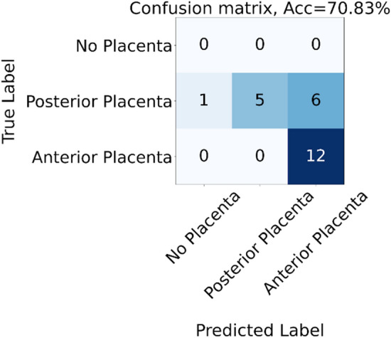
- The anterior/posterior placental-location classifier achieved 71% accuracy and performed better on anterior placentas.
- In a clinician comparison on 24 images, the model’s classification accuracy was comparable to midwives, and when it detected placental tissue it performed better and more robustly than all clinician groups in segmentation; the abstract reports IoU 0.75 for the model versus 0.69, 0.66, and 0.59 for experts, trainees, and midwives, respectively.
- External testing on 100 annotated images from Barcelona, Spain, gave 76% accuracy and IoU 0.68; in the Results section, the authors also report 0.68 ± 0.20 IoU for the 25 images where both prediction and ground truth detected placental tissue.
## Methods
- Retrospective multi-centre study using 7,500 routine obstetric ultrasound images from Copenhagen, Denmark (2018), manually annotated by the lead author for placenta presence/absence and segmentation; 500 images were additionally labeled for anterior/posterior location.
- The main model was Mask R-CNN, initialized with ImageNet-pretrained weights and trained in PyTorch on binary masks derived from annotation polygons; training used 100 epochs, Adam, and optional random augmentation.
- Evaluation included binary image classification, segmentation via Intersection over Union, comparison against 25 clinicians (5 fetal medicine experts, 10 OB-GYN residents, 10 midwives) on 24 images, and external validation on 100 annotated images from Barcelona, Spain.
## Limitations
- The authors state that only 24 images were used for the clinician comparison and 100 images for the cross-country test, which they describe as a limitation because of the small number of images.
- They state that a challenge was heterogeneous, often sub-optimal routine ultrasound data, with placental tissue sometimes visible only in a small part of the image; they note this limited image quality makes the task difficult, especially for clinicians.
- Evident from the text: the main dataset was annotated by a single lead author, so the study does not report inter-annotator reliability for the ground truth annotations.
Model-written summary. Check it against the paper before relying on it.
Integrity signals
Registry
Cites
Statistics
Figures


Verification
Claims the paper makes
8 Valid · 1 Not valid · 10 Not checked · 22 Own results and interpretations
Every source the paper cites for this claim was checked and supports it.
In the paper
“In placental insufficiency and preeclampsia cases, placental tissue often shows histological and macroscopic changes25.”
§ Multi-centre deep learning for placenta segmentation in obstetric ultrasound with multi-observer and cross-country generalization > Discussion
What the sources say
- Supportedcites [25] Burton, G. J. & Jauniaux, E. Pathophysiology of placental-derived fetal growth restriction. Am. J. Obstet. Gynecol. 218, S745–S761 (2018). source
The “Microscopic lesions” section describes histological placental lesions in fetal growth restriction, with or without preeclampsia. The “Macroscopic vascular anomalies” section describes thromboses and infarcts in these pregnancies and notes placental lesions in pregnancies complicated by uteroplacental insufficiency. The supplied passages do not give page numbers. (LLM, conf=0.89)
plainstates an association- Asserted: passes — The authors put it forward as true.
- Checkable: passes — It can be checked against evidence.
- Load-bearing: passes — The paper’s conclusions depend on it.
- Based: passes — The paper gives a reason why it holds.
- Supported
None of the sources cited for this claim have been checked yet.
In the paper
“The model performed better at recognizing the anterior placenta, which is to be expected based on previous research13.”
- Also stated in: § Discussion · plain
“we found that the model performed better on anterior placentas (Fig. 1).”
§ Multi-centre deep learning for placenta segmentation in obstetric ultrasound with multi-observer and cross-country generalization > Results > Classification of anterior/posterior placental tissue
plainstates an association- Asserted: passes — The authors put it forward as true.
- Checkable: passes — It can be checked against evidence.
- Load-bearing: unclear — It is unclear whether the paper’s conclusions depend on it.
- Based: passes — The paper gives a reason why it holds.
- Also stated in: § Discussion · plain
A result the paper reports from its own data — it cites no other source for it, so there is none to check. The quote shows it is in the paper.
In the paper
“The overall image classification accuracy was 81%.”
- Also stated in: § Results based on the entire test set · plain
“The overall image classification accuracy on the entire test set across trimesters, was 81.42%.”
- Also stated in: § Discussion · plain
“Our model identified the presence of placental tissue with an accuracy of 81%”
§ Multi-centre deep learning for placenta segmentation in obstetric ultrasound with multi-observer and cross-country generalization > Abstract
plain- Asserted: passes — The authors put it forward as true.
- Checkable: passes — It can be checked against evidence.
- Load-bearing: passes — The paper’s conclusions depend on it.
- Based: passes — The paper gives a reason why it holds.
- Also stated in: § Results based on the entire test set · plain
A result the paper reports from its own data — it cites no other source for it, so there is none to check. The quote shows it is in the paper.
In the paper
“The average intersection over union score (IoU) reached 0.78.”
- Also stated in: § Discussion · plain
“it reached an average IoU of 0.78.”
§ Multi-centre deep learning for placenta segmentation in obstetric ultrasound with multi-observer and cross-country generalization > Abstract
plain- Asserted: passes — The authors put it forward as true.
- Checkable: passes — It can be checked against evidence.
- Load-bearing: passes — The paper’s conclusions depend on it.
- Based: passes — The paper gives a reason why it holds.
- Also stated in: § Discussion · plain
A result the paper reports from its own data — it cites no other source for it, so there is none to check. The quote shows it is in the paper.
In the paper
“For the 25 images where both the prediction and the ground truth detected placental tissue, the IoU was 0.68 ± 0.20.”
- Also stated in: § Abstract · plain
“The model was cross validated on 100 2nd trimester images from Barcelona, yielding an accuracy of 76%, IoU 0.68.”
§ Multi-centre deep learning for placenta segmentation in obstetric ultrasound with multi-observer and cross-country generalization > Results > Cross-country generalization
plain- Asserted: passes — The authors put it forward as true.
- Checkable: passes — It can be checked against evidence.
- Load-bearing: passes — The paper’s conclusions depend on it.
- Based: passes — The paper gives a reason why it holds.
- Also stated in: § Abstract · plain
13 more claims
Create a free account to see all 41 claims and every source verdict.
- Probably not a substantial claim. 18
- It may state two separate claims as one. 10
Claims other papers make about this one
Each is checked against this paper’s own text: does it say what the citing paper says it does?
The source backs part of the claim, not all of it — or backs it for a different population, condition or comparison.
In the citing paper
“Andreasen et al. and Schilpzand et al. presented an effective AI algorithm for placental localization, including heterogenous data through differences in sonographers’ expertise [109], or using a previous established sweep protocol in low-resource settings [110].”
From Evolving the Era of 5D Ultrasound? A Systematic Literature Review on the Applications for Artificial Intelligence Ultrasound Imaging in Obstetrics and Gynecology (2023) · cites this paper as [109]
Experimental — accuracy not yet measuredThe cited source says what the claim attributes to it.
In the citing paper
“and low accuracy rates in the assessment of posterior wall placentas [109].”
Citations
Cites
He, K., Gkioxari, G., Dollar, P., & Girshick, R. (2017) · 2017 IEEE International Conference on Computer Vision (ICCV) · doi:10.1109/iccv.2017.322 · Cited by 29,137
High-performance medicine: the convergence of human and artificial intelligence
Topol, E. J. (2019) · Nature Medicine · doi:10.1038/s41591-018-0300-7 · Cited by 10,450 · cited 1×
“Within many fields of medical imaging, methods like convolutional neural networks (CNN) are being used to improve visual diagnostics 1 .”
Pathophysiology of placental-derived fetal growth restriction
Burton, G. J., & Jauniaux, E. (2018) · American Journal of Obstetrics and Gynecology · doi:10.1016/j.ajog.2017.11.577 · Cited by 1,070
Uterine artery Doppler at 11 + 0 to 13 + 6 weeks in the prediction of pre‐eclampsia
Plasencia, W., Maiz, N., Bonino, S., Kaihura, C., & Nicolaides, K. H. (2007) · Ultrasound in Obstetrics & Gynecology · doi:10.1002/uog.5157 · Cited by 425 · cited 1×
“Ultrasound can be used to measure Doppler velocimetry in the uterine artery, which is associated with development of preeclampsia and placental pathology 21,22 , but when it comes to morphological markers of impaired placental function, reliable indicators are scarce.”
Burgos-Artizzu, X. P., Coronado-Gutiérrez, D., Valenzuela-Alcaraz, B., Bonet-Carne, E., Eixarch, E., Crispi, F., & Gratacós, E. (2020) · Scientific Reports · doi:10.1038/s41598-020-67076-5 · Cited by 240
Placenta and Fetal Growth Restriction
SALAFIA, C. M., CHARLES, A. K., & MAAS, E. M. (2006) · Clinical Obstetrics and Gynecology · doi:10.1097/00003081-200606000-00007 · Cited by 235 · cited 1×
“In particular, hypertensive pregnancy disorders, compromised fetal growth, and fetal death often stem from placental dysfunction— either due to insufficient initial placentation or diminishing placental function with increasing gestational age 14,15 .”
Allen, V. M., Joseph, K., Murphy, K. E., Magee, L. A., & Ohlsson, A. (2004) · BMC Pregnancy and Childbirth · doi:10.1186/1471-2393-4-17 · Cited by 218 · cited 1×
“In particular, hypertensive pregnancy disorders, compromised fetal growth, and fetal death often stem from placental dysfunction— either due to insufficient initial placentation or diminishing placental function with increasing gestational age 14,15 .”
Sobhaninia, Z., Rafiei, S., Emami, A., Karimi, N., Najarian, K., Samavi, S., & Soroushmehr, S. M. R. (2019) · Annual International Conference of the IEEE Engineering in Medicine and Biology Society (EMBC) · doi:10.1109/embc.2019.8856981 · Cited by 155
Hafner, E., Metzenbauer, M., Höfinger, D., Stonek, F., Schuchter, K., Waldhör, T., & Philipp, K. (2006) · Ultrasound in Obstetrics & Gynecology · doi:10.1002/uog.2641 · Cited by 137 · cited 1×
“A small placenta at the first-trimester scan is associated with an increased risk of impaired fetal growth and preeclampsia 6,7 .”
Machine Learning in Fetal Cardiology: What to Expect
Garcia-Canadilla, P., Sanchez-Martinez, S., Crispi, F., & Bijnens, B. (2020) · Fetal Diagnosis and Therapy · doi:10.1159/000505021 · Cited by 123
Research works printed but not linked: 1
- Paszke, A. Pytorch: An imperative style, high-performance deep learning library. Adv. Neural Inf. Process. Syst. 2019, 8026–8037 (2019).
Cited by
Jost, E., Kosian, P., Jimenez Cruz, J., Albarqouni, S., Gembruch, U., Strizek, B., & Recker, F. (2023) · Journal of Clinical Medicine · doi:10.3390/jcm12216833 · Cited by 44
“Andreasen et al. and Schilpzand et al. presented an effective AI algorithm for placental localization, including heterogenous data through differences in sonographers’ expertise [109], or using a previous established sweep protocol in low-resource settings [110].”
Johnsson, V. L., Svendsen, M. B. S., Kulasegaram, K. M., Sundberg, K., Tiblad, E., Herling, L., Petersen, O. B., & Tolsgaard, M. G. (2023) · Medical Education · doi:10.1111/medu.15190 · Cited by 34
Self-citationPredicting abnormal fetal growth using deep learning
Mikolaj, K. W., Christensen, A. N., Taksøe‐Vester, C., Feragen, A., Petersen, O. B., Lin, M., Nielsen, M., Svendsen, M. B. S., & Tolsgaard, M. G. (2025) · npj Digital Medicine · doi:10.1038/s41746-025-01704-0 · Cited by 26
Self-citationFerreira, I., Simões, J., Pereira, B., Correia, J. V. M., & Areia, A. L. (2024) · Scientific Reports · doi:10.1038/s41598-024-65394-6 · Cited by 16
“This has already instituted clinical applications in fetal imaging, including echocardiography and neurosonography 15,20 .”
Analytical study of the encoder-decoder models for ultrasound image segmentation
Srivastava, S., Vidyarthi, A., & Jain, S. (2023) · Service Oriented Computing and Applications · doi:10.1007/s11761-023-00373-9 · Cited by 14
Metadata
- Authors
- Andreasen, L. A., Feragen, A., Christensen, A. N., Thybo, J. K., Svendsen, M. B. S., Zepf, K., Lekadir, K., & Tolsgaard, M. G.
- Affiliations
- Rigshospitalet (DK)
- Technical University of Denmark (DK)
- Universitat de Barcelona (ES)
- Copenhagen University Hospital (DK)
- Corresponding author
- Andreasen, Lisbeth Anita
- ORCID
- Venue
- Scientific Reports
- Published
- 2023-02-08
- Volume
- 13
- Issue
- 1
- Pages
- 2221-2221
- ISSN
- 2045-2322 (ISSN-L)
- PMC
- PMC9908915
- OpenAlex
- W4319597860
- Semantic Scholar
- 606041dfb065810f706fef7ca2555570fd2c64ce
- Cited by
- 28
- References
- 28
- Citations by year
- 2026: 9 · 2025: 9 · 2024: 7 · 2023: 3
- FWCI
- 2.53
- Type
- journal article
- Language
- en
- Open access
- gold
- doi.org
- Licence
- cc-by
- creativecommons.org/licenses/by/4.0 (tdm)
- creativecommons.org/licenses/by/4.0 (vor)
- Locations
- Scientific Reports (publishedVersion, cc-by)
- PubMed Central (publishedVersion, cc-by)
- PubMed (publishedVersion)
- Research at the University of Copenhagen (University of Copenhagen) (publishedVersion, cc-by)
- DOAJ (DOAJ: Directory of Open Access Journals) (submittedVersion, cc-by-sa)
- Technical University of Denmark, DTU Orbit (Technical University of Denmark, DTU) (submittedVersion, cc-by)
- Funders
- The Capital Region Research Fund
- Topics
- Physical Sciences › Computer Science › Computer Vision and Pattern Recognition › Medical Image Segmentation Techniques
- Health Sciences › Medicine › Health Informatics › Artificial Intelligence in Healthcare and Education
- Physical Sciences › Computer Science › Computer Vision and Pattern Recognition › Advanced Neural Network Applications
- Keywords
- placenta segmentation, prenatal ultrasound, placenta previa, second trimester, hypertensive disorders of pregnancy, placental insufficiency
- MeSH terms
- Deep Learning, Female, Humans, Image Processing, Computer-Assisted, Placenta, Placenta Previa, Pregnancy, Ultrasonography, Prenatal
About these checks
Registry, retraction, Cites and statistics results are computed, not judged, with one exception: a printed reference that no index lists is read once by a model before it counts as a citation error. Claims are extracted by a model and kept only when their quote appears verbatim in the text; each cited source is then checked, with a model’s help, for whether it says what the claim relies on. Those verdicts can be wrong — read the claim’s quote and the cited source before relying on one.
The claim verdicts this report counts are experimental: their accuracy is not yet measured.
A language model drew the claims from the paper on 2026-10-10, under version 3 of the claim definition (model deepseek-v4.1-flash, prompt extract-v3).
A second model labelled each claim on 2026-10-10 (model jev-1.13-20260917, definition revision gold-guideline-v0.2).
Cited sources are checked with a language model’s help (gpt-6-sol).
Statistics and arithmetic are recomputed, not judged.
Confidence bands are shown only for fields calibrated on our annotated test set. None is calibrated yet, so no band is shown.
What counts as a claim ›How we measure confidence ›How we check this ›
Are you an author, or do you see a problem with this page? Request a review
How to cite
Cite CoChat for the checks on this page, and the paper for its own findings.
- This page
- CoChat. (2026). Citation check: Multi-centre deep learning for placenta segmentation in obstetric ultrasound with multi-observer and cross-country generalization. https://app.cochat.ai/papers/10.1038/s41598-023-29105-x
- The paper
- Andreasen, L. A., Feragen, A., Christensen, A. N., Thybo, J. K., Svendsen, M. B. S., Zepf, K., Lekadir, K., & Tolsgaard, M. G. (2023). Multi-centre deep learning for placenta segmentation in obstetric ultrasound with multi-observer and cross-country generalization. Scientific Reports. https://doi.org/10.1038/s41598-023-29105-x
Data sources
Bibliographic data: Semantic Scholar, OpenAlex, Crossref.
Contains information from the Semantic Scholar Academic Graph Datasets, made available under the ODC Attribution License (ODC-BY 1.0).
Semantic Scholar’s data is described in: Kinney, R., et al. (2023). The Semantic Scholar Open Data Platform. arXiv:2301.10140.
