

The DOI matches its Crossref record; 73 of the 74 references it cites were found in a registry or an index.
Combined Inhibition of SHP2 and CXCR1/2 Promotes Antitumor T-cell Response in NSCLC
Open accessPublished version of a preprint
Abstract
SHP2 inhibitors (SHP2i) alone and in various combinations are being tested in multiple tumors with overactivation of the RAS/ERK pathway. SHP2 plays critical roles in normal cell signaling; hence, SHP2is could influence the tumor microenvironment. We found that SHP2i treatment depleted alveolar and M2-like macrophages, induced tumor-intrinsic CCL5/CXCL10 secretion, and promoted B and T lymphocyte infiltration in Kras- and Egfr-mutant non–small cell lung cancer (NSCLC). However, treatment also increased intratumor granulocytic myeloid-derived suppressor cells (gMDSC) via tumor-intrinsic, NFκB-dependent production of CXCR2 ligands. Other RAS/ERK pathway inhibitors also induced CXCR2 ligands and gMDSC influx in mice, and CXCR2 ligands were induced in tumors from patients on KRASG12C inhibitor trials. Combined SHP2 (SHP099)/CXCR1/2 (SX682) inhibition depleted a specific cluster of S100a8/9hi gMDSCs, generated Klrg1+ CD8+ effector T cells with a strong cytotoxic phenotype but expressing the checkpoint receptor NKG2A, and enhanced survival in Kras- and Egfr-mutant models. Our results argue for testing RAS/ERK pathway/CXCR1/2/NKG2A inhibitor combinations in patients with NSCLC. Significance: Our study shows that inhibiting the SHP2/RAS/ERK pathway triggers NFκB-dependent upregulation of CXCR2 ligands and recruitment of S100A8hi gMDSCs, which suppress T cells. Combining SHP2/CXCR2 inhibitors blocks gMDSC immigration, resulting in enhanced Th1 polarization, induced CD8+KLRG1+ effector T cells with high cytotoxic activity, and improved survival in multiple NSCLC models. This article is highlighted in the In This Issue feature, p. 1
Overview
The claims and statistics of this paper haven’t been checked yet.
Run the check — free, no account needed. It reads the open-access text and checks the sources it cites.
Integrity signals
Registry
Cites
Statistics
Figures
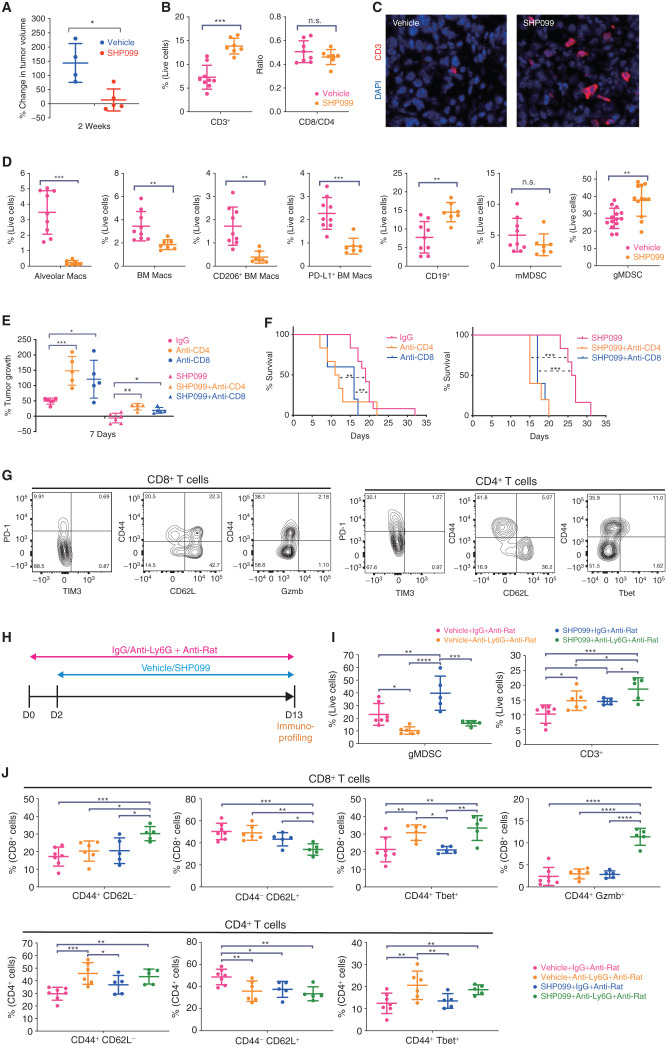
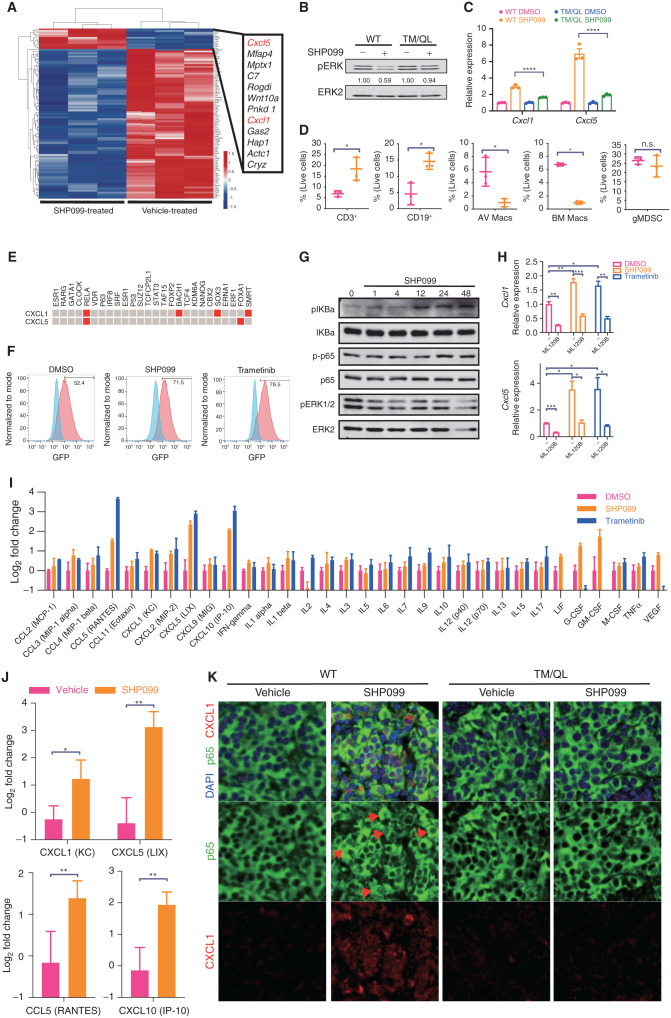
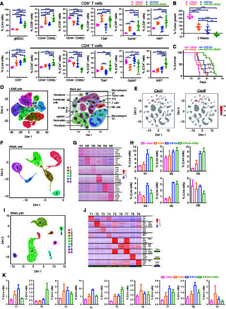
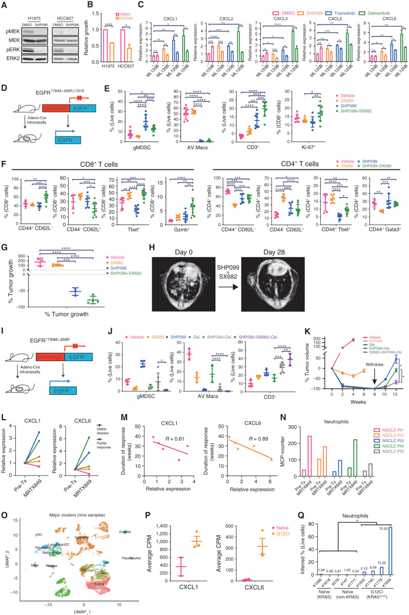
Verification
Claims the paper makes
The claims and statistics of this paper haven’t been checked yet.
Run the check — free, no account needed. It reads the open-access text and checks the sources it cites.
Claims other papers make about this one
Each is checked against this paper’s own text: does it say what the citing paper says it does?
The source backs part of the claim, not all of it — or backs it for a different population, condition or comparison.
In the citing paper
“The mice were not cured, however, potentially owing to T cell exhaustion mediated by PD-1 and/or by two other inhibitory immune-checkpoint receptors, NKG2A and/or KLRG1, which were found to be expressed on the CD8+ T cells.”
From The current state of the art and future trends in RAS-targeted cancer therapies (2022) · cites this paper as [159]
Experimental — accuracy not yet measuredThe cited source says what the claim attributes to it.
In the citing paper
“As noted previously, Tang et al.159 discovered that RAS–MAPK pathway inhibition can result in CXCL1/CXCL5-dependent infiltration of KRAS-mutant NSCLCs by gMDSCs, depletion of which markedly increases accumulation of cytotoxic CD8+ T cells in the TME.”
From The current state of the art and future trends in RAS-targeted cancer therapies (2022) · cites this paper as [159]
In the citing paper
“Using their mouse models, these researchers also demonstrated that tumour control was markedly improved by combining SHP2 and/or EGFR inhibitors with SX-682, a novel oral small-molecule inhibitor of CXCR1 and CXCR2.”
From The current state of the art and future trends in RAS-targeted cancer therapies (2022) · cites this paper as [159]
In the citing paper
“Tang et al.159 also showed that adagrasib increased CXCL1 and CXCL6 mRNA levels (both encoding ligands of CXCR1/2) in KRASG12C-mutant cell lines, as well as tumour infiltration by gMDSCs in preclinical models.”
From The current state of the art and future trends in RAS-targeted cancer therapies (2022) · cites this paper as [159]
In the citing paper
“These findings suggest that oncogenic KRAS promotes immune escape by fostering an immunosuppressive TME that can be mitigated through SHP2 inhibition — albeit subject to resistance mediated by CXCR2 signalling, which could potentially be a target of combinatorial strategies159”
From The current state of the art and future trends in RAS-targeted cancer therapies (2022) · cites this paper as [159]
In the citing paper
“Furthermore, analyses of matched pre-treatment and post-treatment biopsy samples provided evidence of these effects of adagrasib in patients with NSCLC159, highlighting the promise of combining CXCR1/2 inhibitors with KRAS inhibition.”
From The current state of the art and future trends in RAS-targeted cancer therapies (2022) · cites this paper as [159]
In the citing paper
“Tang et al.159 found that in genetically engineered mouse models, SHP2 inhibition in KRAS-mutated or EGFR-mutated NSCLCs depleted alveolar and M2-like macrophage populations, increased T cell and B cell infiltration, but also enhanced accumulation of potently immunosuppressive granulocytic MDSCs (gMDSCs).”
From The current state of the art and future trends in RAS-targeted cancer therapies (2022) · cites this paper as [159]
In the citing paper
“Across all time points in both models, the largest overlap of deregulated genes, which included genes associated with the MAPK family, tumor growth and cancer progression28–30, was identified between adagrasib plus BI-3406 and adagrasib plus TNO155 combination treatments, suggesting similarly induced transcriptomic modulation (Fig. 4a, Extended Data Fig. 4 and Supplementary Tables 6 and 7).”
From Co-targeting SOS1 enhances the antitumor effects of KRASG12C inhibitors by addressing intrinsic and acquired resistance (2024) · cites this paper as [30]
Self-check
Percentages, sums and reported differences re-computed from the paper’s own numbers.
The self-check needs the paper’s full text.
Citations
Cites
Enrichr: a comprehensive gene set enrichment analysis web server 2016 update
Kuleshov, M. V., Jones, M. R., Rouillard, A. D., Fernandez, N. F., Duan, Q., Wang, Z., Koplev, S., Jenkins, S. L., Jagodnik, K. M., Lachmann, A., et al. (2016) · Nucleic Acids Research · doi:10.1093/nar/gkw377 · Cited by 10,799
Enrichr: interactive and collaborative HTML5 gene list enrichment analysis tool
Chen, E. Y., Tan, C. M., Kou, Y., Duan, Q., Wang, Z., Meirelles, G. V., Clark, N. R., & Ma’ayan, A. (2013) · BMC Bioinformatics · doi:10.1186/1471-2105-14-128 · Cited by 7,814
Myeloid-derived-suppressor cells as regulators of the immune system
Gabrilovich, D. I., & Nagaraj, S. (2009) · Nature Reviews Immunology · doi:10.1038/nri2506 · Cited by 6,753
Pembrolizumab plus chemotherapy in metastatic non–small-cell lung cancer
Gandhi, L., Rodríguez-Abreu, D., Gadgeel, S., Esteban, E., Felip, E., De Angelis, F., Domine, M., Clingan, P., Hochmair, M. J., Powell, S. F., et al. (2018) · New England Journal of Medicine · doi:10.1056/nejmoa1801005 · Cited by 6,607
Graph drawing by force‐directed placement
Fruchterman, T. M. J., & Reingold, E. M. (1991) · Software: Practice and Experience · doi:10.1002/spe.4380211102 · Cited by 6,352
Pembrolizumab for the Treatment of Non–Small-Cell Lung Cancer
Garon, E. B., Rizvi, N. A., Hui, R., Leighl, N., Balmanoukian, A. S., Eder, J. P., Patnaik, A., Aggarwal, C., Gubens, M., Horn, L., et al. (2015) · New England Journal of Medicine · doi:10.1056/nejmoa1501824 · Cited by 6,198
Recommendations for myeloid-derived suppressor cell nomenclature and characterization standards
Bronte, V., Brandau, S., Chen, S.-H., Colombo, M. P., Frey, A. B., Greten, T. F., Mandruzzato, S., Murray, P. J., Ochoa, A., Ostrand-Rosenberg, S., et al. (2016) · Nature Communications · doi:10.1038/ncomms12150 · Cited by 2,751
B cells and tertiary lymphoid structures promote immunotherapy response
Helmink, B. A., Reddy, S. M., Gao, J., Zhang, S., Basar, R., Thakur, R., Yizhak, K., Sade-Feldman, M., Blando, J., Han, G., et al. (2020) · Nature · doi:10.1038/s41586-019-1922-8 · Cited by 2,731
Macrophages as regulators of tumour immunity and immunotherapy
DeNardo, D. G., & Ruffell, B. (2019) · Nature reviews. Immunology · doi:10.1038/s41577-019-0127-6 · Cited by 2,619
Tertiary lymphoid structures improve immunotherapy and survival in melanoma
Cabrita, R., Lauss, M., Sanna, A., Donia, M., Skaarup Larsen, M., Mitra, S., Johansson, I., Phung, B., Harbst, K., Vallon-Christersson, J., et al. (2020) · Nature · doi:10.1038/s41586-019-1914-8 · Cited by 2,374
Cited by
Sorted by citations
The current state of the art and future trends in RAS-targeted cancer therapies
Punekar, S. R., Velcheti, V., Neel, B. G., & Wong, K.-K. (2022) · Nature Reviews Clinical Oncology · doi:10.1038/s41571-022-00671-9 · Cited by 549
Self-citationTherapeutic targeting of tumour myeloid cells
Barry, S. T., Gabrilovich, D. I., Sansom, O. J., Campbell, A. D., & Morton, J. P. (2023) · Nature Reviews Cancer · doi:10.1038/s41568-022-00546-2 · Cited by 305
CXCR2 inhibition enables NASH-HCC immunotherapy
Leslie, J., Mackey, J. B. G., Jamieson, T. J., Ramon‐Gil, E., Drake, T. M., Fercoq, F., Clark, W. A., Gilroy, K. L., Hedley, A., Nixon, C., et al. (2022) · Gut · doi:10.1136/gutjnl-2021-326259 · Cited by 230
Myeloid-derived suppressor cells in cancer: therapeutic targets to overcome tumor immune evasion
Lu, J., Luo, Y., Rao, D., Wang, T., Lei, Z., Chen, X., Zhang, B., Li, Y., Liu, B., Xia, L., et al. (2024) · Experimental Hematology and Oncology · doi:10.1186/s40164-024-00505-7 · Cited by 194
Exploiting the therapeutic implications of KRAS inhibition on tumor immunity
Molina-Arcas, M., & Downward, J. (2024) · Cancer Cell · doi:10.1016/j.ccell.2024.02.012 · Cited by 152
Metadata
- Authors
- Tang, K. H., Li, S., Khodadadi-Jamayran, A., Jen, J., Han, H., Guidry, K., Chen, T., Hao, Y., Fedele, C., Zebala, J. A., Maeda, D. Y., Christensen, J. G., Olson, P., Athanas, A., Loomis, C. A., Tsirigos, A., Wong, K.-K., & Neel, B. G.
- Affiliations
- NYU Langone Health (US)
- 2Applied Bioinformatics Laboratories, Office of Science and Research, New York University Grossman School of Medicine, New York, New York.
- New York University (US)
- 3Syntrix Pharmaceuticals, Auburn, Washington.
- Mirati Therapeutics (United States) (US)
- 5Monoceros Biosystems, Inc., San Diego, California.
- Corresponding author
- Tang, Kwan Ho
- Wong, Kwok-Kin
- Neel, Benjamin G.
- ORCID
- Venue
- Cancer Discovery
- Published
- 2021-08-05
- Volume
- 12
- Issue
- 1
- Pages
- 47-61
- ISSN
- 2159-8274 (ISSN-L)
- 2159-8290
- PMC
- PMC8758507
- OpenAlex
- W3191958191
- Cited by
- 105
- References
- 74
- Citations by year
- 2026: 11 · 2025: 25 · 2024: 21 · 2023: 28 · 2022: 18 · 2021: 2
- FWCI
- 6.28
- Type
- journal article
- Language
- en
- Open access
- hybrid
- doi.org
- Licence
- Locations
- Cancer Discovery (publishedVersion, cc-by-nc-nd)
- bioRxiv (Cold Spring Harbor Laboratory) (acceptedVersion, cc-by-nc-nd)
- PubMed Central (publishedVersion, cc-by-nc-nd)
- PubMed (publishedVersion)
- Funders
- National Institutes of Health (CA49152, CA248896, CA016087)
- Topics
- Health Sciences › Medicine › Oncology › Cancer Immunotherapy and Biomarkers
- Life Sciences › Biochemistry, Genetics and Molecular Biology › Molecular Biology › Protein Tyrosine Phosphatases
- Life Sciences › Immunology and Microbiology › Immunology › Immune cells in cancer
- Keywords
- SHP2 inhibition, non-small cell lung cancer, Ras/ERK pathway, M2 macrophages, granulocytic myeloid-derived suppressor cells, S100A8/S100A9, NKG2A, NF-κB signaling, KRAS mutation, EGFR mutation, combination therapy, antitumor T-cell responses, Th1 polarization
- MeSH terms
- Animals, Antineoplastic Agents, Carcinoma, Non-Small-Cell Lung, Enzyme Inhibitors, Humans, Lung Neoplasms, Male, Mice, Inbred C57BL, Xenograft Model Antitumor Assays, Receptors, Interleukin-8B, Cell Line, Tumor, Mice, Protein Tyrosine Phosphatase, Non-Receptor Type 11, Tumor Microenvironment
About these checks
Registry, retraction, Cites and statistics results are computed, not judged, with one exception: a printed reference that no index lists is read once by a model before it counts as a citation error. Claims are extracted by a model and kept only when their quote appears verbatim in the text; each cited source is then checked, with a model’s help, for whether it says what the claim relies on. Those verdicts can be wrong — read the claim’s quote and the cited source before relying on one.
Statistics and arithmetic are recomputed, not judged.
Confidence bands are shown only for fields calibrated on our annotated test set. None is calibrated yet, so no band is shown.
What counts as a claim ›How we measure confidence ›How we check this ›
Are you an author, or do you see a problem with this page? Request a review
