

The DOI matches its Crossref record; 9 of the 9 references it cites were found in a registry or an index.
NI-3201 Is a Bispecific Antibody Mediating PD-L1–Dependent CD28 Co-stimulation on T Cells for Enhanced Tumor Control
Open accessPublished version of a preprint
Abstract
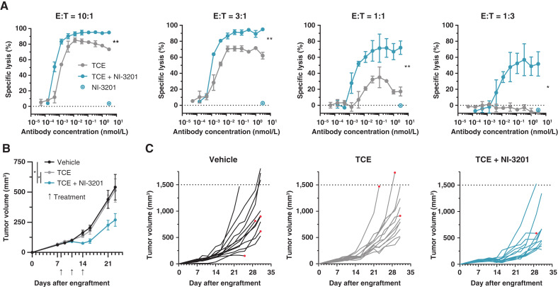
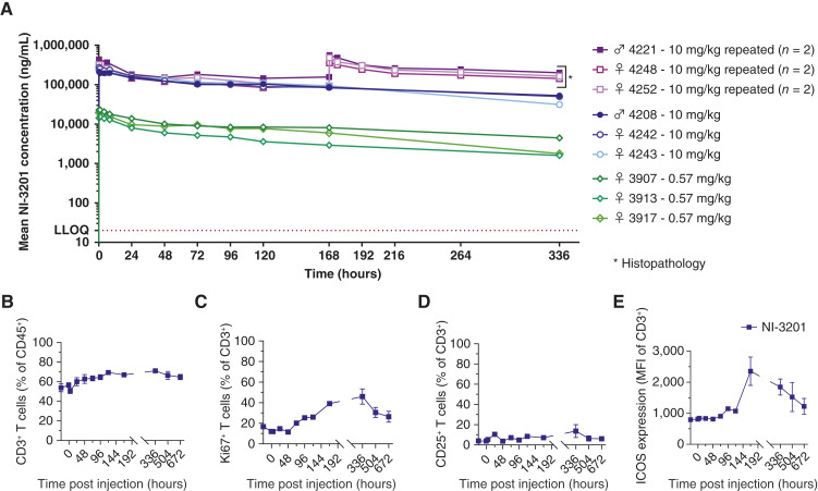
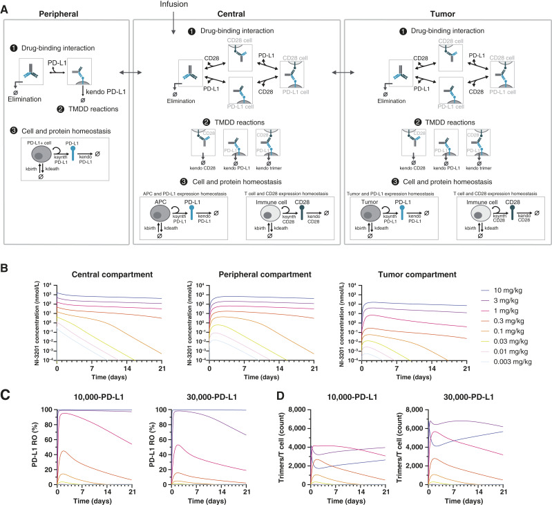
Despite advances in cancer immunotherapy, such as targeting the PD-1/PD-L1 axis, a substantial number of patients harbor tumors that are resistant or relapse. Selective engagement of T-cell co-stimulatory molecules with bispecific antibodies may offer novel therapeutic options by enhancing signal 1–driven activation occurring via T-cell receptor engagement. In this study, we report the development and preclinical characterization of NI-3201, a PD-L1×CD28 bispecific antibody generated on the κλ-body platform that was designed to promote T-cell activity and antitumor function through a dual mechanism of action. We confirmed that NI-3201 blocks the PD-L1/PD-1 immune checkpoint pathway and conditionally provides T-cell co-stimulation via CD28 (signal 2) when engaging PD-L1+ tumors or immune cells. In systems with signal 1–primed T cells, NI-3201 enhanced potent effector functionality: in vitro through antigen-specific recall assays with cytomegalovirus-specific T cells and in vivo by inducing tumor regression and immunologic memory in tumor-associated antigen–expressing MC38 syngeneic mouse models. When T-cell engagers were used to provide synthetic signal 1, the combination with NI-3201 resulted in synergistic T cell–dependent cytotoxicity and potent antitumor activity in two humanized mouse tumor models. Nonhuman primate safety assessments showed favorable tolerability and pharmacokinetics at pharmacologically active doses. Quantitative systems pharmacology modeling predicted that NI-3201 exposure results in antitumor activity in patients, but this remains to be investigated. Overall, this study suggests that by combining PD-L1 blockade with safe and effective CD28 co-stimulation, NI-3201 has the potential to improve cancer immunotherapy outcomes, and the clinical development of NI-3201 for PD-L1+ solid tumors is planned.
Overview
The claims and statistics of this paper haven’t been checked yet.
Run the check — free, no account needed. It reads the open-access text and checks the sources it cites.
Integrity signals
Registry
Cites
Statistics
Figures
Verification
Claims the paper makes
The claims and statistics of this paper haven’t been checked yet.
Run the check — free, no account needed. It reads the open-access text and checks the sources it cites.
Self-check
Percentages, sums and reported differences re-computed from the paper’s own numbers.
The self-check needs the paper’s full text.
Citations
Cites
Highly accurate protein structure prediction with AlphaFold
Jumper, J., Evans, R., Pritzel, A., Green, T., Figurnov, M., Ronneberger, O., Tunyasuvunakool, K., Bates, R., Žídek, A., Potapenko, A., et al. (2021) · Nature · doi:10.1038/s41586-021-03819-2 · Cited by 43,764
Cytokine Storm in a Phase 1 Trial of the Anti-CD28 Monoclonal Antibody TGN1412
Suntharalingam, G., Perry, M. R., Ward, S., Brett, S. J., Castello-Cortes, A., Brunner, M. D., & Panoskaltsis, N. (2006) · New England Journal of Medicine · doi:10.1056/nejmoa063842 · Cited by 2,164 · cited 1×
“The first-in-human study of the high-affinity (1.88 nmol/L) SA anti-CD28 TGN1412 resulted in cata-strophic outcomes with cytokine release syndrome and multiorgan failure in all administered healthy volunteers (12).”
InfluentialT cell costimulatory receptor CD28 is a primary target for PD-1–mediated inhibition
Hui, E., Cheung, J., Zhu, J., Su, X., Taylor, M. J., Wallweber, H. A., Sasmal, D. K., Huang, J., Kim, J. M., Mellman, I., et al. (2017) · Science · doi:10.1126/science.aaf1292 · Cited by 1,689
A decade of immune-checkpoint inhibitors in cancer therapy
Robert, C. (2020) · Nature Communications · doi:10.1038/s41467-020-17670-y · Cited by 1,608
CD28 Costimulation: From Mechanism to Therapy
Esensten, J. H., Helou, Y. A., Chopra, G., Weiss, A., & Bluestone, J. A. (2016) · Immunity · doi:10.1016/j.immuni.2016.04.020 · Cited by 937 · cited 1×
“CD28 is one of the primary co-stimulatory receptors on T cells, and upon engagement with its ligands CD80/CD86 on antigen-presenting cells, CD28 delivers a co-stimulatory signal that synergizes with the TCR signal to promote T-cell cytokine production, survival, differentiation, and expansion (11).”
Stebbings, R., Findlay, L., Edwards, C., Eastwood, D., Bird, C., North, D., Mistry, Y., Dilger, P., Liefooghe, E., Cludts, I., et al. (2007) · The Journal of Immunology · doi:10.4049/jimmunol.179.5.3325 · Cited by 316 · cited 3×
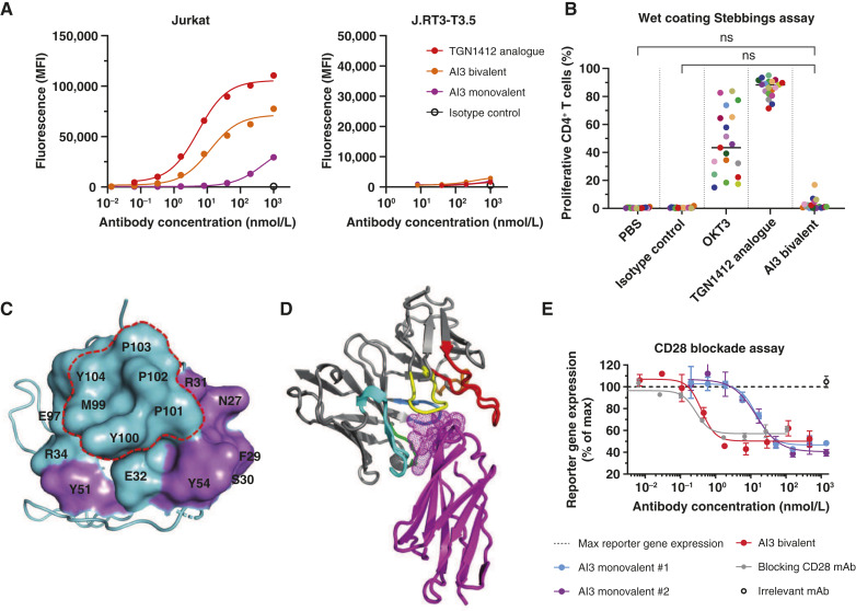
“1B ), NI-3201 had no SA in the Stebbings assay (25), independent of the way the antibody was coated onto plates (Supplementary Fig.”
InfluentialEastwood, D., Findlay, L., Poole, S., Bird, C., Wadhwa, M., Moore, M., Burns, C., Thorpe, R., & Stebbings, R. (2010) · British Journal of Pharmacology · doi:10.1111/j.1476-5381.2010.00922.x · Cited by 300 · cited 1×
“Despite documented limitations in using cynomolgus monkeys to assess the risks of CD28-driven cytokine release syndrome (66), recent NHP studies have shown a significant induction of IL6 (and a consequent increase in CRP levels) in animals that were administered a TGN1412 analogue at 10 mg/kg (14).”
Crystal structure of a soluble CD28-Fab complex
Evans, E. J., Esnouf, R. M., Manso-Sancho, R., Gilbert, R. J. C., James, J. R., Yu, C., Fennelly, J. A., Vowles, C., Hanke, T., Walse, B., et al. (2005) · Nature Immunology · doi:10.1038/ni1170 · Cited by 180
Römer, P. S., Berr, S., Avota, E., Na, S.-Y., Battaglia, M., ten Berge, I., Einsele, H., & Hünig, T. (2011) · Blood · doi:10.1182/blood-2010-12-319780 · Cited by 153
Research works printed but not linked: 58
- Sharma, 2015, The future of immune checkpoint therapy, Science, 348, 56, 10.1126/science.aaa8172 · DOI · Paper page
- Zou, 2016, PD-L1 (B7-H1) and PD-1 pathway blockade for cancer therapy: mechanisms, response biomarkers, and combinations, Sci Transl Med, 8, 328rv4, 10.1126/scitranslmed.aad7118 · DOI
- Freeman, 2000, Engagement of the PD-1 immunoinhibitory receptor by a novel B7 family member leads to negative regulation of lymphocyte activation, J Exp Med, 192, 1027, 10.1084/jem.192.7.1027 · DOI · Paper page
- Shiravand, 2022, Immune checkpoint inhibitors in cancer therapy, Curr Oncol, 29, 3044, 10.3390/curroncol29050247 · DOI · Paper page
- Topalian, 2015, Immune checkpoint blockade: a common denominator approach to cancer therapy, Cancer Cell, 27, 450, 10.1016/j.ccell.2015.03.001 · DOI · Paper page
- Zhang, 2023, Immune-checkpoint inhibitor resistance in cancer treatment: current progress and future directions, Cancer Lett, 562, 216182, 10.1016/j.canlet.2023.216182 · DOI
- Lotz, 2024, Characterization of anti-drug antibody responses to the T-cell engaging bispecific antibody cibisatamab to understand the impact on exposure, Front Immunol, 15, 1406353, 10.3389/fimmu.2024.1406353 · DOI
- Lim, 2024, Agonist antibodies for cancer immunotherapy: history, hopes, and challenges, Clin Cancer Res, 30, 1712, 10.1158/1078-0432.CCR-23-1014 · DOI
- Lotze, 2024, CD28 co-stimulation: novel insights and applications in cancer immunotherapy, Nat Rev Immunol, 24, 878, 10.1038/s41577-024-01061-1 · DOI
- Skokos, 2020, A class of costimulatory CD28-bispecific antibodies that enhance the antitumor activity of CD3-bispecific antibodies, Sci Transl Med, 12, eaaw7888, 10.1126/scitranslmed.aaw7888 · DOI
- Waite, 2020, Tumor-targeted CD28 bispecific antibodies enhance the antitumor efficacy of PD-1 immunotherapy, Sci Transl Med, 12, eaba2325, 10.1126/scitranslmed.aba2325 · DOI
- Wei, 2022, CD22-targeted CD28 bispecific antibody enhances antitumor efficacy of odronextamab in refractory diffuse large B cell lymphoma models, Sci Transl Med, 14, eabn1082, 10.1126/scitranslmed.abn1082 · DOI
- Sam, 2024, CD19-CD28: an affinity-optimized CD28 agonist for combination with glofitamab (CD20-TCB) as off-the-shelf immunotherapy, Blood, 143, 2152, 10.1182/blood.2023023381 · DOI
- Stein, 2023, Preliminary results from a phase 1/2 study of co-stimulatory bispecific PSMAxCD28 antibody REGN5678 in patients (pts) with metastatic castration-resistant prostate cancer (mCRPC), J Clin Oncol, 41, 154, 10.1200/JCO.2023.41.6_suppl.154 · DOI
- Johnson, 2022, Phase 1/2 study of REGN7075 (EGFRxCD28 costimulatory bispecific antibody) in combination with cemiplimab (anti–PD-1) in patients with advanced solid tumors: initial dose-escalation results, 10.1136/jitc-2022-SITC2022.0735 · DOI
- Winer, 2023, REGN5668 (MUC16xCD28 bispecific antibody) with cemiplimab (anti-PD-1 antibody) in recurrent ovarian cancer: phase 1 dose-escalation study, Immuno Oncol Technol, 20, 100589
- Fischer, 2015, Exploiting light chains for the scalable generation and platform purification of native human bispecific IgG, Nat Commun, 6, 6113, 10.1038/ncomms7113 · DOI
- Malinge, 2024, Structural analysis of light chain-driven bispecific antibodies targeting CD47 and PD-L1, MAbs, 16, 2362432, 10.1080/19420862.2024.2362432 · DOI
- Seckinger, 2023, Development and characterization of NILK-2301, a novel CEACAM5xCD3 κλ bispecific antibody for immunotherapy of CEACAM5-expressing cancers, J Hematol Oncol, 16, 117, 10.1186/s13045-023-01516-3 · DOI
- Vonrhein, 2011, Data processing and analysis with the autoPROC toolbox, Acta Crystallogr D Biol Crystallogr, 67, 293, 10.1107/S0907444911007773 · DOI
- McCoy, 2007, Phaser crystallographic software, J Appl Crystallogr, 40, 658, 10.1107/S0021889807021206 · DOI · Paper page
- Kovalevskiy, 2018, Overview of refinement procedures within REFMAC 5: utilizing data from different sources, Acta Crystallogr D Struct Biol, 74, 215, 10.1107/S2059798318000979 · DOI
- Emsley, 2010, Features and development of Coot, Acta Crystallogr D Biol Crystallogr, 66, 486, 10.1107/S0907444910007493 · DOI · Paper page
- Smart, 2012, Exploiting structure similarity in refinement: automated NCS and target-structure restraints in BUSTER, Acta Crystallogr D Biol Crystallogr, 68, 368, 10.1107/S0907444911056058 · DOI · Paper page
- Lee, 1971, The interpretation of protein structures: estimation of static accessibility, J Mol Biol, 55, 379, 10.1016/0022-2836(71)90324-X · DOI
- Kabat, 1991, Sequences of proteins of immunological interest
- McLean, 2004, A BALB/c murine lung alveolar carcinoma used to establish a surgical spontaneous metastasis model, Clin Exp Metastasis, 21, 363, 10.1023/B:CLIN.0000046176.33867.c5 · DOI
- Stroh, 2021, Model-informed drug development of the masked anti-PD-L1 antibody CX-072, Clin Pharmacol Ther, 109, 383, 10.1002/cpt.1985 · DOI
- Stroh, 2019, Quantitative systems pharmacology model of a masked, tumor-activated antibody, CPT Pharmacometrics Syst Pharmacol, 8, 676, 10.1002/psp4.12448 · DOI
- Herbst, 2014, Predictive correlates of response to the anti-PD-L1 antibody MPDL3280A in cancer patients, Nature, 515, 563, 10.1038/nature14011 · DOI · Paper page
- Center for Drug Evaluation and Research, 2016, Durvalumab pharmacology/toxicology BLA review and evaluation [internet]
- Davies, 1993, Physiological parameters in laboratory animals and humans, Pharm Res, 10, 1093, 10.1023/A:1018943613122 · DOI
- Ryman, 2017, Pharmacokinetics of monoclonal antibodies, CPT Pharmacometrics Syst Pharmacol, 6, 576, 10.1002/psp4.12224 · DOI
- Center for Drug Evaluation and Research, 2016, Atezolizumab pharmacology/toxicology BLA review and evaluation [internet]
- Thurber, 2012, A mechanistic compartmental model for total antibody uptake in tumors, J Theor Biol, 314, 57, 10.1016/j.jtbi.2012.08.034 · DOI
- Zhao, 2018, Antigen-presenting cell-intrinsic PD-1 neutralizes PD-L1 in cis to attenuate PD-1 signaling in T cells, Cell Rep, 24, 379, 10.1016/j.celrep.2018.06.054 · DOI
- Li, 2016, Glycosylation and stabilization of programmed death ligand-1 suppresses T-cell activity, Nat Commun, 7, 12632, 10.1038/ncomms12632 · DOI
- Schlosshauer, 2004, Realistic protein–protein association rates from a simple diffusional model neglecting long-range interactions, free energy barriers, and landscape ruggedness, Protein Sci, 13, 1660, 10.1110/ps.03517304 · DOI
- Flowers, 2023, A next generation mathematical model for the in vitro to clinical translation of T-cell engagers, J Pharmacokinet Pharmacodyn, 50, 215, 10.1007/s10928-023-09846-y · DOI
- Singh, 2015, Quantitative prediction of human pharmacokinetics for mAbs exhibiting target-mediated disposition, AAPS J, 17, 389, 10.1208/s12248-014-9690-8 · DOI
- Del Monte, 2009, Does the cell number 109 still really fit one gram of tumor tissue?, Cell Cycle, 8, 505, 10.4161/cc.8.3.7608 · DOI
- Cao, 2014, Survey of monoclonal antibody disposition in man utilizing a minimal physiologically-based pharmacokinetic model, J Pharmacokinet Pharmacodyn, 41, 571, 10.1007/s10928-014-9374-0 · DOI
- Kather, 2018, Topography of cancer-associated immune cells in human solid tumors, Elife, 7, e36967, 10.7554/eLife.36967 · DOI
- Kalina, 2019, CD maps—dynamic profiling of CD1–CD100 surface expression on human leukocyte and lymphocyte subsets, Front Immunol, 10, 2434, 10.3389/fimmu.2019.02434 · DOI
- Egen, 2002, Cytotoxic T lymphocyte antigen-4 accumulation in the immunological synapse is regulated by TCR signal strength, Immunity, 16, 23, 10.1016/S1074-7613(01)00259-X · DOI
- Goebeler, 2020, T cell-engaging therapies —BiTEs and beyond, Nat Rev Clin Oncol, 17, 418, 10.1038/s41571-020-0347-5 · DOI
- van Meer, 2013, Immunogenicity of mAbs in non-human primates during nonclinical safety assessment, MAbs, 5, 810, 10.4161/mabs.25234 · DOI
- Betts, 2020, Mechanistic quantitative pharmacology strategies for the early clinical development of bispecific antibodies in oncology, Clin Pharmacol Ther, 108, 528, 10.1002/cpt.1961 · DOI
- Lühder, 2003, Topological requirements and signaling properties of T cell–activating, anti-CD28 antibody superagonists, J Exp Med, 197, 955, 10.1084/jem.20021024 · DOI · Paper page
- Axelrod, 2022, T cells specific for α-myosin drive immunotherapy-related myocarditis, Nature, 611, 818, 10.1038/s41586-022-05432-3 · DOI · Paper page
- Davar, 2024, Phase I studies of davoceticept (ALPN-202), a PD-L1-dependent CD28 co-stimulator and dual PD-L1/CTLA-4 inhibitor, as monotherapy and in combination with pembrolizumab in advanced solid tumors (NEON-1 and NEON-2), J Immunother Cancer, 12, e009474, 10.1136/jitc-2024-009474 · DOI
- Dheilly, 2017, Selective blockade of the ubiquitous checkpoint receptor CD47 is enabled by dual-targeting bispecific antibodies, Mol Ther, 25, 523, 10.1016/j.ymthe.2016.11.006 · DOI
- Xu, 2000, Diversity in the CDR3 region of V(H) is sufficient for most antibody specificities, Immunity, 13, 37, 10.1016/S1074-7613(00)00006-6 · DOI · Paper page
- Tsuchiya, 2016, The diversity of H3 loops determines the antigen-binding tendencies of antibody CDR loops, Protein Sci, 25, 815, 10.1002/pro.2874 · DOI · Paper page
- Garcia-Diaz, 2017, Interferon receptor signaling pathways regulating PD-L1 and PD-L2 expression, Cell Rep, 19, 1189, 10.1016/j.celrep.2017.04.031 · DOI · Paper page
- Mimura, 2018, PD-L1 expression is mainly regulated by interferon gamma associated with JAK-STAT pathway in gastric cancer, Cancer Sci, 109, 43, 10.1111/cas.13424 · DOI
- Topp, 2014, Phase II trial of the anti-CD19 bispecific T cell–engager blinatumomab shows hematologic and molecular remissions in patients with relapsed or refractory B-precursor acute lymphoblastic leukemia, J Clin Oncol, 32, 4134, 10.1200/JCO.2014.56.3247 · DOI
- Budde, 2018, Mosunetuzumab, a full-length bispecific CD20/CD3 antibody, displays clinical activity in relapsed/refractory B-cell non-hodgkin lymphoma (NHL): interim safety and efficacy results from a phase 1 study, Blood, 132, 399, 10.1182/blood-2018-99-118344 · DOI
Metadata
- Authors
- Majocchi, S., Lloveras, P., Nouveau, L., Legrand, M., Viandier, A., Malinge, P., Charreton, M., Raymond, C., Pace, E. A., Millard, B. L., Svensson, L. A., Kelpšas, V., Anceriz, N., Salgado-Pires, S., Daubeuf, B., Magistrelli, G., Gueneau, F., Moine, V., Masternak, K., Shang, L., Fischer, N., & Ferlin, W. G.
- Affiliations
- Light Chain Bioscience (Switzerland) (CH)
- 1Light Chain Bioscience – Novimmune SA, Geneva, Switzerland.
- 2Residual Dynamics, Camano Island, Washington.
- SARomics Biostructures (Sweden) (SE)
- Corresponding author
- Ferlin, Walter G.
- ORCID
- Majocchi, Sara
- Lloveras, Pauline
- Nouveau, Lise
- Legrand, Margaux
- Viandier, Alizee
- Malinge, Pauline
- Charreton, Maud
- Raymond, Cecile
- Pace, Emily A.
- Millard, Bjorn L.
- Svensson, L. Anders
- Kelpšas, Vinardas
- Anceriz, Nadia
- Salgado-Pires, Susana
- Daubeuf, Bruno
- Magistrelli, Giovanni
- Gueneau, Franck
- Moine, Valéry
- Masternak, Krzysztof
- Shang, Limin
- Fischer, Nicolas
- Ferlin, Walter G.
- Venue
- Cancer Immunology Research
- Published
- 2025-01-06
- Volume
- 13
- Issue
- 3
- Pages
- 365-383
- ISSN
- 2326-6066 (ISSN-L)
- 2326-6074
- PMC
- PMC11876958
- OpenAlex
- W4406086774
- Cited by
- 12
- References
- 9
- Citations by year
- 2026: 7 · 2025: 5
- FWCI
- 4.94
- Type
- journal article
- Language
- en
- Open access
- hybrid
- doi.org
- Licence
- Locations
- Cancer Immunology Research (publishedVersion, cc-by-nc-nd)
- PubMed Central (publishedVersion, cc-by-nc-nd)
- PubMed (publishedVersion)
- Funders
- VINNOVA (2019‐02496, 2018-07152, 2018-04969)
- Svenska Forskningsrådet Formas (2018-07152, 2019-02496)
- Vetenskapsrådet (2018–04969, 07152, 2019–02496, 201807152, 2018-)
- Topics
- Health Sciences › Medicine › Oncology › Cancer Immunotherapy and Biomarkers
- Life Sciences › Immunology and Microbiology › Immunology › Monoclonal and Polyclonal Antibodies Research
- Life Sciences › Immunology and Microbiology › Immunology › Immunotherapy and Immune Responses
- Keywords
- bispecific antibodies, PD-L1, CD28, T cell costimulation, PD-1/PD-L1 blockade, T-cell engagers, T cell activation, antitumor immunity, MC38 colon carcinoma, pharmacokinetics, quantitative systems pharmacology, solid tumors
- MeSH terms
- Animals, Female, Humans, Immunotherapy, Lymphocyte Activation, Neoplasms, T-Lymphocytes, Antibodies, Bispecific, CD28 Antigens, Xenograft Model Antitumor Assays, Cell Line, Tumor, Mice, B7-H1 Antigen
About these checks
Registry, retraction, Cites and statistics results are computed, not judged, with one exception: a printed reference that no index lists is read once by a model before it counts as a citation error. Claims are extracted by a model and kept only when their quote appears verbatim in the text; each cited source is then checked, with a model’s help, for whether it says what the claim relies on. Those verdicts can be wrong — read the claim’s quote and the cited source before relying on one.
Statistics and arithmetic are recomputed, not judged.
Confidence bands are shown only for fields calibrated on our annotated test set. None is calibrated yet, so no band is shown.
What counts as a claim ›How we measure confidence ›How we check this ›
Are you an author, or do you see a problem with this page? Request a review
