

The DOI matches its Crossref record; 8 of the 8 references it cites were found in a registry or an index.
Contemporary Insights Into the Genetics of Hypertrophic Cardiomyopathy: Toward a New Era in Clinical Testing?
Abstract
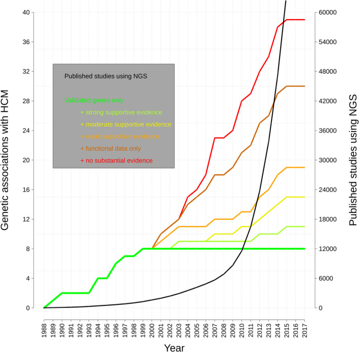
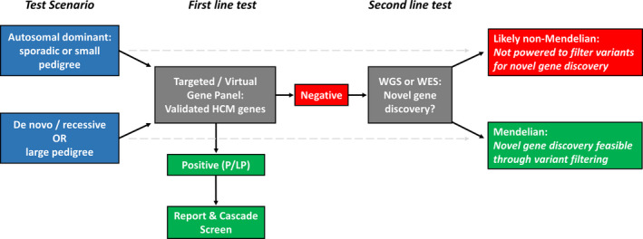
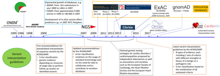
Genetic testing for hypertrophic cardiomyopathy ( HCM ) is an established clinical technique, supported by 30 years of research into its genetic etiology. Although pathogenic variants are often detected in patients and used to identify at‐risk relatives, the effectiveness of genetic testing has been hampered by ambiguous genetic associations (yielding uncertain and potentially false‐positive results), difficulties in classifying variants, and uncertainty about genotype‐negative patients. Recent case‐control studies on rare variation, improved data sharing, and meta‐analysis of case cohorts contributed to new insights into the genetic basis of HCM . In particular, although research into new genes and mechanisms remains essential, reassessment of Mendelian genetic associations in HCM argues that current clinical genetic testing should be limited to a small number of validated disease genes that yield informative and interpretable results. Accurate and consistent variant interpretation has benefited from new standardized variant interpretation guidelines and innovative approaches to improve classification. Most cases lacking a pathogenic variant are now believed to indicate non‐Mendelian HCM , with more benign prognosis and minimal risk to relatives. Here, we discuss recent advances in the genetics of HCM and their application to clinical genetic testing together with practical issues regarding implementation. Although this review focuses on HCM , many of the issues discussed are also relevant to other inherited cardiac diseases.
Overview
The claims and statistics of this paper haven’t been checked yet.
Run the check — free, no account needed. It reads the open-access text and checks the sources it cites.
Integrity signals
Registry
Cites
Statistics
Figures
Verification
Claims the paper makes
The claims and statistics of this paper haven’t been checked yet.
Run the check — free, no account needed. It reads the open-access text and checks the sources it cites.
Claims other papers make about this one
Each is checked against this paper’s own text: does it say what the citing paper says it does?
The cited source says what the claim attributes to it.
In the citing paper
“The most common of the sarcomeric gene variants in hypertrophic cardiomyopathy are those affecting the thick filament proteins cardiac myosin binding protein C (MYBPC3) and β-myosin heavy chain (MYH7).”
From Current management of hypertrophic cardiomyopathy (2025) · cites this paper as [77]
The cited source says what the claim attributes to it.
In the citing paper
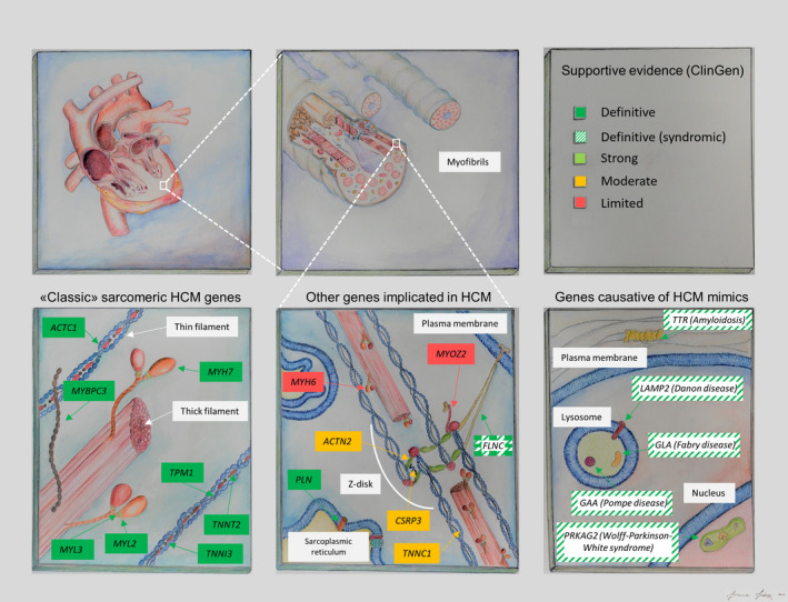
“To date, variants in eight different sarcomeric genes have been identified as definitively causative of hypertrophic cardiomyopathy, and variants in several other genes show at least moderate evidence of pathogenicity (table 4; fig 2).[75][76][77]”
From Current management of hypertrophic cardiomyopathy (2025) · cites this paper as [77]
Self-check
Percentages, sums and reported differences re-computed from the paper’s own numbers.
The self-check needs the paper’s full text.
Citations
Cites
Richards, S., Aziz, N., Bale, S., Bick, D., Das, S., Gastier-Foster, J., Grody, W. W., Hegde, M., Lyon, E., Spector, E., et al. (2015) · Genetics in Medicine · doi:10.1038/gim.2015.30 · Cited by 32,594
Clinical use of current polygenic risk scores may exacerbate health disparities
Martin, A. R., Kanai, M., Kamatani, Y., Okada, Y., Neale, B. M., & Daly, M. J. (2019) · Nature Genetics · doi:10.1038/s41588-019-0379-x · Cited by 2,821
ClinGen — The Clinical Genome Resource
Rehm, H. L., Berg, J. S., Brooks, L. D., Bustamante, C. D., Evans, J. P., Landrum, M. J., Ledbetter, D. H., Maglott, D. R., Martin, C. L., Nussbaum, R. L., et al. (2015) · New England Journal of Medicine · doi:10.1056/nejmsr1406261 · Cited by 1,496
Geisterfer-Lowrance, A. A. T., Kass, S., Tanigawa, G., Vosberg, H.-P., McKenna, W., Seidman, C. E., & Seidman, J. G. (1990) · Cell · doi:10.1016/0092-8674(90)90274-i · Cited by 1,380
O'Mahony, C., Jichi, F., Pavlou, M., Monserrat, L., Anastasakis, A., Rapezzi, C., Biagini, E., Gimeno, J. R., Limongelli, G., McKenna, W. J., et al. (2014) · European Heart Journal · doi:10.1093/eurheartj/eht439 · Cited by 1,251
Genetic Misdiagnoses and the Potential for Health Disparities
Manrai, A. K., Funke, B. H., Rehm, H. L., Olesen, M. S., Maron, B. A., Szolovits, P., Margulies, D. M., Loscalzo, J., & Kohane, I. S. (2016) · New England Journal of Medicine · doi:10.1056/nejmsa1507092 · Cited by 808
Modeling the ACMG/AMP variant classification guidelines as a Bayesian classification framework
Tavtigian, S. V., Greenblatt, M. S., Harrison, S. M., Nussbaum, R. L., Prabhu, S. A., Boucher, K. M., & Biesecker, L. G. (2018) · Genetics in Medicine · doi:10.1038/gim.2017.210 · Cited by 590
Using high-resolution variant frequencies to empower clinical genome interpretation
Whiffin, N., Minikel, E., Walsh, R., O’Donnell-Luria, A. H., Karczewski, K., Ing, A. Y., Barton, P. J. R., Funke, B., Cook, S. A., MacArthur, D., et al. (2017) · Genetics in Medicine · doi:10.1038/gim.2017.26 · Cited by 441 · cited 1×
“TNNT2 Cardiac troponin T Locus 1q3 1993 11 AD 3 (97) 8.”
Self-citation
Research works printed but not linked: 130
- 10.1016/S0140-6736(12)60397-3 · DOI · Paper page
- 10.1093/eurheartj/ehu284 · DOI · Paper page
- 10.1161/01.CIR.92.4.785 · DOI
- 10.1016/j.jacc.2015.01.019 · DOI
- 10.1136/hrt.20.1.1 · DOI
- Braunwald E, Lambrew CT, Rockoff SD, Ross J, Morrow AG. Idiopathic hypertrophic subaortic stenosis. I. A description of the disease based upon an analysis of 64 patients. Circulation. 1964;30(suppl 4):3–119.
- 10.1056/NEJM198911163212005 · DOI
- Solomon SD, Geisterfer‐Lowrance AA, Vosberg HP, Hiller G, Jarcho JA, Morton CC, McBride WO, Mitchell AL, Bale AE, McKenna WJ. A locus for familial hypertrophic cardiomyopathy is closely linked to the cardiac myosin heavy chain genes, CRI‐L436, and CRI‐L329 on chromosome 14 at q11‐q12. Am J Hum Genet. 1990;47:389–394.
- 10.1172/JCI114802 · DOI
- 10.1038/ng0493-333 · DOI
- 10.1016/0092-8674(94)90054-X · DOI
- 10.1038/ng0793-311 · DOI
- 10.1038/ng1295-434 · DOI
- 10.1073/pnas.90.13.6270 · DOI
- 10.1038/ng0596-63 · DOI
- 10.1038/ng0897-379 · DOI
- 10.1007/s001090050210 · DOI
- 10.1172/JCI6460 · DOI
- 10.1074/jbc.M009823200 · DOI
- 10.1038/sj.ejhg.5200872 · DOI
- 10.1093/hmg/ddi146 · DOI
- 10.1007/s40291-018-0324-1 · DOI
- 10.1073/pnas.83.24.9641 · DOI
- Van der Ploeg AT, Hoefsloot LH, Hoogeveen‐Westerveld M, Petersen EM, Reuser AJ. Glycogenosis type II: protein and DNA analysis in five South African families from various ethnic origins. Am J Hum Genet. 1989;44:787–793.
- Martiniuk F, Bodkin M, Tzall S, Hirschhorn R. Identification of the base‐pair substitution responsible for a human acid alpha glucosidase allele with lower “affinity” for glycogen (GAA 2) and transient gene expression in deficient cells. Am J Hum Genet. 1990;47:440–445.
- 10.1172/JCI114027 · DOI
- 10.1093/hmg/2.7.1051 · DOI
- 10.1002/humu.1380030204 · DOI
- 10.1136/hrt.2003.029504 · DOI
- 10.1056/NEJMoa033349 · DOI
- 10.1007/s10038-007-0184-8 · DOI
- 10.1056/NEJM200106143442403 · DOI · Paper page
- 10.1136/jmg.28.2.73 · DOI
- 10.1002/humu.1380050302 · DOI
- 10.1002/humu.1132 · DOI
- 10.1056/NEJMoa013354 · DOI
- Zellweger H, Brown BI, McCormick WF, Tu JB. A mild form of muscular glycogenosis in two brothers with alpha‐1, 4‐glucosidase deficiency. Ann Paediatr. 1965;205:413–437.
- 10.1002/j.1460-2075.1988.tb02998.x · DOI
- 10.1007/BF02185782 · DOI
- 10.1126/science.170.3954.180 · DOI
- 10.1093/nar/17.8.3301 · DOI
- 10.1212/WNL.31.1.51 · DOI
- 10.1038/35022604 · DOI · Paper page
- 10.1016/S0021-9258(19)42128-5 · DOI
- 10.1073/pnas.90.6.2375 · DOI · Paper page
- 10.1007/BF01870754 · DOI
- 10.1161/CIRCGEN.119.002460 · DOI
- Walsh R, Buchan R, Wilk A, John S, Felkin LE, Thomson KL, Chiaw TH, Loong CCW, Pua CJ, Raphael C, et al. Defining the genetic architecture of hypertrophic cardiomyopathy: re‐evaluating the role of non‐sarcomeric genes. Eur Heart J. 2017;38:3461–3468.
- 10.1002/humu.1143 · DOI
- 10.1172/JCI17892 · DOI · Paper page
- 10.1161/01.cir.0000056522.82563.5f · DOI
- 10.1016/j.yjmcc.2007.04.006 · DOI
- 10.1016/j.jacc.2009.11.016 · DOI
- 10.1038/ncomms6326 · DOI
- 10.1016/j.jacc.2015.10.093 · DOI
- 10.1016/j.jacc.2018.10.001 · DOI
- 10.1038/nature11632 · DOI · Paper page
- 10.1161/CIRCGENETICS.112.962928 · DOI
- 10.1038/ejhg.2012.283 · DOI
- 10.1038/nature19057 · DOI · Paper page
- 10.1038/gim.2016.90 · DOI
- 10.1161/CIRCULATIONAHA.119.037661 · DOI
- 10.1038/s41436-018-0046-0 · DOI
- 10.1093/eurheartj/ehq067 · DOI
- 10.1136/heartjnl-2011-300368 · DOI
- 10.1038/gim.2014.205 · DOI · Paper page
- 10.1007/s10897-014-9774-8 · DOI
- 10.1161/CIR.0b013e318223e2bd · DOI
- 10.1161/CIRCRESAHA.117.310987 · DOI
- 10.15420/aer.2013.2.1.36 · DOI
- 10.1186/s40246‐015‐0031‐5 · DOI
- 10.1038/gim.2017.247 · DOI
- 10.1161/CIRCGENETICS.117.001768 · DOI
- 10.1016/j.ejmg.2015.10.001 · DOI
- 10.1371/journal.pone.0181465 · DOI
- 10.1002/mgg3.187 · DOI
- 10.1016/j.jacc.2018.04.078 · DOI
- 10.1007/s10974-011-9268-3 · DOI
- 10.1007/s12265-016-9673-5 · DOI
- 10.1373/clinchem.2016.254698 · DOI
- 10.1186/s12920-014-0067-8 · DOI
- 10.1016/j.mayocp.2016.08.008 · DOI
- 10.1097/00125817-200009000-00009 · DOI
- 10.1038/gim.2017.14 · DOI · Paper page
- 10.1002/humu.23643 · DOI
- 10.1038/gim.2017.258 · DOI
- 10.1002/humu.23665 · DOI
- 10.1016/j.ajhg.2016.04.003 · DOI
- 10.1002/humu.23626 · DOI
- 10.1186/s13073-019-0616-z · DOI
- 10.1073/pnas.1606950113 · DOI
- 10.1038/gim.2018.3 · DOI
- 10.1038/gim.2017.218 · DOI
- 10.1161/CIRCULATIONAHA.117.033200 · DOI · Paper page
- 10.1038/ng.3703 · DOI
- 10.1016/j.ajhg.2016.08.016 · DOI · Paper page
- 10.1042/BCJ20180685 · DOI
- 10.1093/eurheartj/ehy249 · DOI
- 10.1016/j.jacbts.2018.08.008 · DOI
- 10.1161/CIRCULATIONAHA.117.032273 · DOI
- 10.1161/CIRCULATIONAHA.118.035028 · DOI
- 10.1038/s41586-018-0461-z · DOI · Paper page
- 10.1001/jamacardio.2017.5333 · DOI
- 10.1038/gim.2016.190 · DOI
- 10.1038/s41436-018-0039-z · DOI
- 10.1056/NEJM199504203321603 · DOI
- 10.1056/NEJM199804303381802 · DOI
- 10.1016/j.jacc.2014.09.059 · DOI
- 10.1161/CIRCGENETICS.113.000331 · DOI
- 10.1161/CIRCIMAGING.116.005311 · DOI
- 10.1016/j.mayocp.2014.01.025 · DOI
- 10.1136/heartjnl-2014-306387 · DOI · Paper page
- 10.1161/CIRCGENETICS.116.001660 · DOI
- 10.1161/CIRCGENETICS.116.001620 · DOI
- 10.1136/heartjnl-2013-303939 · DOI
- 10.1016/j.amjcard.2016.06.038 · DOI · Paper page
- 10.1038/gim.2013.44 · DOI
- 10.1038/gim.2017.79 · DOI · Paper page
- 10.1161/CIRCGEN.118.002368 · DOI
- 10.1016/j.jacc.2012.02.068 · DOI
- 10.1161/CIRCRESAHA.110.223834 · DOI · Paper page
- 10.1161/CIRCGENETICS.112.965277 · DOI
- 10.1093/nar/gkt1211 · DOI
- 10.1371/journal.pmed.1001779 · DOI · Paper page
- 10.1136/bmj.k1687 · DOI
- 10.1186/s12967-015-0704-9 · DOI
- 10.1161/CIRCGENETICS.116.001666 · DOI
- 10.1161/CIRCGENETICS.116.001685 · DOI
- 10.1161/CIRCGENETICS.117.001749 · DOI
- 10.1038/s41588-018-0192-y · DOI · Paper page
Cited by
Sorted by citations
Current management of hypertrophic cardiomyopathy
Sikand, N., Stendahl, J., Sen, S., Lampert, R., & Day, S. (2025) · BMJ · doi:10.1136/bmj-2023-077274 · Cited by 26
Metadata
- Authors
- Mazzarotto, F., Olivotto, I., Boschi, B., Girolami, F., Poggesi, C., Barton, P. J. R., & Walsh, R.
- Venue
- Journal of the American Heart Association
- Published
- 2020-04-21
- Volume
- 9
- Issue
- 8
- ISSN
- 2047-9980
- PMC
- PMC7428545
- Cited by
- 63
- References
- 8
- Language
- en
About these checks
Registry, retraction, Cites and statistics results are computed, not judged, with one exception: a printed reference that no index lists is read once by a model before it counts as a citation error. Claims are extracted by a model and kept only when their quote appears verbatim in the text; each cited source is then checked, with a model’s help, for whether it says what the claim relies on. Those verdicts can be wrong — read the claim’s quote and the cited source before relying on one.
Statistics and arithmetic are recomputed, not judged.
Confidence bands are shown only for fields calibrated on our annotated test set. None is calibrated yet, so no band is shown.
What counts as a claim ›How we measure confidence ›How we check this ›
Are you an author, or do you see a problem with this page? Request a review
How to cite
Cite CoChat for the checks on this page, and the paper for its own findings.
- This page
- CoChat. (2026). Citation check: Contemporary Insights Into the Genetics of Hypertrophic Cardiomyopathy: Toward a New Era in Clinical Testing? https://app.cochat.ai/papers/10.1161/jaha.119.015473
- The paper
- Mazzarotto, F., Olivotto, I., Boschi, B., Girolami, F., Poggesi, C., Barton, P. J. R., & Walsh, R. (2020). Contemporary Insights Into the Genetics of Hypertrophic Cardiomyopathy: Toward a New Era in Clinical Testing? Journal of the American Heart Association. https://doi.org/10.1161/jaha.119.015473
