Cited by 13Acute Myeloid Leukemia ResearchHematopoietic Stem Cell TransplantationChronic Myeloid Leukemia Treatments

The DOI matches its Crossref record; 58 of the 58 references it cites were found in a registry or an index; none of the 72 sources its claims cite is checked for support yet.

Loss of Dnmt3a impairs hematopoietic homeostasis and myeloid cell skewing via the PI3Kinase pathway

Open access

Abstract

Loss-of-function mutations in the DNA methyltransferase 3A (DNMT3A ) are seen in a large number of patients with acute myeloid leukemia (AML) with normal cytogenetics and are frequently associated with poor prognosis. DNMT3A mutations are an early preleukemic event, which — when combined with other genetic lesions — result in full-blown leukemia. Here, we show that loss of Dnmt3a in hematopoietic stem and progenitor cells (HSC/Ps) results in myeloproliferation, which is associated with hyperactivation of the phosphatidylinositol 3-kinase (PI3K) pathway. PI3Kα/β or the PI3Kα/δ inhibitor treatment partially corrects myeloproliferation, although the partial rescue is more efficient in response to the PI3Kα/β inhibitor treatment. In vivo RNA-Seq analysis on drug-treated Dnmt3a –/– HSC/Ps showed a reduction in the expression of genes associated with chemokines, inflammation, cell attachment, and extracellular matrix compared with controls. Remarkably, drug-treated leukemic mice showed a reversal in the enhanced fetal liver HSC-like gene signature observed in vehicle-treated Dnmt3a –/– LSK cells as well as a reduction in the expression of genes involved in regulating actin cytoskeleton-based functions, including the RHO/RAC GTPases. In a human PDX model bearing DNMT3A mutant AML, PI3Kα/β inhibitor treatment prolonged their survival and rescued the leukemic burden. Our results identify a potentially new target for treating DNMT3A mutation–driven myeloid malignancies.

Overview

CoChat Summary

Written by AI from the paper’s full text.

TL;DR

Loss of Dnmt3a in hematopoietic stem/progenitor cells drives myeloproliferation and myeloid skewing through hyperactivation of PI3K signaling, and PI3Kα/β inhibition partially reverses these abnormalities in mice and prolongs survival in a human DNMT3A-mutant AML PDX model. The findings support targeting PI3K, and downstream RAC/CDC42, in DNMT3A mutation–driven myeloid malignancies.

Key findings

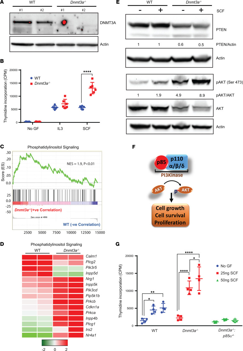
  • Dnmt3a–/– BM mononuclear cells showed increased SCF-induced proliferation, PI3K pathway enrichment by GSEA, increased AKT Ser473 activation, and reduced PTEN protein; genetic loss of p85α reduced SCF-induced hyperproliferation.
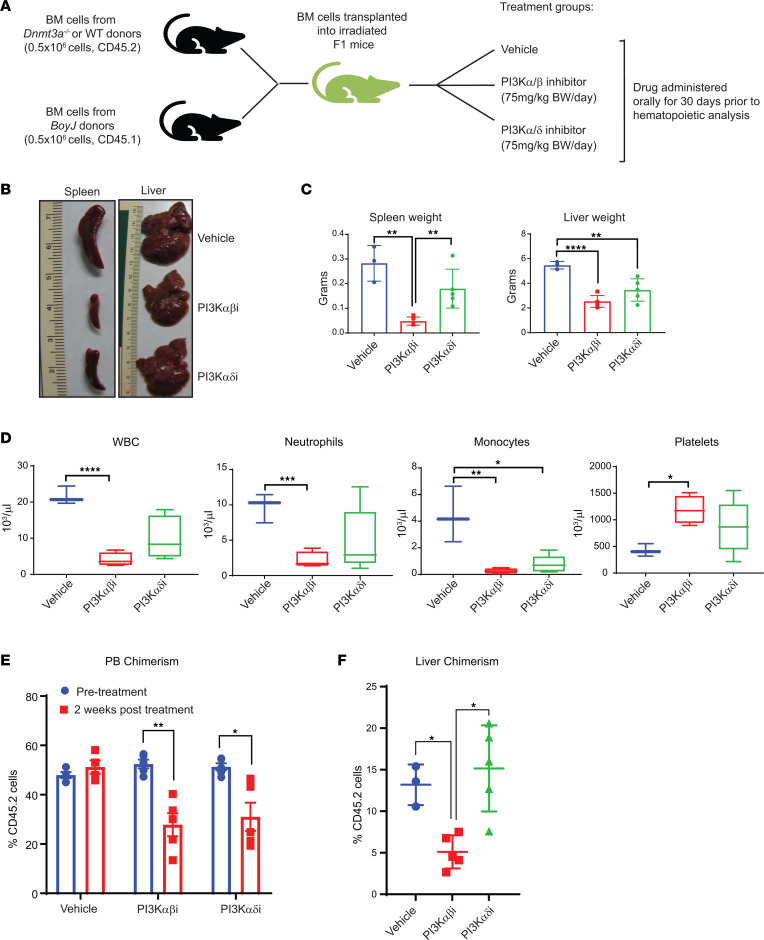
  • In transplanted Dnmt3a–/– cells, PI3Kα/β inhibitor (Bay1082439) or PI3Kα/δ inhibitor (GDC-0941) partially corrected hepatosplenomegaly, blood counts, thrombocytopenia, and engraftment; the PI3Kα/β inhibitor produced more dramatic restoration.
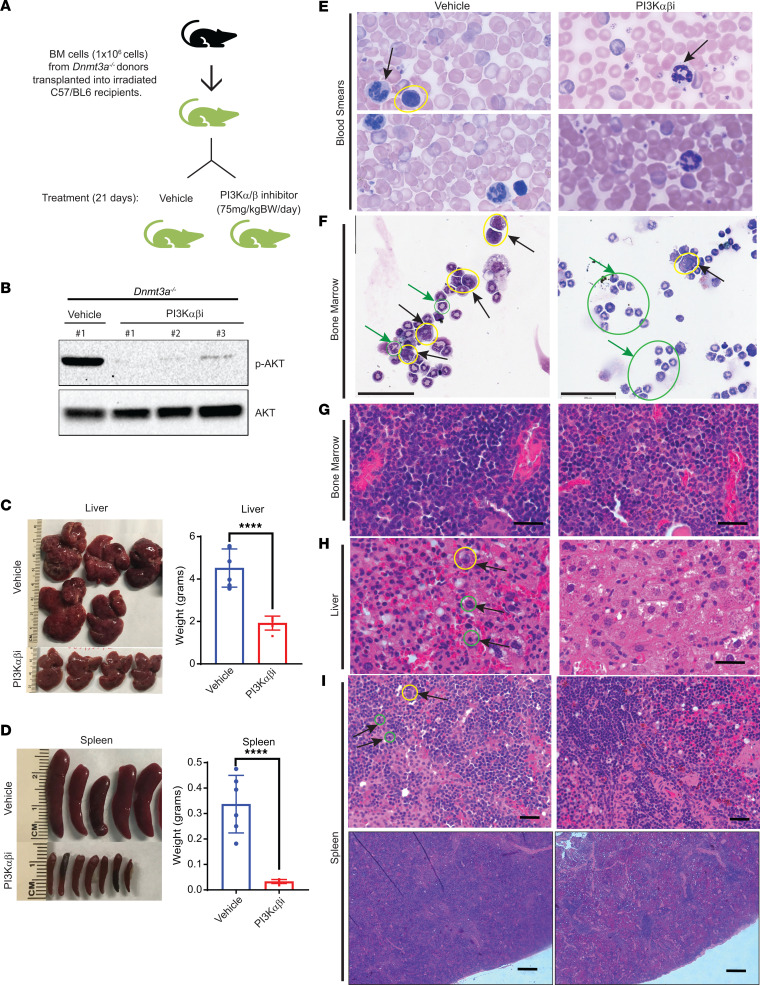
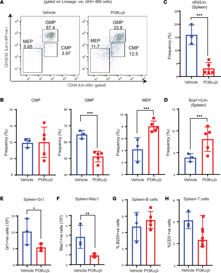
  • PI3Kα/β inhibition reduced AKT activation, liver/spleen size, myeloid progenitors, and dysplastic myeloid cells, while increasing erythroid progenitors and platelets; RNA-Seq showed repression of chemokine, inflammation, cell motility, attachment, extracellular matrix, and RHO/RAC/CDC42 pathway genes, and reversal of a fetal liver HSC-like signature.
  • In a human PDX model bearing DNMT3A-mutant AML, PI3Kα/β inhibitor treatment prolonged survival: all vehicle-treated mice died by day 45, whereas none of the drug-treated mice were dead at that time point; splenic and PB human leukemic CD45 engraftment were reduced. RAC/CDC42 inhibition with MBQ-167 also reduced hepatosplenomegaly and improved blood counts in Dnmt3a-null mice.

Methods

  • Used Mx1-Cre inducible conditional Dnmt3a-KO mice, p85α/Dnmt3a double-KO mice, BM transplantation into irradiated recipients, and in vivo treatment with PI3Kα/β inhibitor Bay1082439 (75 mg/kg for 30 or 21 days), PI3Kα/δ inhibitor GDC-0941 (75 mg/kg for 30 days), or RAC/CDC42 inhibitor MBQ-167 (20 mg/kg for 14 days).
  • Assessed proliferation by thymidine incorporation, PI3K/AKT signaling by Western blot and flow cytometry, hematopoietic populations by flow cytometry and colony-forming assays, tissue pathology by histology, and migration by transwell assays.
  • Performed RNA-Seq and GSEA on WT versus Dnmt3a–/– BMNCs (3 WT and 3 Dnmt3a–/– mice) and on vehicle- versus PI3Kα/β inhibitor–treated BM cells; also transduced human cord blood CD34+ cells with empty vector, WT DNMT3A-GFP, or DNMT3A R882H-GFP for RNA-Seq, and used a human AML PDX model in NSGS mice with a multi-mutational sample bearing FLT3-ITD, DNMT3A R882H, NPM1, and CHEK2 mutations.

Limitations

  • The paper does not include an explicit limitations section.
  • Evident from the text: most mechanistic and treatment studies use the Mx1-Cre conditional Dnmt3a-KO mouse model rather than native human DNMT3A-mutant AML, and PI3K inhibitor treatment produced partial, not complete, correction of hematologic abnormalities.
  • Evident from the text: the human PDX model used a multi-mutational AML sample bearing FLT3-ITD, DNMT3A R882H, NPM1, and CHEK2 mutations, so the observed survival benefit cannot be attributed solely to DNMT3A mutation; toxicity and long-term safety of PI3K or RAC/CDC42 inhibition are not reported.

Model-written summary. Check it against the paper before relying on it.

Integrity signals

Registry

Crossref · 10.1172/jci.insight.163864

Cites

58 of 58 references verified0 citation errors · 0 not checkable

Statistics

Nothing to recompute.

Figures

Figure 1 Loss of Dnmt3a in BMNC results in increased cell proliferation via the PI3K pathway. (A) WT and Dnmt3afl/fl:Mx-Cre mice were administered poly I:C at 12–16 weeks of age, and BMNCs were subjected to Western blot analysis to detect the presence of DNMT3a protein. Two independent WT and Dnmt3a–/– mouse–derived BMNCs were utilized for these experiments. (B) BMNCs from WT or Dnmt3a–/– mice were cultured in media supplemented with SCF (50 ng/mL) or IL-3 (10 ng/mL) or no growth factor for 48 hours. Cell proliferation was evaluated by [3H] thymidine incorporation. Counts per minute (CPM) are shown. Three independent experiments, n = 6, mean ± SD, ****P < 0.00005. BM cells collected from WT or Dnmt3a–/– mice were subjected to RNA isolation and, subsequently, next-generation sequencing. (C and D) GSEA revealed an enrichment for genes in the PI3K signaling pathway (C), and upregulation of specific genes in the PI3K pathway are shown in the heatmap (D). (E) BMNCs from WT or Dnmt3a–/– mice were starved of growth factors and stimulated with SCF, followed by Western blot analysis. (F) Class I PI3K complex is composed of p85 regulatory subunit and p110α, p110β, and p110δ catalytic subunits. Activated PI3K signaling regulates AKT phosphorylation, which in turn promotes cell grow, cell survival, and proliferation. (G) WT, Dnmt3afl/fl:Mx-Cre, and Dnmt3afl/fl:p85αfl/fl:Mx-Cre mice were administered poly I:C at 12–16 weeks of age. BMNCs were collected from 3 different mice of each genotype and stimulated with SCF (25 ng/mL or 50 ng/mL) or in the absence of growth factors for 48 hours. Cell proliferation was evaluated by [3H] thymidine incorporation. Counts per minute (CPM) are shown. Experiment performed 3 times, and representative experiment shown with n = 4, mean ± SD, *P < 0.05, **P < 0.005, ****P < 0.00005. Two-way ANOVA, Tukey’s multiple-comparison test (B and C).
Figure 1 Loss of Dnmt3a in BMNC results in increased cell proliferation via the PI3K pathway. (A) WT and Dnmt3afl/fl:Mx-Cre mice were administered poly I:C at 12–16 weeks of age, and BMNCs were subjected to Western blot analysis to detect the presence of DNMT3a protein. Two independent WT and Dnmt3a–/– mouse–derived BMNCs were utilized for these experiments. (B) BMNCs from WT or Dnmt3a–/– mice were cultured in media supplemented with SCF (50 ng/mL) or IL-3 (10 ng/mL) or no growth factor for 48 hours. Cell proliferation was evaluated by [3H] thymidine incorporation. Counts per minute (CPM) are shown. Three independent experiments, n = 6, mean ± SD, ****P < 0.00005. BM cells collected from WT or Dnmt3a–/– mice were subjected to RNA isolation and, subsequently, next-generation sequencing. (C and D) GSEA revealed an enrichment for genes in the PI3K signaling pathway (C), and upregulation of specific genes in the PI3K pathway are shown in the heatmap (D). (E) BMNCs from WT or Dnmt3a–/– mice were starved of growth factors and stimulated with SCF, followed by Western blot analysis. (F) Class I PI3K complex is composed of p85 regulatory subunit and p110α, p110β, and p110δ catalytic subunits. Activated PI3K signaling regulates AKT phosphorylation, which in turn promotes cell grow, cell survival, and proliferation. (G) WT, Dnmt3afl/fl:Mx-Cre, and Dnmt3afl/fl:p85αfl/fl:Mx-Cre mice were administered poly I:C at 12–16 weeks of age. BMNCs were collected from 3 different mice of each genotype and stimulated with SCF (25 ng/mL or 50 ng/mL) or in the absence of growth factors for 48 hours. Cell proliferation was evaluated by [3H] thymidine incorporation. Counts per minute (CPM) are shown. Experiment performed 3 times, and representative experiment shown with n = 4, mean ± SD, *P < 0.05, **P < 0.005, ****P < 0.00005. Two-way ANOVA, Tukey’s multiple-comparison test (B and C).CC BY · PubMed Central
Figure 2 PI3K αβ inhibition modulates Dnmt3a loss–induced myeloid leukemia development. (A) Donor cells from Dnmt3a–/– mice were mixed with BoyJ cells in 1:1 ratio (0.5 × 10⁶ versus 0.5 × 10⁶) for a competitive transplantation assay. Six weeks after transplantation, mice were treated with vehicle or the PI3K αβ inhibitor (Bay1082439; 7 mg/kg body weight) or PI3K αδ inhibitor (GDC-0941; 75 mg/kg body weight) for 30 days, and mice were analyzed. (B) Representative images of liver and spleen from vehicle- and drug-treated mice are shown. (C) Quantitative assessment of spleen and liver weights from the indicated groups. n = 3–5, mean ± SD, **P < 0.005, ****P < 0.00005. (D) PB counts from mice described in B and C before they were sacrificed. n = 3–5, *P < 0.05, **P < 0.005, ***P < 0.0005, ****P < 0.00005. The boxes shown with lower and upper quartiles separated by the median (horizontal line), and the whiskers extend to the minimum and maximum values. (E and F) Mice described in A were analyzed for PB chimerism 2 weeks after drug treatment and after 30 days after drug treatment for liver chimerism. Chimerism was assessed by staining the cells using an anti-CD45.2 antibody and flow cytometry. n = 3–5, mean ± SEM, *P < 0.05, **P < 0.005. One-way ANOVA in C, D, and F; 2-way ANOVA in E with Tukey’s multiple comparison test performed.
Figure 2 PI3K αβ inhibition modulates Dnmt3a loss–induced myeloid leukemia development. (A) Donor cells from Dnmt3a–/– mice were mixed with BoyJ cells in 1:1 ratio (0.5 × 10⁶ versus 0.5 × 10⁶) for a competitive transplantation assay. Six weeks after transplantation, mice were treated with vehicle or the PI3K αβ inhibitor (Bay1082439; 7 mg/kg body weight) or PI3K αδ inhibitor (GDC-0941; 75 mg/kg body weight) for 30 days, and mice were analyzed. (B) Representative images of liver and spleen from vehicle- and drug-treated mice are shown. (C) Quantitative assessment of spleen and liver weights from the indicated groups. n = 3–5, mean ± SD, **P < 0.005, ****P < 0.00005. (D) PB counts from mice described in B and C before they were sacrificed. n = 3–5, *P < 0.05, **P < 0.005, ***P < 0.0005, ****P < 0.00005. The boxes shown with lower and upper quartiles separated by the median (horizontal line), and the whiskers extend to the minimum and maximum values. (E and F) Mice described in A were analyzed for PB chimerism 2 weeks after drug treatment and after 30 days after drug treatment for liver chimerism. Chimerism was assessed by staining the cells using an anti-CD45.2 antibody and flow cytometry. n = 3–5, mean ± SEM, *P < 0.05, **P < 0.005. One-way ANOVA in C, D, and F; 2-way ANOVA in E with Tukey’s multiple comparison test performed.CC BY · PubMed Central
Figure 3 PI3K αβ inhibition rescues tissue myeloid cell infiltration as well as hepatosplenomegaly due to loss of Dnmt3a in HSC/Ps. (A) BM cells from Dnmt3a–/– mice were transplanted into lethally irradiated C57BL/6 mice, and 6 weeks after transplantation, mice were treated with vehicle or the PI3K αβ inhibitor (Bay1082439; 75 mg/kg body weight) for 21 days. (B) BM bearing Dnmt3a–/– cells from mice treated with vehicle or the PI3K αβ inhibitor were subjected to Western blot analysis to analyze pAKT and total AKT protein levels. Numbers at the top represent the number of mice used for these experiments. (C and D) Images of spleens and livers from vehicle- and drug-treated mice and respective quantitative assessment of liver and spleen weights from vehicle- or drug-treated mice. n = 5–6, mean ± SEM, unpaired t test (2-tailed), ****P < 0.00005. (E and F) Peripheral blood smears and BM cytospin preparations showing reduced dysplasia of myeloid and erythroid lineage cells in PI3K αβ inhibitor–treated mice compared with vehicle group. Arrows in blood smears indicate segmented neutrophils. Yellow circle indicates myeloblasts, and green circles/green arrows indicate metamyelocytes. PB smear images shown at 100× magnification (E). Scale bar: 60 μm (F). (G) Representative images for H&E-stained sections of BM. Scale bar: 50μm (H) H&E-stained liver sections showing increased sinusoidal spaces with reduced immature myeloid cell infiltration in in PI3Kαβ inhibitor treatment condition compared with vehicle treatment. Immature myeloid precursor Metamyelocytes (yellow circle) and proliferating myeloid cells (green circles) in vehicle-treated mice are indicated. Scale bar: 50 μm (I) H&E-stained spleen sections showing reduced immature myeloid cell infiltration in the PI3Kαβ inhibitor treatment condition compared with vehicle treatment. Yellow circle indicates dysplastic megakaryocyte. More proliferating myeloid cells in vehicle-treated mice (green circles indicate mitotic figures of myeloproliferation). Scale bar: 50 μm (upper panel), 1,000 μm (lower panel).
Figure 3 PI3K αβ inhibition rescues tissue myeloid cell infiltration as well as hepatosplenomegaly due to loss of Dnmt3a in HSC/Ps. (A) BM cells from Dnmt3a–/– mice were transplanted into lethally irradiated C57BL/6 mice, and 6 weeks after transplantation, mice were treated with vehicle or the PI3K αβ inhibitor (Bay1082439; 75 mg/kg body weight) for 21 days. (B) BM bearing Dnmt3a–/– cells from mice treated with vehicle or the PI3K αβ inhibitor were subjected to Western blot analysis to analyze pAKT and total AKT protein levels. Numbers at the top represent the number of mice used for these experiments. (C and D) Images of spleens and livers from vehicle- and drug-treated mice and respective quantitative assessment of liver and spleen weights from vehicle- or drug-treated mice. n = 5–6, mean ± SEM, unpaired t test (2-tailed), ****P < 0.00005. (E and F) Peripheral blood smears and BM cytospin preparations showing reduced dysplasia of myeloid and erythroid lineage cells in PI3K αβ inhibitor–treated mice compared with vehicle group. Arrows in blood smears indicate segmented neutrophils. Yellow circle indicates myeloblasts, and green circles/green arrows indicate metamyelocytes. PB smear images shown at 100× magnification (E). Scale bar: 60 μm (F). (G) Representative images for H&E-stained sections of BM. Scale bar: 50μm (H) H&E-stained liver sections showing increased sinusoidal spaces with reduced immature myeloid cell infiltration in in PI3Kαβ inhibitor treatment condition compared with vehicle treatment. Immature myeloid precursor Metamyelocytes (yellow circle) and proliferating myeloid cells (green circles) in vehicle-treated mice are indicated. Scale bar: 50 μm (I) H&E-stained spleen sections showing reduced immature myeloid cell infiltration in the PI3Kαβ inhibitor treatment condition compared with vehicle treatment. Yellow circle indicates dysplastic megakaryocyte. More proliferating myeloid cells in vehicle-treated mice (green circles indicate mitotic figures of myeloproliferation). Scale bar: 50 μm (upper panel), 1,000 μm (lower panel).CC BY · PubMed Central
Figure 4 PI3K αβ inhibition decreases myeloid progenitors and improves megakaryocyte-erythrocyte progenitors in Dnmt3a–/– malignant mice. (A and B) Flow cytometry analysis was performed on BM cells from mice transplanted with Dnmt3a–/– cells and treated with the PI3K αβ inhibitor. Representative dot plots and quantitative data showing the frequency of Lin–/c-KIT+ myeloid progenitors: CMPs, GMPs, and MEPs. n = 3–5, mean ± SEM, ***P = 0.0005. (C and D) Spleen cells collected from vehicle- or PI3Kαβ inhibitor–treated mice as in A were subjected to flow cytometry analysis to detect Lin–c-KIT+Sca-1+ cells. Quantitative data showing reduced c-KIT+ (Lin–) myeloid progenitor cells (C) and increased differentiated c-KIT– spleen cells (D) in PI3K αβ inhibitor–treated group compared with controls. n = 3–5, mean ± SEM, ***P = 0.0005. (E–H) Flow cytometry was performed on spleen cells from vehicle or PI3K αβ inhibitor–treated mice as in A. Quantitative data showing a reduction in myeloid cell burden in the spleen from drug treated Dnmt3a–/– mice compared with controls. n = 3–5, mean ± SEM, *P = 0.05, **P = 0.005, unpaired t test (2-tailed) performed (B–H).
Figure 4 PI3K αβ inhibition decreases myeloid progenitors and improves megakaryocyte-erythrocyte progenitors in Dnmt3a–/– malignant mice. (A and B) Flow cytometry analysis was performed on BM cells from mice transplanted with Dnmt3a–/– cells and treated with the PI3K αβ inhibitor. Representative dot plots and quantitative data showing the frequency of Lin–/c-KIT+ myeloid progenitors: CMPs, GMPs, and MEPs. n = 3–5, mean ± SEM, ***P = 0.0005. (C and D) Spleen cells collected from vehicle- or PI3Kαβ inhibitor–treated mice as in A were subjected to flow cytometry analysis to detect Lin–c-KIT+Sca-1+ cells. Quantitative data showing reduced c-KIT+ (Lin–) myeloid progenitor cells (C) and increased differentiated c-KIT– spleen cells (D) in PI3K αβ inhibitor–treated group compared with controls. n = 3–5, mean ± SEM, ***P = 0.0005. (E–H) Flow cytometry was performed on spleen cells from vehicle or PI3K αβ inhibitor–treated mice as in A. Quantitative data showing a reduction in myeloid cell burden in the spleen from drug treated Dnmt3a–/– mice compared with controls. n = 3–5, mean ± SEM, *P = 0.05, **P = 0.005, unpaired t test (2-tailed) performed (B–H).CC BY · PubMed Central
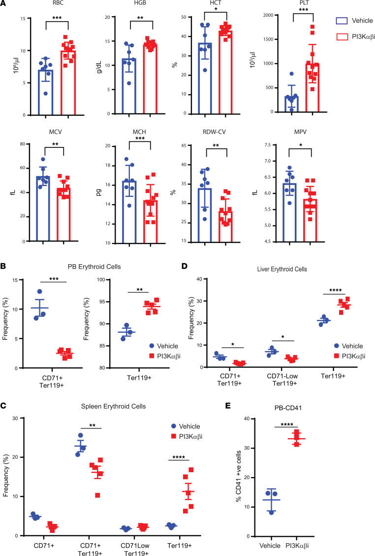
Figure 5 PI3K αβ inhibition improves erythroid cell maturation in mice bearing Dnmt3a–/– malignant cells. (A) Peripheral red cell parameters in mice transplanted with Dnmt3a–/– BM cells treated with vehicle or the PI3K αβ inhibitor (Bay1082439) for 21 days. n = 7–11, mean ± SEM, *P = 0.05, **P = 0.005, ***P = 0.0005. (B–D) Flow cytometry analysis was performed on PB (B), spleen (C), or liver cells (D) from mice transplanted with Dnmt3a–/– cells and treated with the PI3K αβ inhibitor as in A. Quantitative data showing CD71+Ter119+ immature erythroid cells, CD71loTer119+ immature erythroid cells, and Ter119+ mature erythroid cells in vehicle- and PI3K αβ inhibitor–treated mice. n = 3–5, mean ± SEM, *P = 0.05, **P = 0.005, ***P = 0.0005, ****P = 0.00005. (E) Quantitative data showing increased presence of mature platelets in the PB of drug-treated mice compared with controls. n = 3–5, mean ± SEM, ****P = 0.00005. Unpaired t test (2-tailed) was performed in A, B, and E. Two-way ANOVA (Sidak’s multiple comparison) was performed in D and E.
Figure 5 PI3K αβ inhibition improves erythroid cell maturation in mice bearing Dnmt3a–/– malignant cells. (A) Peripheral red cell parameters in mice transplanted with Dnmt3a–/– BM cells treated with vehicle or the PI3K αβ inhibitor (Bay1082439) for 21 days. n = 7–11, mean ± SEM, *P = 0.05, **P = 0.005, ***P = 0.0005. (B–D) Flow cytometry analysis was performed on PB (B), spleen (C), or liver cells (D) from mice transplanted with Dnmt3a–/– cells and treated with the PI3K αβ inhibitor as in A. Quantitative data showing CD71+Ter119+ immature erythroid cells, CD71loTer119+ immature erythroid cells, and Ter119+ mature erythroid cells in vehicle- and PI3K αβ inhibitor–treated mice. n = 3–5, mean ± SEM, *P = 0.05, **P = 0.005, ***P = 0.0005, ****P = 0.00005. (E) Quantitative data showing increased presence of mature platelets in the PB of drug-treated mice compared with controls. n = 3–5, mean ± SEM, ****P = 0.00005. Unpaired t test (2-tailed) was performed in A, B, and E. Two-way ANOVA (Sidak’s multiple comparison) was performed in D and E.CC BY · PubMed Central
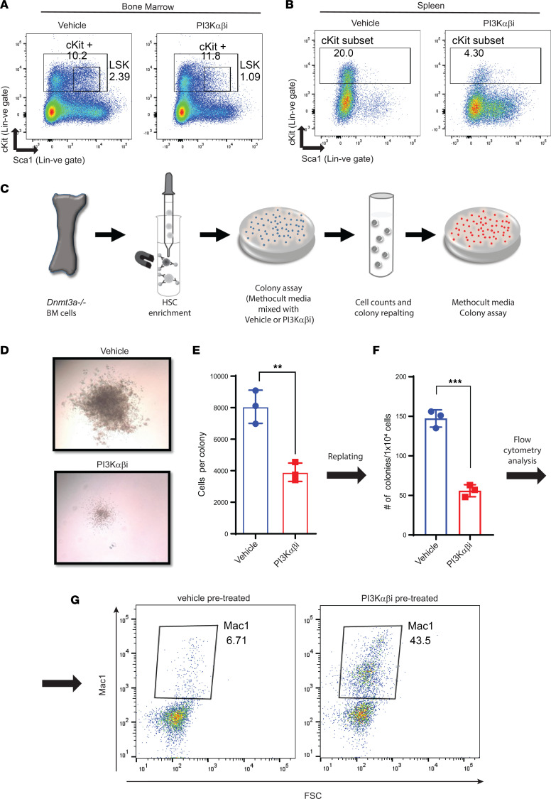
Figure 6 PI3K inhibition promotes cell differentiation in Dnmt3a-deleted hematopoietic stem cells. (A and B) BM and spleen cells from mice transplanted with Dnmt3a–/– cells and treated with the PI3K αβ inhibitor were subjected to flow cytometry analysis to detect Lin–c-KIT+Sca-1+ cells. Representative dot plots showing Lin–c-KIT+Sca-1+ cells in BM (A) and spleen (B). (C) A schematic depicting our strategy to assess the impact of PI3K inhibitor on the ability of Dnmt3a–/– HSPCs to give rise to colonies in a methylcellulose-based assay in vitro. Briefly, HSPCs were enriched from the BM of Dnmt3a–/– mice and platted in a methylcellulose-based media, along with the PI3K inhibitor and cytokines (50 ng/mL rmSCF, 10 ng/mL rmIL-3, 10 ng/mL rhIL-6, 3 U/mL rhEPO, 10 ng/mL Flt-3, and 10 ng/mL thrombopoietin (TPO). Colonies were enumerated on day 7, and cells were replatted in methylcellulose media along with growth factors and colonies were scored again after secondary platting. (D) Equal number of cells (10,000 cells) were plated in methocult media and cultured for 1 week in the presence of PI3Kαβ inhibitor (250 nM) or vehicle. Representative colony images depicting reduced colony size under conditions of PI3K αβ inhibitor treatment compared with vehicle conditions. Experiment was performed in triplicate. Number of colonies and total number of cells per plate were quantified. (E) Quantitative data show average number of cells per colony in drug and vehicle treatment groups. n = 3, mean ± SD, **P = 0.005. (F) Quantitative data showing number of colonies after replatting cells derived from the vehicle- and drug-treated groups. n = 3, mean ± SD, ***P = 0.0005. (G) Flow cytometry analysis of secondary replatted colonies using an antibody against Mac1. Dot plots show the level of Mac1 expression in drug-treated versus vehicle-treated groups. Experiment was performed in triplicate. Unpaired t test (2-tailed) was performed in E and F.
Figure 6 PI3K inhibition promotes cell differentiation in Dnmt3a-deleted hematopoietic stem cells. (A and B) BM and spleen cells from mice transplanted with Dnmt3a–/– cells and treated with the PI3K αβ inhibitor were subjected to flow cytometry analysis to detect Lin–c-KIT+Sca-1+ cells. Representative dot plots showing Lin–c-KIT+Sca-1+ cells in BM (A) and spleen (B). (C) A schematic depicting our strategy to assess the impact of PI3K inhibitor on the ability of Dnmt3a–/– HSPCs to give rise to colonies in a methylcellulose-based assay in vitro. Briefly, HSPCs were enriched from the BM of Dnmt3a–/– mice and platted in a methylcellulose-based media, along with the PI3K inhibitor and cytokines (50 ng/mL rmSCF, 10 ng/mL rmIL-3, 10 ng/mL rhIL-6, 3 U/mL rhEPO, 10 ng/mL Flt-3, and 10 ng/mL thrombopoietin (TPO). Colonies were enumerated on day 7, and cells were replatted in methylcellulose media along with growth factors and colonies were scored again after secondary platting. (D) Equal number of cells (10,000 cells) were plated in methocult media and cultured for 1 week in the presence of PI3Kαβ inhibitor (250 nM) or vehicle. Representative colony images depicting reduced colony size under conditions of PI3K αβ inhibitor treatment compared with vehicle conditions. Experiment was performed in triplicate. Number of colonies and total number of cells per plate were quantified. (E) Quantitative data show average number of cells per colony in drug and vehicle treatment groups. n = 3, mean ± SD, **P = 0.005. (F) Quantitative data showing number of colonies after replatting cells derived from the vehicle- and drug-treated groups. n = 3, mean ± SD, ***P = 0.0005. (G) Flow cytometry analysis of secondary replatted colonies using an antibody against Mac1. Dot plots show the level of Mac1 expression in drug-treated versus vehicle-treated groups. Experiment was performed in triplicate. Unpaired t test (2-tailed) was performed in E and F.CC BY · PubMed Central
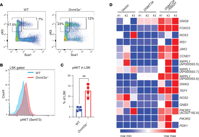
Figure 7 Increased PI3K signaling in Dnmt3a-deleted hematopoietic LSK cells and DNMT3A-mutated hCD34+ HSPCs. (A) Flow cytometry analysis performed on BM cells collected from malignant Dnmt3a–/– and age-matched WT mice. Representative plots for Lin–Sca-1+cKit+ cells from the indicated genotypes are shown. (B) Histogram showing intracellular activation of Akt (Ser473) in LSK cells from indicated genotypes. (C) Quantitative data showing percent phospho-AKT+ LSKs relative to IgG controls. n = 4, mean ± SEM, unpaired t test (2-tailed), **P = 0.005. (D) CB CD34+ HSC/Ps were transduced with empty vector, DNMT3a-GFP, or DNMT3aR882H-GFP, and sorted GFP+ cells were subjected to RNA-Seq. Heatmap showing PI3K pathway related gene expression.
Figure 7 Increased PI3K signaling in Dnmt3a-deleted hematopoietic LSK cells and DNMT3A-mutated hCD34+ HSPCs. (A) Flow cytometry analysis performed on BM cells collected from malignant Dnmt3a–/– and age-matched WT mice. Representative plots for Lin–Sca-1+cKit+ cells from the indicated genotypes are shown. (B) Histogram showing intracellular activation of Akt (Ser473) in LSK cells from indicated genotypes. (C) Quantitative data showing percent phospho-AKT+ LSKs relative to IgG controls. n = 4, mean ± SEM, unpaired t test (2-tailed), **P = 0.005. (D) CB CD34+ HSC/Ps were transduced with empty vector, DNMT3a-GFP, or DNMT3aR882H-GFP, and sorted GFP+ cells were subjected to RNA-Seq. Heatmap showing PI3K pathway related gene expression.CC BY · PubMed Central
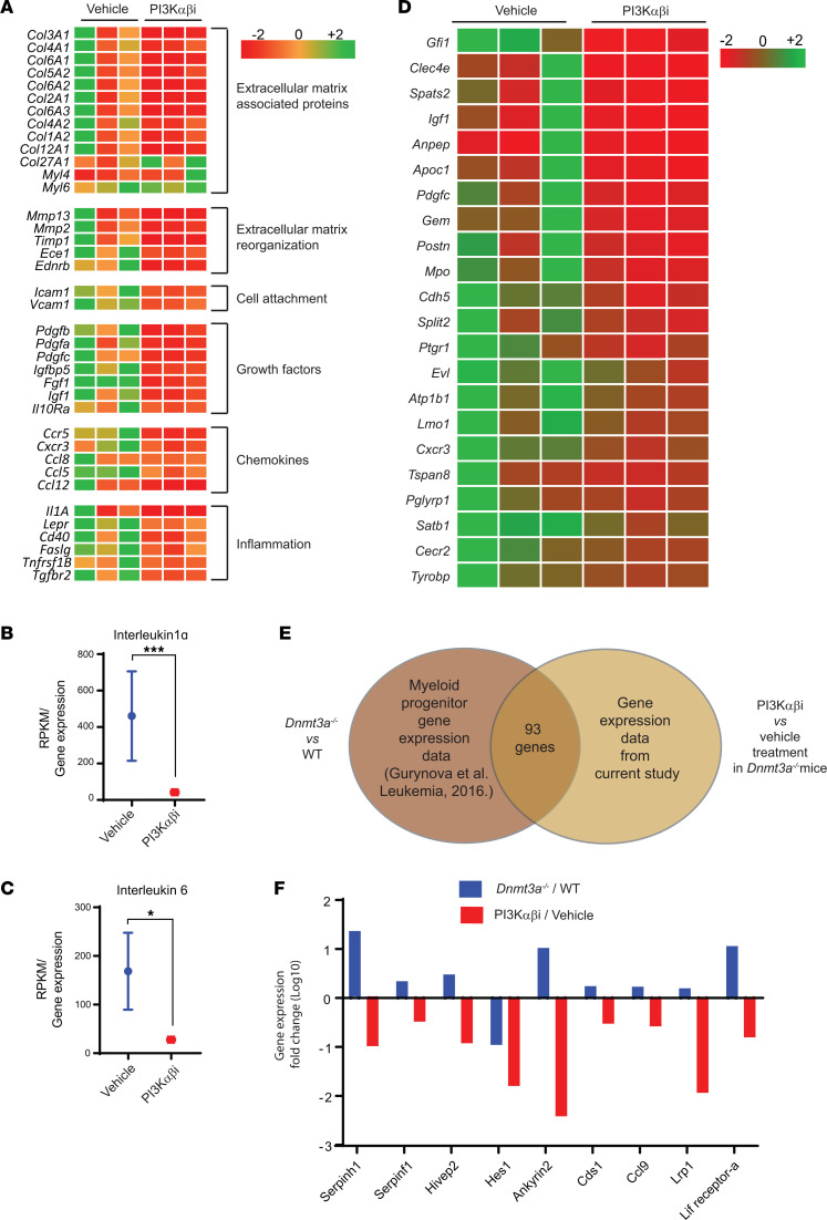
Figure 8 PI3K inhibition reverts Dnmt3a loss–induced changes in the expression of genes involved in cell migration and inflammation and alters the expression of fetal liver HSC genes. BM cells were collected from vehicle or PI3K αβ inhibitor–treated mice bearing Dnmt3a–/– malignant cells. RNA was isolated and subjected to next-generation sequencing, and gene expression analysis was performed. (A) Heatmap showing that PI3K αβ inhibitor treatment reduces the expression of genes involved in cell motility, cell attachment, inflammatory cytokines, and chemokines. (B and C) Quantitative assessment of the level of expression of IL-1α and IL-6 in drug-treated mice versus controls. n = 3, mean ± SEM, EdgeR DE analysis, *P = 0.05, ***P = 0.0005. (D) Heatmap showing gene expression involved in the development of fetal liver HSCs downregulated as a result of PI3K αβ inhibitor treatment in the Dnmt3a–/– cells versus controls. (E and F) Analysis of GMP progenitor gene expression data from a study involving Dnmt3a–/– and WT mice (13) was compared with the BM-derived gene expression data in the current study from vehicle- and PI3K αβ inhibitor–treated mice bearing Dnmt3a–/– cells. Venn diagram shows that 93 genes were found to be differentially regulated in both data sets. Comparative assessment of gene expression fold changes of indicated genes (F) in Dnmt3a–/–/WT and PI3Kαβ/vehicle treatment groups.
Figure 8 PI3K inhibition reverts Dnmt3a loss–induced changes in the expression of genes involved in cell migration and inflammation and alters the expression of fetal liver HSC genes. BM cells were collected from vehicle or PI3K αβ inhibitor–treated mice bearing Dnmt3a–/– malignant cells. RNA was isolated and subjected to next-generation sequencing, and gene expression analysis was performed. (A) Heatmap showing that PI3K αβ inhibitor treatment reduces the expression of genes involved in cell motility, cell attachment, inflammatory cytokines, and chemokines. (B and C) Quantitative assessment of the level of expression of IL-1α and IL-6 in drug-treated mice versus controls. n = 3, mean ± SEM, EdgeR DE analysis, *P = 0.05, ***P = 0.0005. (D) Heatmap showing gene expression involved in the development of fetal liver HSCs downregulated as a result of PI3K αβ inhibitor treatment in the Dnmt3a–/– cells versus controls. (E and F) Analysis of GMP progenitor gene expression data from a study involving Dnmt3a–/– and WT mice (13) was compared with the BM-derived gene expression data in the current study from vehicle- and PI3K αβ inhibitor–treated mice bearing Dnmt3a–/– cells. Venn diagram shows that 93 genes were found to be differentially regulated in both data sets. Comparative assessment of gene expression fold changes of indicated genes (F) in Dnmt3a–/–/WT and PI3Kαβ/vehicle treatment groups.CC BY · PubMed Central
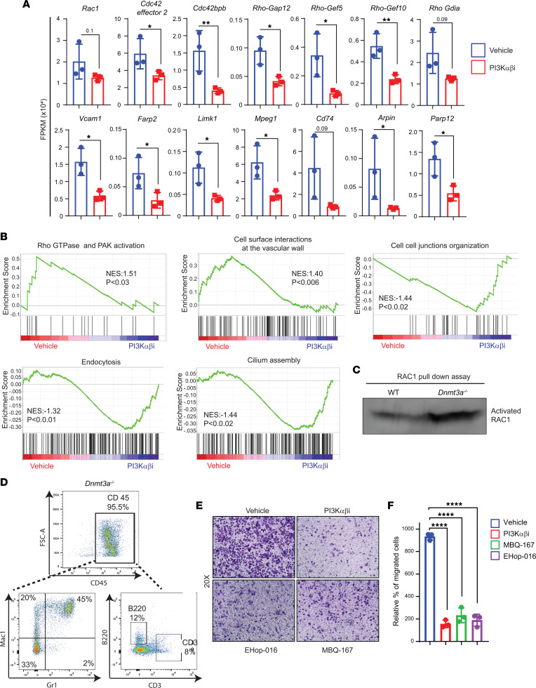
Figure 9 PI3K signaling promotes Dnmt3a loss–induced cell migration via RAC1 GTPase. (A) Quantitative assessment of the level of expression of genes that belong to the RAC1/CDC42 pathway that are significantly reduced in PI3K drug–treated group compared with vehicle group. n = 3, mean ± SD, unpaired t test (2-tailed), *P = 0.05, **P = 0.005. (B) Gene set enrichment plots showing reduced RHO GTPase and PAK signaling associated with RAC1 pathway and altered biological processes in PI3K αβ inhibitor–treated Dnmt3a–/– BM cells compared with vehicle-treated group. (C) BM cells collected from WT or Dnmt3a–/– mice were subjected to activated RAC1 pulldown assay using PAK binding domain beads. Western blot analysis was performed to detect activated RAC1 using an anti-RAC1 specific antibody. Representative of 3 independent experiments shown. (D) Liver tissues harvested from hepatomegaly presenting malignant Dnmt3a–/– mice were subjected to CD45+ cell enrichment using magnetic cell separation, and percent CD45 enrichment was assessed using flow cytometry. Figure shows the percent of Mac1+, Gr1+, B220+, and CD3+ cells within the CD45+ enriched fraction of Dnmt3a–/– cells. (E and F) CD45+ cells (2.5 × 10⁵) enriched from Dnmt3a–/– mice livers as in D were subjected to transwell migration assay for 20 hours at 37°C in the presence or absence of 100 nM PI3K αβ inhibitor (Bay1082439), 0.5 μM RAC1/3 inhibitor (EHop-016), or 200 nM RAC/CDC42 inhibitor (MBQ-167). Migrated cells were stained with 0.1% crystal violet, and representative images (20× magnification) for treatments are shown. Quantitation of percent migrated cells is shown in F. n = 3, mean ± SD, 1-way ANOVA (Tukey’s multiple comparison test), ****P < 0.00001.
Figure 9 PI3K signaling promotes Dnmt3a loss–induced cell migration via RAC1 GTPase. (A) Quantitative assessment of the level of expression of genes that belong to the RAC1/CDC42 pathway that are significantly reduced in PI3K drug–treated group compared with vehicle group. n = 3, mean ± SD, unpaired t test (2-tailed), *P = 0.05, **P = 0.005. (B) Gene set enrichment plots showing reduced RHO GTPase and PAK signaling associated with RAC1 pathway and altered biological processes in PI3K αβ inhibitor–treated Dnmt3a–/– BM cells compared with vehicle-treated group. (C) BM cells collected from WT or Dnmt3a–/– mice were subjected to activated RAC1 pulldown assay using PAK binding domain beads. Western blot analysis was performed to detect activated RAC1 using an anti-RAC1 specific antibody. Representative of 3 independent experiments shown. (D) Liver tissues harvested from hepatomegaly presenting malignant Dnmt3a–/– mice were subjected to CD45+ cell enrichment using magnetic cell separation, and percent CD45 enrichment was assessed using flow cytometry. Figure shows the percent of Mac1+, Gr1+, B220+, and CD3+ cells within the CD45+ enriched fraction of Dnmt3a–/– cells. (E and F) CD45+ cells (2.5 × 10⁵) enriched from Dnmt3a–/– mice livers as in D were subjected to transwell migration assay for 20 hours at 37°C in the presence or absence of 100 nM PI3K αβ inhibitor (Bay1082439), 0.5 μM RAC1/3 inhibitor (EHop-016), or 200 nM RAC/CDC42 inhibitor (MBQ-167). Migrated cells were stained with 0.1% crystal violet, and representative images (20× magnification) for treatments are shown. Quantitation of percent migrated cells is shown in F. n = 3, mean ± SD, 1-way ANOVA (Tukey’s multiple comparison test), ****P < 0.00001.CC BY · PubMed Central
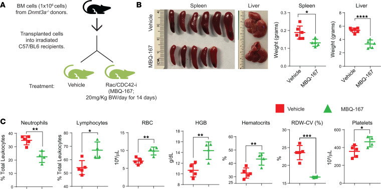
Figure 10 RAC/CDC42 pathway inhibition partially rescues hepatosplenomegaly and decreases Dnmt3a loss–induced myeloid malignancy characteristics in mice. (A) BM cells from Dnmt3a–/– mice were transplanted into lethally irradiated C57BL/6 mice, and 6 weeks after transplantation, mice were treated with vehicle or the RAC/CDC42 inhibitor (MBQ-167; 20 mg/kg body weight/day) for 14 days. (B) Images of spleens and livers from the vehicle- and drug-treated mice. Quantitative assessment of liver and spleen weights from vehicle- or drug-treated mice. n = 5–6, mean ± SEM, *P < 0.05, ****P < 0.00005. (C) Peripheral red cell parameters in mice in A. n = 4–6, mean ± SEM, *P = 0.05, **P = 0.005, ***P = 0.0005, unpaired t test (2-tailed) performed (B and C).
Figure 10 RAC/CDC42 pathway inhibition partially rescues hepatosplenomegaly and decreases Dnmt3a loss–induced myeloid malignancy characteristics in mice. (A) BM cells from Dnmt3a–/– mice were transplanted into lethally irradiated C57BL/6 mice, and 6 weeks after transplantation, mice were treated with vehicle or the RAC/CDC42 inhibitor (MBQ-167; 20 mg/kg body weight/day) for 14 days. (B) Images of spleens and livers from the vehicle- and drug-treated mice. Quantitative assessment of liver and spleen weights from vehicle- or drug-treated mice. n = 5–6, mean ± SEM, *P < 0.05, ****P < 0.00005. (C) Peripheral red cell parameters in mice in A. n = 4–6, mean ± SEM, *P = 0.05, **P = 0.005, ***P = 0.0005, unpaired t test (2-tailed) performed (B and C).CC BY · PubMed Central
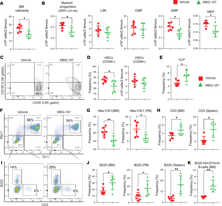
Figure 11 RAC/CDC42 pathway inhibition decreases Dnmt3a loss–induced myeloid skewing and improves B cells and T cells in mice. (A) BM cellularity quantitation for 2 femurs in each mouse from mice transplanted with Dnmt3a–/– cells and treated with the vehicle or MBQ-167. n = 5–6, mean ± SEM, *P = 0.05. (B) Flow cytometry analysis was performed on BM cells from mice transplanted with Dnmt3a–/– cells and treated with vehicle or MBQ-167. Quantitative data showing the absolute number of Lin–cKit+ myeloid progenitors, Lin–cKit+Sca-1+ (LSKs), CMPs, GMPs, and MEPs. n = 5–6, mean ± SEM, *P = 0.05. (C and D) Flow cytometry analysis was performed on BM cells from mice in A, and representative dot plots for CD150- and CD48-stained (LSK gated) HSCs as shown in F; quantitative data plotted for the frequency and absolute number of CD150–CD48+ cells is shown in G. n = 5–6, mean ± SEM, *P = 0.05. (E) Representative data showing the frequency of CLPs in BM cells from mice transplanted with Dnmt3a–/– cells and treated with vehicle or MBQ-167. n = 5–6, mean ± SEM, *P = 0.05. Flow cytometry was performed on BM cells from vehicle- or MBQ-167–treated mice as in Figure 10A, and a representative dot plot for Mac1/Gr1 expression is shown in F and B220/CD3 cells in I. Quantitative data showing decreased Gr1+Mac1+ in BM and PB fractions (G); increased CD3+ T cells in BM and spleen fractions (H); increased B220+ B cells in BM, spleen, and in PB fractions (J); and increased B220hiCD19hi mature B cells in BM fractions (K) of drug-treated mice compared with controls. n = 5–6, mean ± SEM, *P = 0.05, **P = 0.005, unpaired t test (2-tailed) performed (B, D, E, G, H, J, and K).
Figure 11 RAC/CDC42 pathway inhibition decreases Dnmt3a loss–induced myeloid skewing and improves B cells and T cells in mice. (A) BM cellularity quantitation for 2 femurs in each mouse from mice transplanted with Dnmt3a–/– cells and treated with the vehicle or MBQ-167. n = 5–6, mean ± SEM, *P = 0.05. (B) Flow cytometry analysis was performed on BM cells from mice transplanted with Dnmt3a–/– cells and treated with vehicle or MBQ-167. Quantitative data showing the absolute number of Lin–cKit+ myeloid progenitors, Lin–cKit+Sca-1+ (LSKs), CMPs, GMPs, and MEPs. n = 5–6, mean ± SEM, *P = 0.05. (C and D) Flow cytometry analysis was performed on BM cells from mice in A, and representative dot plots for CD150- and CD48-stained (LSK gated) HSCs as shown in F; quantitative data plotted for the frequency and absolute number of CD150–CD48+ cells is shown in G. n = 5–6, mean ± SEM, *P = 0.05. (E) Representative data showing the frequency of CLPs in BM cells from mice transplanted with Dnmt3a–/– cells and treated with vehicle or MBQ-167. n = 5–6, mean ± SEM, *P = 0.05. Flow cytometry was performed on BM cells from vehicle- or MBQ-167–treated mice as in Figure 10A, and a representative dot plot for Mac1/Gr1 expression is shown in F and B220/CD3 cells in I. Quantitative data showing decreased Gr1+Mac1+ in BM and PB fractions (G); increased CD3+ T cells in BM and spleen fractions (H); increased B220+ B cells in BM, spleen, and in PB fractions (J); and increased B220hiCD19hi mature B cells in BM fractions (K) of drug-treated mice compared with controls. n = 5–6, mean ± SEM, *P = 0.05, **P = 0.005, unpaired t test (2-tailed) performed (B, D, E, G, H, J, and K).CC BY · PubMed Central
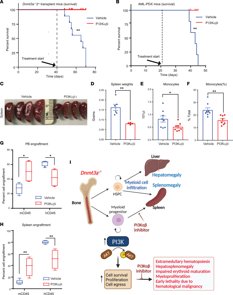
Figure 12 PI3K inhibitor treatment of mice bearing human AML cells bearing DNMT3A mutation enhances their survival. (A) Secondary recipients of Dnmt3a–/– BM cells were prepared for the survival study by injecting 2 × 10⁶ of Dnmt3a–/– cells from primary BM transplants of Dnmt3a–/– cells. Six weeks after transplantation, these mice were treated with the PI3K inhibitor (Bay1082439) for 21 days (75 mg/kg body weight). As seen in A, all mice belonging to the vehicle group succumbed by day 70 after transplant, whereas none of the drug-treated mice died within this time period. n = 9, log-rank (Mantel-Cox) test, P < 0.003. (B) Patient-derived AML cells (1 million) bearing DNMT3A mutation were transplanted into NSG-cyto mice to generate AML-PDX. Starting day 20 after transplant, these mice were treated with vehicle or PI3K αβ inhibitor for 21 days. As seen in B, all mice belonging to the vehicle group succumbed by day 45 after transplant, whereas none of the drug-treated mice died within this time period. n = 5, log-rank (Mantel-Cox) test, P < 0.001. (C) Images of spleens derived from drug- and vehicle-treated mice. (D) Quantitative analysis of spleen weights from vehicle- and drug-treated AML-PDX mice. n = 5, mean ± SEM, unpaired t test (2-tailed), P < 0.001. (E and F) Quantitative analysis of the number of monocytes in vehicle- versus drug-treated mice. n = 8, mean ± SEM, unpaired t test (2-tailed), *P = 0.05, **P = 0.005. (G and H) Quantitative assessment of percent engraftment of murine and human CD45 cells in the PB and spleen of vehicle- and drug-treated mice as assessed by flow cytometry. n = 5–8, 2-way ANOVA, *P = 0.05, **P = 0.005. The boxes shown with lower and upper quartiles separated by the median (horizontal line), and the whiskers extend to the minimum and maximum values. (I) Model illustrating the role of PI3K signaling in Dnmt3a loss–induced myeloid malignancy. PI3Kαβ specific inhibitor blocks the Dnmt3a loss–induced malignant characteristics and improves survival.
Figure 12 PI3K inhibitor treatment of mice bearing human AML cells bearing DNMT3A mutation enhances their survival. (A) Secondary recipients of Dnmt3a–/– BM cells were prepared for the survival study by injecting 2 × 10⁶ of Dnmt3a–/– cells from primary BM transplants of Dnmt3a–/– cells. Six weeks after transplantation, these mice were treated with the PI3K inhibitor (Bay1082439) for 21 days (75 mg/kg body weight). As seen in A, all mice belonging to the vehicle group succumbed by day 70 after transplant, whereas none of the drug-treated mice died within this time period. n = 9, log-rank (Mantel-Cox) test, P < 0.003. (B) Patient-derived AML cells (1 million) bearing DNMT3A mutation were transplanted into NSG-cyto mice to generate AML-PDX. Starting day 20 after transplant, these mice were treated with vehicle or PI3K αβ inhibitor for 21 days. As seen in B, all mice belonging to the vehicle group succumbed by day 45 after transplant, whereas none of the drug-treated mice died within this time period. n = 5, log-rank (Mantel-Cox) test, P < 0.001. (C) Images of spleens derived from drug- and vehicle-treated mice. (D) Quantitative analysis of spleen weights from vehicle- and drug-treated AML-PDX mice. n = 5, mean ± SEM, unpaired t test (2-tailed), P < 0.001. (E and F) Quantitative analysis of the number of monocytes in vehicle- versus drug-treated mice. n = 8, mean ± SEM, unpaired t test (2-tailed), *P = 0.05, **P = 0.005. (G and H) Quantitative assessment of percent engraftment of murine and human CD45 cells in the PB and spleen of vehicle- and drug-treated mice as assessed by flow cytometry. n = 5–8, 2-way ANOVA, *P = 0.05, **P = 0.005. The boxes shown with lower and upper quartiles separated by the median (horizontal line), and the whiskers extend to the minimum and maximum values. (I) Model illustrating the role of PI3K signaling in Dnmt3a loss–induced myeloid malignancy. PI3Kαβ specific inhibitor blocks the Dnmt3a loss–induced malignant characteristics and improves survival.CC BY · PubMed Central

Verification

Claims the paper makes

82 Own result · 32 Check pending · 7 No full text of the sources · 7 Own interpretation · 2 Not checked yet · 1 Sources not found · 1 Own method

  • It may state two separate claims as one. 37
  • Probably not a substantial claim. 32

Create a free account to see these claims.

Create a free account to see these assumptions.

72 more claims

Create a free account to see all 132 claims and to verify the 50 references still open.

See all claims — free account

Self-check

Percentages, sums and reported differences re-computed from the paper’s own numbers.

Each contradiction between two passages listed here was confirmed by an independent check.

1 of 1

Citations

Cites

Show all 58 references

Cited by

Metadata

Authors
Palam, L. R., Ramdas, B., Pickerell, K., Pasupuleti, S. K., Kanumuri, R., Cesarano, A., Szymanski, M., Selman, B., Dave, U. P., Sandusky, G., Perna, F., Paczesny, S., & Kapur, R.
Affiliations
  • Indiana University Indianapolis (US)
  • Division of Hematology/Oncology, Department of Medicine, Indiana University School of Medicine, Indianapolis, Indiana, USA.
  • Medical University of South Carolina (US)
Corresponding author
Kapur, Reuben
Venue
JCI Insight
Published
2023-05-08
Volume
8
Issue
9
ISSN
2379-3708 (ISSN-L)
OpenAlex
W4361190850
Cited by
13
References
58
Citations by year
2026: 5 · 2025: 5 · 2024: 2 · 2023: 1
FWCI
2.44
Type
journal article
Language
en
Open access
Locations
  • JCI Insight (publishedVersion, cc-by)
  • PubMed Central (publishedVersion, cc-by)
  • PubMed (publishedVersion)
  • IUScholarWorks (Indiana University) (submittedVersion, cc-by)
  • DOAJ (DOAJ: Directory of Open Access Journals) (submittedVersion, cc-by-sa)
Funders
NIH (R01CA173852,R01CA134777,R01HL146137,R01HL140961,R21CA263470,R21CA263239)
Topics
  • Health Sciences › Medicine › Hematology › Acute Myeloid Leukemia Research
  • Health Sciences › Medicine › Hematology › Hematopoietic Stem Cell Transplantation
  • Health Sciences › Medicine › Hematology › Chronic Myeloid Leukemia Treatments
Keywords
DNMT3A, acute myeloid leukemia, hematopoietic stem and progenitor cells, PI3K signaling, myeloproliferation, hematopoietic homeostasis, actin cytoskeleton, extracellular matrix, patient-derived xenografts, DNA methylation
MeSH terms
DNA Methyltransferase 3A, Animals, DNA (Cytosine-5-)-Methyltransferases, Homeostasis, Humans, Leukemia, Myeloid, Acute, Phosphatidylinositol 3-Kinases, Myeloid Cells, Mice

About these checks

Registry, retraction, Cites and statistics results are computed, not judged, with one exception: a printed reference that no index lists is read once by a model before it counts as a citation error. Claims are extracted by a model and kept only when their quote appears verbatim in the text; each cited source is then checked, with a model’s help, for whether it says what the claim relies on. Those verdicts can be wrong — read the claim’s quote and the cited source before relying on one.

A language model drew the claims from the paper on 2026-11-12, under version 3 of the claim definition (model deepseek-v4.1-flash, prompt extract-v3).

A second model labelled each claim on 2026-11-12 (model jev-1.13-20260917, definition revision gold-guideline-v0.2).

Statistics and arithmetic are recomputed, not judged.

Confidence bands are shown only for fields calibrated on our annotated test set. None is calibrated yet, so no band is shown.

What counts as a claim ›How we measure confidence ›How we check this ›

Are you an author, or do you see a problem with this page? Request a review