

The DOI matches its Crossref record; 157 of the 157 references it cites were found in a registry or an index.
The Angular Gyrus: Multiple Functions and Multiple Subdivisions
Open accessAbstract
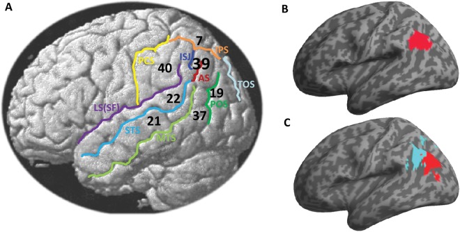
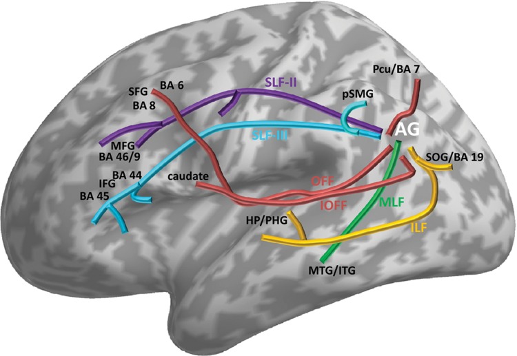
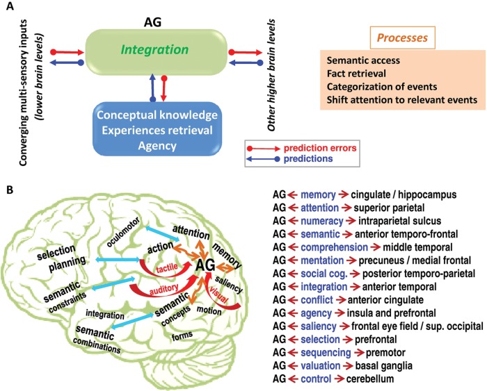
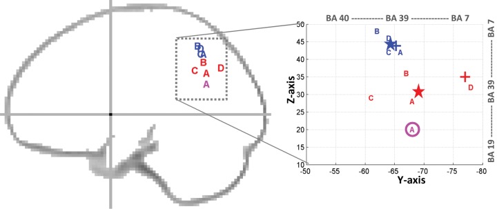
There is considerable interest in the structural and functional properties of the angular gyrus (AG). Located in the posterior part of the inferior parietal lobule, the AG has been shown in numerous meta-analysis reviews to be consistently activated in a variety of tasks. This review discusses the involvement of the AG in semantic processing, word reading and comprehension, number processing, default mode network, memory retrieval, attention and spatial cognition, reasoning, and social cognition. This large functional neuroimaging literature depicts a major role for the AG in processing concepts rather than percepts when interfacing perception-to-recognition-to-action. More specifically, the AG emerges as a cross-modal hub where converging multisensory information is combined and integrated to comprehend and give sense to events, manipulate mental representations, solve familiar problems, and reorient attention to relevant information. In addition, this review discusses recent findings that point to the existence of multiple subdivisions in the AG. This spatial parcellation can serve as a framework for reporting AG activations with greater definition. This review also acknowledges that the role of the AG cannot comprehensibly be identified in isolation but needs to be understood in parallel with the influence from other regions. Several interesting questions that warrant further investigations are finally emphasized.
Overview
The claims and statistics of this paper haven’t been checked yet.
Free: it reads the open-access text and checks the first 5 claims that cite a source we can check. Create a free account to check every claim.
Integrity signals
Registry
Cites
Statistics
Figures
Verification
Claims the paper makes
The claims and statistics of this paper haven’t been checked yet.
Free: it reads the open-access text and checks the first 5 claims that cite a source we can check. Create a free account to check every claim.
Claims other papers make about this one
Each is checked against this paper’s own text: does it say what the citing paper says it does?
In the citing paper
“Furthermore, it was previously thought that these areas were involved in self-referential processes [64–66], perspective taking [67], cognitive distancing [68–71], and sustained attention [72].”
From The meditative mind: a comprehensive meta-analysis of MRI studies. (2015) · cites this paper as [64]
Self-check
Percentages, sums and reported differences re-computed from the paper’s own numbers.
The self-check needs the paper’s full text.
Citations
Cites
A default mode of brain function
Raichle, M. E., MacLeod, A. M., Snyder, A. Z., Powers, W. J., Gusnard, D. A., & Shulman, G. L. (2001) · Proceedings of the National Academy of Sciences · doi:10.1073/pnas.98.2.676 · Cited by 12,190
The Brain's Default Network: Anatomy, Function, and Relevance to Disease
Buckner, R. L., Andrews‐Hanna, J. R., & Schacter, D. L. (2008) · Annals of the New York Academy of Sciences · doi:10.1196/annals.1440.011 · Cited by 9,761
The organization of the human cerebral cortex estimated by intrinsic functional connectivity
Thomas Yeo, B. T., Krienen, F. M., Sepulcre, J., Sabuncu, M. R., Lashkari, D., Hollinshead, M., Roffman, J. L., Smoller, J. W., Zöllei, L., Polimeni, J. R., et al. (2011) · Journal of Neurophysiology · doi:10.1152/jn.00338.2011 · Cited by 9,182
The free-energy principle: a unified brain theory?
Friston, K. (2010) · Nature Reviews Neuroscience · doi:10.1038/nrn2787 · Cited by 8,120 · cited 1×
“This framework is borrowed from the popular predictive coding framework (as reviewed in Friston 2010) that models the brain as a hierarchical inference engine that is trying to optimize probabilistic representations of what caused its sensory input.”
The organization of the human cerebellum estimated by intrinsic functional connectivity
Buckner, R. L., Krienen, F. M., Castellanos, A., Diaz, J. C., & Yeo, B. T. T. (2011) · Journal of Neurophysiology · doi:10.1152/jn.00339.2011 · Cited by 7,304
Functional connectivity in the resting brain: a network analysis of the default mode hypothesis
Greicius, M. D., Krasnow, B., Reiss, A. L., & Menon, V. (2003) · Proceedings of the National Academy of Sciences · doi:10.1073/pnas.0135058100 · Cited by 6,358
Dynamic mapping of human cortical development during childhood through early adulthood
Gogtay, N., Giedd, J. N., Lusk, L., Hayashi, K. M., Greenstein, D., Vaituzis, A. C., Nugent, T. F., Herman, D. H., Clasen, L. S., Toga, A. W., et al. (2004) · Proceedings of the National Academy of Sciences · doi:10.1073/pnas.0402680101 · Cited by 5,604 · cited 1×
“Its cortical volume is estimated at around 13.2 cm3 and 11.7 cm3 in the left and right hemisphere, respectively (Rademacher and others 1992), suggesting a relative leftgreater-than-right structural asymmetry (see also Eidelberg and Galaburda 1984).”
BackgroundInfluentialCorrespondence of the brain's functional architecture during activation and rest
Smith, S. M., Fox, P. T., Miller, K. L., Glahn, D. C., Fox, P. M., Mackay, C. E., Filippini, N., Watkins, K. E., Toro, R., Laird, A. R., et al. (2009) · Proceedings of the National Academy of Sciences · doi:10.1073/pnas.0905267106 · Cited by 5,150
Mapping the structural core of human cerebral cortex
Hagmann, P., Cammoun, L., Gigandet, X., Meuli, R., Honey, C. J., Wedeen, V. J., & Sporns, O. (2008) · PLoS Biology · doi:10.1371/journal.pbio.0060159 · Cited by 4,173
Binder, J. R., Desai, R. H., Graves, W. W., & Conant, L. L. (2009) · Cerebral Cortex · doi:10.1093/cercor/bhp055 · Cited by 4,060
Research works printed but not linked: 4
- Barnes KA, 2011, Cereb Cortex
- Dejerine J, 1891, Societe de Biologie, 43, 197
- Kelly C, 2010, Eur J Neurol, 32, 383
- Mar RA, 2011, Annu Rev Neurosci, 62, 103
Cited by
Andrews‐Hanna, J. R., Smallwood, J., & Spreng, R. N. (2014) · Annals of the New York Academy of Sciences · doi:10.1111/nyas.12360 · Cited by 2,102
The default mode network in cognition: a topographical perspective
Smallwood, J., Bernhardt, B. C., Leech, R., Bzdok, D., Jefferies, E., & Margulies, D. S. (2021) · Nature Reviews Neuroscience · doi:10.1038/s41583-021-00474-4 · Cited by 935
Guell, X., Gabrieli, J. D. E., & Schmahmann, J. D. (2018) · NeuroImage · doi:10.1016/j.neuroimage.2018.01.082 · Cited by 460
Origins of the brain networks for advanced mathematics in expert mathematicians
Amalric, M., & Dehaene, S. (2016) · Proceedings of the National Academy of Sciences · doi:10.1073/pnas.1603205113 · Cited by 413
Re-evaluating the role of TPJ in attentional control: Contextual updating?
Geng, J. J., & Vossel, S. (2013) · Neuroscience & Biobehavioral Reviews · doi:10.1016/j.neubiorev.2013.08.010 · Cited by 411
Metadata
- Authors
- Seghier, M. L.
- Affiliations
- Wellcome Centre for Human Neuroimaging (GB)
- Corresponding author
- Seghier, Mohamed L.
- ORCID
- Seghier, Mohamed L.
- Venue
- The Neuroscientist
- Publisher
- SAGE Publications
- Published
- 2012-04-30
- Volume
- 19
- Issue
- 1
- Pages
- 43-61
- ISSN
- 1073-8584 (ISSN-L)
- 1089-4098
- PMC
- PMC4107834
- OpenAlex
- W2134248637
- Cited by
- 1,585
- References
- 157
- Citations by year
- 2026: 95 · 2025: 166 · 2024: 127 · 2023: 127 · 2022: 180 · 2021: 149 · 2020: 131 · 2019: 130 · 2018: 107 · 2017: 115 · 2016: 90 · 2015: 71 · 2014: 53 · 2013: 38 · 2012: 2
- FWCI
- 26.83
- Type
- journal article
- Language
- en
- Open access
- hybrid
- doi.org
- Licence
- cc-by
- journals.sagepub.com/page/policies/text-and-data-mining-license (tdm)
- Locations
- The Neuroscientist (publishedVersion, cc-by)
- PubMed Central (publishedVersion, cc-by)
- PubMed (publishedVersion)
- UCL Discovery (University College London) (submittedVersion)
- Funders
- Wellcome Trust
- Topics
- Life Sciences › Neuroscience › Cognitive Neuroscience › Neurobiology of Language and Bilingualism
- Social Sciences › Psychology › Developmental and Educational Psychology › Reading and Literacy Development
- Social Sciences › Psychology › Developmental and Educational Psychology › Cognitive and developmental aspects of mathematical skills
- Keywords
- angular gyrus, inferior parietal lobule, semantic processing, word reading, number processing, default mode network, memory retrieval, attention, spatial cognition, reasoning, social cognition, cross-modal integration, multisensory integration, parcellation, functional specialization
- MeSH terms
- Brain Mapping, Cognition, Diagnostic Imaging, Humans, Image Processing, Computer-Assisted, Neural Pathways, Parietal Lobe, Reading, Semantics, Meta-Analysis as Topic
About these checks
Registry, retraction, Cites and statistics results are computed, not judged, with one exception: a printed reference that no index lists is read once by a model before it counts as a citation error. Claims are extracted by a model and kept only when their quote appears verbatim in the text; each cited source is then checked, with a model’s help, for whether it says what the claim relies on. Those verdicts can be wrong — read the claim’s quote and the cited source before relying on one.
Statistics and arithmetic are recomputed, not judged.
Confidence bands are shown only for fields calibrated on our annotated test set. None is calibrated yet, so no band is shown.
What counts as a claim ›How we measure confidence ›How we check this ›
Are you an author, or do you see a problem with this page? Request a review
How to cite
Cite CoChat for the checks on this page, and the paper for its own findings.
- This page
- CoChat. (2026). Citation check: The Angular Gyrus: Multiple Functions and Multiple Subdivisions. https://app.cochat.ai/papers/10.1177/1073858412440596
- The paper
- Seghier, M. L. (2013). The Angular Gyrus: Multiple Functions and Multiple Subdivisions. The Neuroscientist. https://doi.org/10.1177/1073858412440596
Data sources
Bibliographic data: Semantic Scholar, OpenAlex, Crossref.
Contains information from the Semantic Scholar Academic Graph Datasets, made available under the ODC Attribution License (ODC-BY 1.0).
Semantic Scholar’s data is described in: Kinney, R., et al. (2023). The Semantic Scholar Open Data Platform. arXiv:2301.10140.
